. 2023 Mar 15;3(1):37.
doi: 10.1038/s43856-023-00264-2.

Evaluation and deployment of isotype-specific salivary antibody assays for detecting previous SARS-CoV-2 infection in children and adults

Collaborators, Affiliations

Evaluation and deployment of isotype-specific salivary antibody assays for detecting previous SARS-CoV-2 infection in children and adults

Amy C Thomas et al. Commun Med (Lond). .

Abstract

Background: Saliva is easily obtainable non-invasively and potentially suitable for detecting both current and previous SARS-CoV-2 infection, but there is limited evidence on the utility of salivary antibody testing for community surveillance.

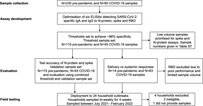
Methods: We established 6 ELISAs detecting IgA and IgG antibodies to whole SARS-CoV-2 spike protein, to its receptor binding domain region and to nucleocapsid protein in saliva. We evaluated diagnostic performance, and using paired saliva and serum samples, correlated mucosal and systemic antibody responses. The best-performing assays were field-tested in 20 household outbreaks.

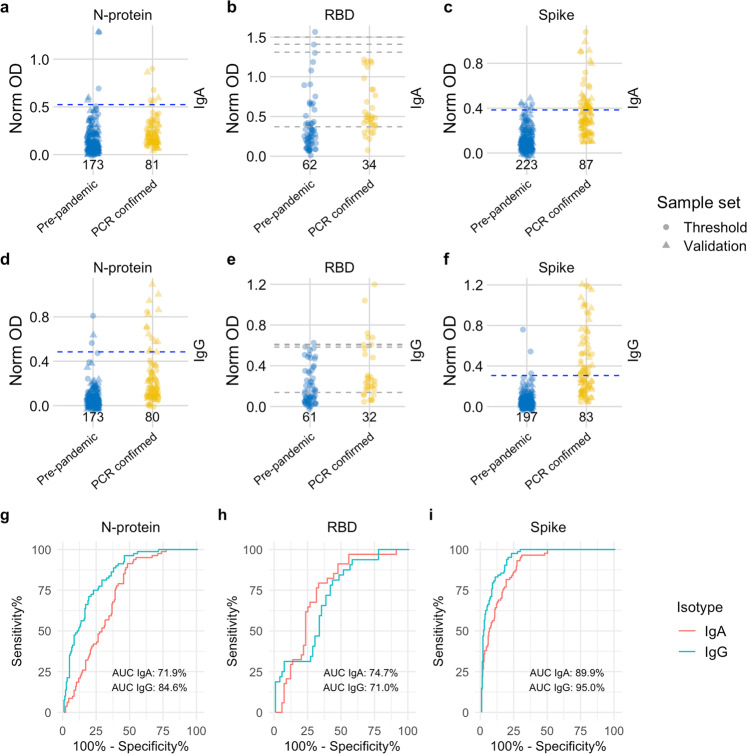
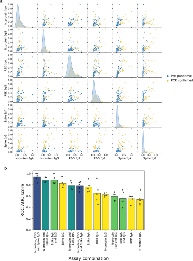
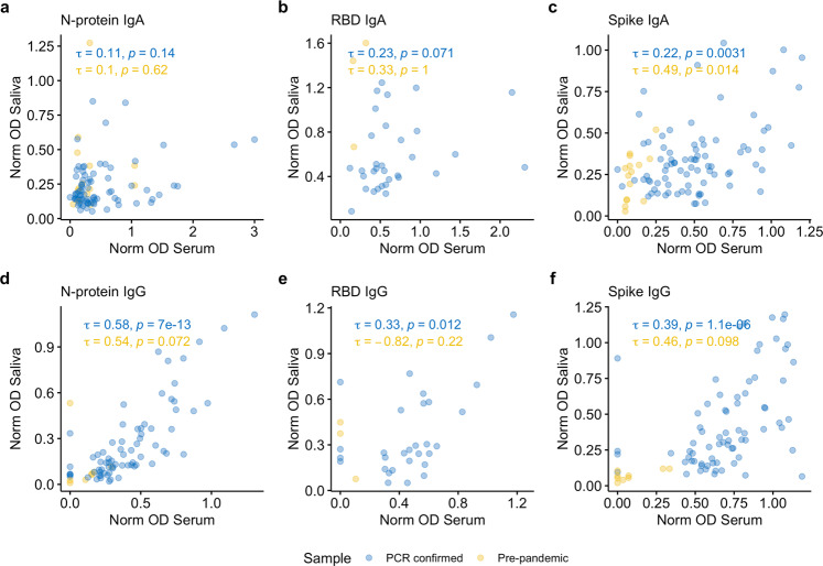
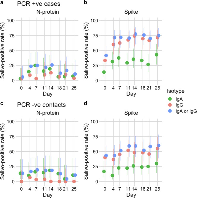
Results: We demonstrate in test accuracy (N = 320), spike IgG (ROC AUC: 95.0%, 92.8-97.3%) and spike IgA (ROC AUC: 89.9%, 86.5-93.2%) assays to discriminate best between pre-pandemic and post COVID-19 saliva samples. Specificity was 100% in younger age groups (0-19 years) for spike IgA and IgG. However, sensitivity was low for the best-performing assay (spike IgG: 50.6%, 39.8-61.4%). Using machine learning, diagnostic performance was improved when a combination of tests was used. As expected, salivary IgA was poorly correlated with serum, indicating an oral mucosal response whereas salivary IgG responses were predictive of those in serum. When deployed to household outbreaks, antibody responses were heterogeneous but remained a reliable indicator of recent infection. Intriguingly, unvaccinated children without confirmed infection showed evidence of exposure almost exclusively through specific IgA responses.

Conclusions: Through robust standardisation, evaluation and field-testing, this work provides a platform for further studies investigating SARS-CoV-2 transmission and mucosal immunity with the potential for expanding salivo-surveillance to other respiratory infections in hard-to-reach settings.

Plain language summary

If a person has been previously infected with SARS-CoV-2 they will produce specific proteins, called antibodies. These are present in the saliva and blood. Saliva is easier to obtain than blood, so we developed and evaluated six tests that detect SARS-CoV-2 antibodies in saliva in children and adults. Some tests detected antibodies to a particular protein made by SARS-CoV-2 called the spike protein, and these tests worked best. The most accurate results were obtained by using a combination of tests. Similar tests could also be developed to detect other respiratory infections which will enable easier identification of infected individuals.


Conflict of interest statement

The authors declare the following competing interests: AF is a member of the Joint Committee on Vaccination and Immunisation, the UK national immunisation technical advisory group and is chair of the WHO European regional technical advisory group of experts (ETAGE) on immunisation and ex officio a member of the WHO SAGE working group on COVID vaccines. He is investigator on studies and trials funded by Pfizer, Sanofi, Valneva, the Gates Foundation and the UK government.

Figures

Fig. 1
Fig. 1. Study flow diagram describing samples and the processes used in immunoassay development, evaluation and field-testing.
Saliva and serum samples were collected pre-SARS-CoV-2 emergence from children and adults (known negatives, N = 230), and from individuals with PCR-confirmed SARS-CoV-2 infection (known positives, N = 90). Known negative and positive samples were used to set thresholds for positivity and evaluate performance. Assays were field tested using samples (N = 510) from 20 household outbreaks.
Fig. 2
Fig. 2. Distribution of semi-quantitative titres for each assay and corresponding ROC curves.
ac Dotplots show the scatter of values for SARS-CoV-2 specific IgA responses against N-protein (N = 254), RBD (N = 96) and spike (N = 310), specific IgG responses against N-protein (N = 253), RBD (N = 93) and spike (N = 280) are shown in (df). Data are presented for known negative and known positive samples, the number of samples tested in each group is also shown under the corresponding data points. For N-protein and spike, proposed thresholds (97th–99th percentile and Youden’s index) are shown as dashed lines, with the final selected threshold in blue; thresholds were derived in the threshold setting phase (threshold samples shown as circles, validation samples shown as triangles). RBD was not taken forward to full evaluation, so performance in the threshold set and corresponding proposed thresholds are shown as dashed grey lines. g, i ROC curves for N-protein and spike represent assay performance determined on all threshold and validation set samples combined. h The ROC curve for RBD represents assay performance in threshold sample set. ROC = receiver operating characteristic curve. AUC = area under the curve. Norm OD = Normalised OD (a relative ratio to the serum standard). N-protein = Nucleocapsid protein. RBD = Receptor binding domain.
Fig. 3
Fig. 3. Combining assays improves discriminatory performance.
a Pairwise scatter plots and kernel density estimates of antibody responses for N = 229 pre-pandemic (blue) and N = 89 SARS-CoV-2 PCR-confirmed (orange) samples assayed at a single dilution in each of the 6 assays: spike, N-protein, RBD IgA (1 in 10) and IgG (1 in 5). The kernel density estimates along the diagonal represent the distribution of responses measured for a single assay, whilst the scatter plots compare the responses measured across two different assays. b Comparison of the performance (measured via ROC AUC score) of AdaBoost models trained either with one of the 6 individual assays (yellow bars and dots), or with a selected combination of those assays (green, turquoise, blue bars and dots). The models were trained using 5-fold cross-validation: the dots represent the ROC AUC scores measured for the individual folds, whilst the bars represent the mean of these 5 scores. ROC AUC score = area under the receiver operating characteristic curve.
Fig. 4
Fig. 4. Correlation between mucosal and systemic antibody.
af Correlation between serum and salivary IgA and IgG responses to spike protein, nucleocapsid protein (N-protein) and receptor binding domain (RBD) a, d N-protein IgA (N = 91) and IgG (N = 80) assays. b, e RBD IgA (N = 35) and IgG (N = 33), c, f spike IgA (N = 97) and IgG (N = 81). PCR-confirmed samples are shown in blue and pre-pandemic samples are shown in yellow. Correlations (Kendall’s tau) were performed for paired saliva and serum samples collected on the same day.
Fig. 5
Fig. 5. Application of assays to household outbreaks.
a-d Salivo-positive rate for 20 households with SARS-CoV-2 outbreaks (N = 20 index cases and 51 contacts) based on detection of anti-nucleocapsid (N-protein) or anti-spike, IgA (green points), IgG (red points), IgA or IgG (blue points). Salivo-conversion for household members with PCR-confirmed infection (N = 31) during the study is shown in (a, b), household members remaining PCR negative (N = 36) are shown in (c, d). Error bars represent 95% confidence intervals for a single proportion (Wilson method).

References

    1. Amanat F, et al. A serological assay to detect SARS-CoV-2 seroconversion in humans. Nat. Med. 2020;26:1033–1036. doi: 10.1038/s41591-020-0913-5. - DOI - PMC - PubMed
    1. Vusirikala A, et al. Seroprevalence of SARS-CoV-2 antibodies in university students: Cross-sectional study, December 2020, England. J. Infection. 2021;83:104–111. doi: 10.1016/j.jinf.2021.04.028. - DOI - PMC - PubMed
    1. Earle KA, et al. Evidence for antibody as a protective correlate for COVID-19 vaccines. Vaccine. 2021;39:4423–4428. doi: 10.1016/j.vaccine.2021.05.063. - DOI - PMC - PubMed
    1. Khoury DS, et al. Neutralizing antibody levels are highly predictive of immune protection from symptomatic SARS-CoV-2 infection. Nat. Med. 2021;27:1205–1211. doi: 10.1038/s41591-021-01377-8. - DOI - PubMed
    1. Ju B, et al. Human neutralizing antibodies elicited by SARS-CoV-2 infection. Nature. 2020;584:115–119. doi: 10.1038/s41586-020-2380-z. - DOI - PubMed