. 2023 Mar 16;21(1):56.
doi: 10.1186/s12916-023-02759-0.

Exogenous mitochondrial transplantation improves survival and neurological outcomes after resuscitation from cardiac arrest

Affiliations

Exogenous mitochondrial transplantation improves survival and neurological outcomes after resuscitation from cardiac arrest

Kei Hayashida et al. BMC Med. .

Abstract

Background: Mitochondrial transplantation (MTx) is an emerging but poorly understood technology with the potential to mitigate severe ischemia-reperfusion injuries after cardiac arrest (CA). To address critical gaps in the current knowledge, we test the hypothesis that MTx can improve outcomes after CA resuscitation.

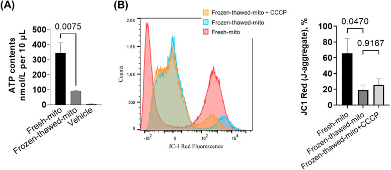
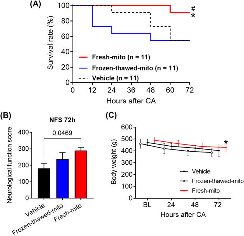
Methods: This study consists of both in vitro and in vivo studies. We initially examined the migration of exogenous mitochondria into primary neural cell culture in vitro. Exogenous mitochondria extracted from the brain and muscle tissues of donor rats and endogenous mitochondria in the neural cells were separately labeled before co-culture. After a period of 24 h following co-culture, mitochondrial transfer was observed using microscopy. In vitro adenosine triphosphate (ATP) contents were assessed between freshly isolated and frozen-thawed mitochondria to compare their effects on survival. Our main study was an in vivo rat model of CA in which rats were subjected to 10 min of asphyxial CA followed by resuscitation. At the time of achieving successful resuscitation, rats were randomly assigned into one of three groups of intravenous injections: vehicle, frozen-thawed, or fresh viable mitochondria. During 72 h post-CA, the therapeutic efficacy of MTx was assessed by comparison of survival rates. The persistence of labeled donor mitochondria within critical organs of recipient animals 24 h post-CA was visualized via microscopy.

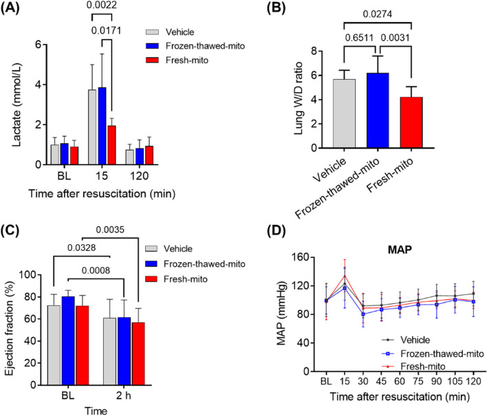
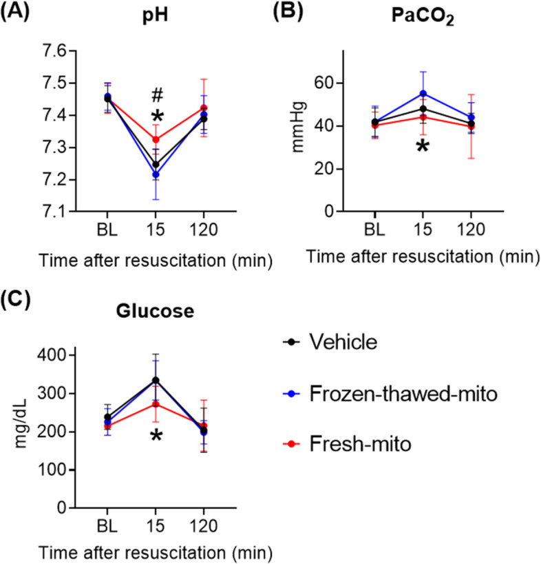
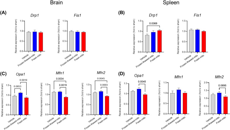
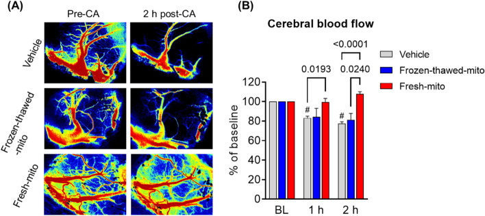
Results: The donated mitochondria were successfully taken up into cultured neural cells. Transferred exogenous mitochondria co-localized with endogenous mitochondria inside neural cells. ATP content in fresh mitochondria was approximately four times higher than in frozen-thawed mitochondria. In the in vivo survival study, freshly isolated functional mitochondria, but not frozen-thawed mitochondria, significantly increased 72-h survival from 55 to 91% (P = 0.048 vs. vehicle). The beneficial effects on survival were associated with improvements in rapid recovery of arterial lactate and glucose levels, cerebral microcirculation, lung edema, and neurological function. Labeled mitochondria were observed inside the vital organs of the surviving rats 24 h post-CA.

Conclusions: MTx performed immediately after resuscitation improved survival and neurological recovery in post-CA rats. These results provide a foundation for future studies to promote the development of MTx as a novel therapeutic strategy to save lives currently lost after CA.

Keywords: Cardiac arrest; Ischemia and reperfusion; Mitochondria; Mitochondrial transplantation.


Conflict of interest statement

The authors declare that they have no competing interests.

Figures

Fig. 1
Fig. 1
Transfer of exogenous brain- and muscle-derived mitochondria into neural cell cultures. Representative images of exogenous mitochondria stained with MitoTracker Deep Red and co-cultured with brain cells, whose endogenous mitochondria were stained with MitoTracker Green. The exogenous mitochondria (red) were extracted from A the brain or B the pectoral muscle of the donor rat. Scale bar indicates 20 µm
Fig. 2
Fig. 2
Freshly isolated mitochondria are more functional than frozen-thawed mitochondria. A Mitochondrial ATP contents in vehicle, frozen-thawed mitochondria, and fresh mitochondria immediately after mitochondrial isolation. One-way analysis of variance (ANOVA) with Šidák’s correction for post hoc comparisons was used. n = 3–4 per group. B Flow cytometry analysis of the mitochondrial membrane potential of the isolated mitochondria stained with JC-1 verified that the percentage of J-aggregates was markedly higher in the fresh-mito group than in the frozen-thawed-mito group. Carbonyl cyanide 3-chlorophenylhydrazone (CCCP) did not alter the ΔψM in the frozen-thawed-mito group. The one-way ANOVA with Šidák’s correction for post hoc comparisons was used. n = 4 per group
Fig. 3
Fig. 3
Mitochondrial transplantation using freshly isolated mitochondria improves 72-h neurological function and survival after cardiac arrest and resuscitation. A Survival rates during the first 72 h after cardiac arrest (CA) and resuscitation. n = 11 per group. *P = 0.048 vs. vehicle group; #P = 0.038 vs. frozen-thawed-mito group. Kaplan–Meier analysis using the Gehan–Breslow–Wilcoxon test was used. B Neurological functional scores (NFS) at 72 h post-CA in surviving animals in the vehicle, frozen-thawed mitochondria, or freshly isolated mitochondria groups. A score of 0 indicates brain death or dead rats. Dead animals were excluded from the analyses. The Kruskal–Wallis test followed by Dunn’s multiple comparison test was used. C Daily changes in body weight in post-CA animals treated with vehicle, frozen-thawed mitochondria, or fresh mitochondria. A mixed-effects model for repeated-measures analyses, followed by one-way ANOVA with Šidák’s correction for post hoc comparisons was used. *P = 0.044 vs. vehicle group. Data represent the mean ± standard deviation
Fig. 4
Fig. 4
Mitochondrial transplantation enhances early lactate normalization and mitigates lung injury after cardiac arrest and resuscitation. A Arterial lactate levels at pre-arrest baseline and 15- and 120-min post-resuscitation. n = 11 per group. A mixed-effects model for repeated-measures analyses, followed by one-way analysis of variance (ANOVA) with Šidák’s correction for post hoc comparisons, was used. B Cardiac arrest-induced lung edema at 72 h post-resuscitation was diminished by delivering freshly isolated mitochondria. C Left ventricular ejection fraction at pre-arrest baseline and 2 h post-resuscitation in post-arrest rats treated with vehicle, frozen-thawed mitochondria (frozen-thawed-mito), or freshly isolated mitochondria (fresh-mito). A mixed-effects model for repeated-measures analysis, followed by ANOVA with Šidák’s correction for post hoc comparisons, was employed. D Changes in the mean arterial pressure (MAP). A mixed-effects model for repeated-measures analysis, followed by ANOVA with Šidák’s correction for post hoc comparisons, was used. Data represent the mean ± standard deviation
Fig. 5
Fig. 5
Mitochondrial transplantation normalizes metabolic parameters early after cardiac arrest and resuscitation. Arterial A pH, B partial pressure of carbon dioxide (PaCO2), and C glucose levels at pre-CA baseline and at 15 min and 2 h after resuscitation among groups. *P < 0.05; fresh-mito vs. vehicle group, #P < 0.05; fresh-mito vs. frozen-thawed-mito group. n = 11 per group. A mixed-effects model for repeated-measures analysis, followed by ANOVA with Šidák’s correction for post hoc comparisons, was used. Data represent the mean ± standard deviation
Fig. 6
Fig. 6
Gene expression changes in the brain and spleen in survived rats at 72 h after cardiac arrest and resuscitation with or without mitochondrial transplantation. Gene expression of molecules related with mitochondrial fission proteins (dynamin-related protein 1 [Drp1], mitochondrial fission 1 protein [Fis1]) in the A brain and B spleen and mitochondrial fusion proteins (optic atrophy-1 [Opa1], Mitofusin-1 [Mfn-1], Mitofusin-2 [Mfn-2]) in the C brain and D spleen. ANOVA with Sidak’s correction for post hoc comparisons was used. n = 6, 6, and 10 for the vehicle, frozen-thawed-mito, and fresh-mito groups, respectively. Data represent the mean ± standard deviation
Fig. 7
Fig. 7
Mitochondrial transplantation enhances cerebral perfusion early after cardiac arrest and resuscitation. A Representative photographs of laser speckle contrast imaging at pre-CA baseline and at 2 h after CA in three groups. B Changes in the mean relative cerebral blood flow (rCBF) of ROIs in post-CA rats treated with vehicle, frozen-thawed mitochondria (frozen–thawed-mito), or freshly isolated mitochondria (fresh-mito). A mixed-effects model for repeated-measures analysis, followed by ANOVA with Šidák’s correction for post hoc comparisons, was used. n = 6 per group. Data represent the mean ± standard deviation
Fig. 8
Fig. 8
Confocal fluorescence imaging reveals the persistence of the donor mitochondria within the organs at 1 and 24 h after cardiac arrest and resuscitation. Transplanted mitochondria (red) were observed in the brain, kidney, and spleen at 1 and 24 h after cardiac arrest. Arrowheads indicate the donated mitochondrial particles labeled using MitoTracker Deep Red dye before the injection. The cell nuclei were counterstained with DAPI (blue). Scale bar indicates 20 µm

References

    1. Neumar RW, Nolan JP, Adrie C, Aibiki M, Berg RA, Bottiger BW, Callaway C, Clark RS, Geocadin RG, Jauch EC, et al. Post-cardiac arrest syndrome: epidemiology, pathophysiology, treatment, and prognostication. A consensus statement from the International Liaison Committee on Resuscitation (American Heart Association, Australian and New Zealand Council on Resuscitation, European Resuscitation Council, Heart and Stroke Foundation of Canada, InterAmerican Heart Foundation, Resuscitation Council of Asia, and the Resuscitation Council of Southern Africa); the American Heart Association Emergency Cardiovascular Care Committee; the Council on Cardiovascular Surgery and Anesthesia; the Council on Cardiopulmonary, Perioperative, and Critical Care; the Council on Clinical Cardiology; and the Stroke Council. Circulation. 2008;118(23):2452–2483. doi: 10.1161/CIRCULATIONAHA.108.190652. - DOI - PubMed
    1. Gazmuri RJ, Radhakrishnan J. Protecting mitochondrial bioenergetic function during resuscitation from cardiac arrest. Crit Care Clin. 2012;28(2):245–270. doi: 10.1016/j.ccc.2012.02.001. - DOI - PMC - PubMed
    1. Sharp WW. Dynamin-related protein 1 as a therapeutic target in cardiac arrest. J Mol Med (Berl) 2015;93(3):243–252. doi: 10.1007/s00109-015-1257-3. - DOI - PMC - PubMed
    1. Tait SW, Green DR. Mitochondria and cell signalling. J Cell Sci. 2012;125(Pt 4):807–815. doi: 10.1242/jcs.099234. - DOI - PMC - PubMed
    1. Kaur MM, Sharma DS. Mitochondrial repair as potential pharmacological target in cerebral ischemia. Mitochondrion. 2022;63:23–31. doi: 10.1016/j.mito.2022.01.001. - DOI - PubMed

Publication types

Substances