. 2023 Mar 15;24(1):80.
doi: 10.1186/s12931-023-02380-y.

Time-resolved transcriptomic profiling of the developing rabbit's lungs: impact of premature birth and implications for modelling bronchopulmonary dysplasia

Affiliations

Time-resolved transcriptomic profiling of the developing rabbit's lungs: impact of premature birth and implications for modelling bronchopulmonary dysplasia

Matteo Storti et al. Respir Res. .

Abstract

Background: Premature birth, perinatal inflammation, and life-saving therapies such as postnatal oxygen and mechanical ventilation are strongly associated with the development of bronchopulmonary dysplasia (BPD); these risk factors, alone or combined, cause lung inflammation and alter programmed molecular patterns of normal lung development. The current knowledge on the molecular regulation of lung development mainly derives from mechanistic studies conducted in newborn rodents exposed to postnatal hyperoxia, which have been proven useful but have some limitations.

Methods: Here, we used the rabbit model of BPD as a cost-effective alternative model that mirrors human lung development and, in addition, enables investigating the impact of premature birth per se on the pathophysiology of BPD without further perinatal insults (e.g., hyperoxia, LPS-induced inflammation). First, we characterized the rabbit's normal lung development along the distinct stages (i.e., pseudoglandular, canalicular, saccular, and alveolar phases) using histological, transcriptomic and proteomic analyses. Then, the impact of premature birth was investigated, comparing the sequential transcriptomic profiles of preterm rabbits obtained at different time intervals during their first week of postnatal life with those from age-matched term pups.

Results: Histological findings showed stage-specific morphological features of the developing rabbit's lung and validated the selected time intervals for the transcriptomic profiling. Cell cycle and embryo development, oxidative phosphorylation, and WNT signaling, among others, showed high gene expression in the pseudoglandular phase. Autophagy, epithelial morphogenesis, response to transforming growth factor β, angiogenesis, epithelium/endothelial cells development, and epithelium/endothelial cells migration pathways appeared upregulated from the 28th day of gestation (early saccular phase), which represents the starting point of the premature rabbit model. Premature birth caused a significant dysregulation of the inflammatory response. TNF-responsive, NF-κB regulated genes were significantly upregulated at premature delivery and triggered downstream inflammatory pathways such as leukocyte activation and cytokine signaling, which persisted upregulated during the first week of life. Preterm birth also dysregulated relevant pathways for normal lung development, such as blood vessel morphogenesis and epithelial-mesenchymal transition.

Conclusion: These findings establish the 28-day gestation premature rabbit as a suitable model for mechanistic and pharmacological studies in the context of BPD.

Keywords: Bronchopulmonary dysplasia; Lung development; Premature birth; Proteomics; Transcriptomics.


Conflict of interest statement

MS, MLF, CC, GV, BP, FS and FRC are employees of Chiesi Farmaceutici S.p.A.; XM served as a consultant for this study.

Figures

Fig. 1
Fig. 1
Scheme of the experimental design. Samples were collected at different lung developmental phases (i.e., pseudoglandular, canalicular, saccular and alveolar phases). Fetal (F) rabbit pups were extracted via C-section on the 20th, 23rd, 25th, 27th, 28th and 29th day of gestation (F20–F29); lung samples were harvested immediately after delivery, trying to avoid that pups took their first breath. Term (T) pups were naturally delivered on the 31st day of gestation and maintained with their mothers in room air until lung collection: immediately after birth (T31), and 4 (T35), 6 (T37), and 11 days (T42) after natural birth. Preterm (P) rabbits were delivered through C-section on the 28th day of gestation and maintained in room air until lung collection: immediately after birth (P0), and 3 (P3) and 7 (P7) days after preterm delivery
Fig. 2
Fig. 2
Histological characterization and histomorphometry data of the rabbit’s normal lung development. Representative microphotographs of the lung parenchyma during the pseudoglandular (AC), canalicular (DF), saccular (GI) and alveolar phases (JO) of lung development. The first two columns from the left, show sections stained with hematoxylin and eosin obtained at each lung developmental stage. In the right column, Masson’s trichrome staining (C) shows the scarce presence of intercellular matrix during the pseudoglandular phase, and orcein staining (F, I, L, O) shows fine elastic fibers (dotted arrows). The evolution of tissue density (TD), radial alveolar count (RAC) and the medial thickness of pre- and intra-acinar arteries (MT%) are shown in P, Q and R (as mean ± standard error of the mean of at least six lungs per phase of lung development. Asterisks above horizontal lines indicate a significant difference in the comparisons between different lung developmental phases (ANOVA followed by Tukey’s test; *P < 0.05; **P < 0.01; ***P < 0.001)
Fig. 3
Fig. 3
A Principal component analysis (PCA) highlighting specific clustering of lung samples belonging to the same time-point. The last number in sample names indicates in which RNA-sequencing run the sample was sequenced. B Histogram representation of differentially expressed genes (DEG) identified by comparative transcriptomic profiling of different developmental phases (P = pseudoglandular, C = canalicular, S = saccular, and A = alveolar phases). Up-regulated (UP) and down-regulated (DN) genes are shown in red and blue, respectively
Fig. 4
Fig. 4
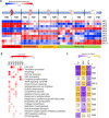
Developmental phase-dependent gene expression analysis. A Module eigengene (ME) heatmap representation of gene expression data derived from lung samples collected at the indicated (from fetal to post-natal) developmental time-points. Median-normalized expression levels are shown on a low-to-high scale (blue–white–red). B Results of pathway enrichment analysis performed on distinct modules of co-expressed genes. Representative terms, among the most significantly enriched in one or more modules, are reported (modules 8, 9 and 10 are not shown as they are enriched only in marginally statistically significant pathways; the full list of terms and modules is available in Additional file 1). Color saturation corresponds to enrichment significance (− Log q-values). C: Correlation analysis between MEs and histomorphometry parameters (negative correlations are shown in purple and positive correlations in yellow, − Log p-values are indicated in parenthesis)
Fig. 5
Fig. 5
Comparison of the expression at mRNA and protein levels for selected genes of interest. Z-score-normalized expression levels are indicated on a low-to-high scale (blue–white–red)
Fig. 6
Fig. 6
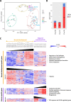
The impact of premature birth on the molecular pathways of lung development. A Principal component analysis (PCA) shows preterm pups clustering closely to, yet well separated from, term pups of the same gestational age. Samples were sequenced in three different RNA-sequencing runs and batch-effect correction was applied. B Number of differentially expressed genes between preterm rabbit pups and age-matched term pups at different time points. Upregulated and downregulated genes are shown in red and blue, respectively. C Heatmap representing expression levels during normal development or after premature birth. Gene set 1 and gene set 2 refer to genes and pathways respectively upregulated and downregulated in term pups but not modulated in preterm pups; gene set 3 and gene set 4 refer to genes and pathways respectively upregulated and downregulated in preterm pups but not modulated in term pups. The last number in sample names indicates in which RNA-sequencing run the sample was sequenced. Z-score-normalized expression level is indicated on a low-to-high scale (blue–white–red). Main pathways enriched in each gene set are indicated on the right
Fig. 7
Fig. 7
Developmental gene expression comparison between rabbits and mice. A Comparison between rabbit and mouse physiological lung development. Rabbits are born at term (T31) in the alveolar phase, whereas mice are born at term (PND0) in the saccular phase. Mice enter the alveolar phase only 4–5 days after birth. B Up-regulated pathways in rabbits (blue line) and mice (grey line) at preterm birth (F28 and E18.5 time points, respectively). C Up-regulated pathways in rabbits (blue line) and mice (grey line) at term birth (T31 and PND0 time points, respectively). The identification of processes enrichment for each comparison was performed using the Metascape software. Only processes with q-values ≤ 10–4 were considered significantly enriched

References

    1. Baker CD, Alvira CM. Disrupted lung development and bronchopulmonary dysplasia: opportunities for lung repair and regeneration. Curr Opin Pediatr. 2014;26:306–314. doi: 10.1097/MOP.0000000000000095. - DOI - PMC - PubMed
    1. Day CL, Ryan RM. Bronchopulmonary dysplasia: new becomes old again! Pediatr Res. 2017;81:210–213. doi: 10.1038/pr.2016.201. - DOI - PubMed
    1. Bancalari E. Molecular bases for lung development, injury, and repair. In: Polin RA, editor. Newborn lung neonatol quest controv. Saunders; 2012. pp. 3–27.
    1. Sweet DG, Carnielli V, Greisen G, Hallman M, Ozek E, Te Pas A, et al. European consensus guidelines on the management of respiratory distress syndrome—2019 update. Neonatology. 2019;2019:432–450. doi: 10.1159/000499361. - DOI - PMC - PubMed
    1. Thébaud B, Goss KN, Laughon M, Whitsett JA, Abman SH, Steinhorn RH, et al. Bronchopulmonary dysplasia. Nat Rev Dis Prim. 2019;5:78. doi: 10.1038/s41572-019-0127-7. - DOI - PMC - PubMed