

Kala-Azar: A Case Report
- PMID: 36923201
- PMCID: PMC10010315
- DOI: 10.7759/cureus.34864
Kala-Azar: A Case Report
Abstract
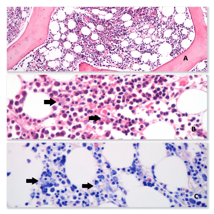
Leishmaniasis is a zoonosis caused by unicellular protozoans Leishmania. The transmission can be zoonotic or anthroponotic, depending on the species, and the main vector is the phlebotomine sandfly. The disease is endemic in the tropics of Asia and Africa but is considered rare in Portugal, especially in immunocompetent hosts. Its main clinical syndromes constitute cutaneous leishmaniasis, mucocutaneous disease, and visceral leishmaniasis. The latter is also known as kala-azar and is caused by the infection of the phagocytes of the reticuloendothelial system, causing the typical symptoms: fever, hepatosplenomegaly, and pancytopenia. The clinical manifestations are non-specific, frequently causing a delay in the diagnosis, especially in nonendemic areas and immunocompetent hosts. Early diagnosis and treatment are essential, given the high mortality rate in untreated patients. The diagnosis is based on the direct visualization of the protozoan and molecular methods, such as polymerase chain reaction tests. Amphotericin B is considered the first-line treatment. We present a case of visceral leishmaniasis in an immunocompetent patient with fever, hepatosplenomegaly, and pancytopenia.
Keywords: high fever; immunocompetent adult; unintentional weight loss; visceral leishmaniasis (vl); “pancytopenia”.
Copyright © 2023, Jancar et al.
Conflict of interest statement
The authors have declared that no competing interests exist.
Figures

References
-
- Diagnosis and treatment of leishmaniasis: Clinical practice guidelines by the Infectious Diseases Society of America (IDSA) and the American Society of Tropical Medicine and Hygiene (ASTMH) Aronson N, Herwaldt BL, Libman M, et al. Clin Infect Dis. 2016;63:1539–1557. - PubMed
-
- Visceral leishmaniasis: what are the needs for diagnosis, treatment and control? Chappuis F, Sundar S, Hailu A, et al. Nat Rev Microbiol. 2007;5:873–882. - PubMed
-
- Holiday souvenirs from the Mediterranean: three instructive cases of visceral leishmaniasis. Buonomano R, Brinkmann F, Leupin N, Boscacci R, Zimmermann A, Müller N, Fux CA. Scand J Infect Dis. 2009;41:777–781. - PubMed
Publication types
LinkOut - more resources
Full Text Sources