Review
. 2023 Feb 27:14:1094845.
doi: 10.3389/fphys.2023.1094845. eCollection 2023.

SHock-INduced Endotheliopathy (SHINE): A mechanistic justification for viscoelastography-guided resuscitation of traumatic and non-traumatic shock

Affiliations
Review

SHock-INduced Endotheliopathy (SHINE): A mechanistic justification for viscoelastography-guided resuscitation of traumatic and non-traumatic shock

Connor M Bunch et al. Front Physiol. .

Abstract

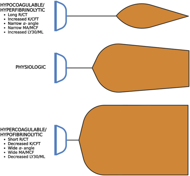
Irrespective of the reason for hypoperfusion, hypocoagulable and/or hyperfibrinolytic hemostatic aberrancies afflict up to one-quarter of critically ill patients in shock. Intensivists and traumatologists have embraced the concept of SHock-INduced Endotheliopathy (SHINE) as a foundational derangement in progressive shock wherein sympatho-adrenal activation may cause systemic endothelial injury. The pro-thrombotic endothelium lends to micro-thrombosis, enacting a cycle of worsening perfusion and increasing catecholamines, endothelial injury, de-endothelialization, and multiple organ failure. The hypocoagulable/hyperfibrinolytic hemostatic phenotype is thought to be driven by endothelial release of anti-thrombogenic mediators to the bloodstream and perivascular sympathetic nerve release of tissue plasminogen activator directly into the microvasculature. In the shock state, this hemostatic phenotype may be a counterbalancing, yet maladaptive, attempt to restore blood flow against a systemically pro-thrombotic endothelium and increased blood viscosity. We therefore review endothelial physiology with emphasis on glycocalyx function, unique biomarkers, and coagulofibrinolytic mediators, setting the stage for understanding the pathophysiology and hemostatic phenotypes of SHINE in various etiologies of shock. We propose that the hyperfibrinolytic phenotype is exemplified in progressive shock whether related to trauma-induced coagulopathy, sepsis-induced coagulopathy, or post-cardiac arrest syndrome-associated coagulopathy. Regardless of the initial insult, SHINE appears to be a catecholamine-driven entity which early in the disease course may manifest as hyper- or hypocoagulopathic and hyper- or hypofibrinolytic hemostatic imbalance. Moreover, these hemostatic derangements may rapidly evolve along the thrombohemorrhagic spectrum depending on the etiology, timing, and methods of resuscitation. Given the intricate hemochemical makeup and changes during these shock states, macroscopic whole blood tests of coagulative kinetics and clot strength serve as clinically useful and simple means for hemostasis phenotyping. We suggest that viscoelastic hemostatic assays such as thromboelastography (TEG) and rotational thromboelastometry (ROTEM) are currently the most applicable clinical tools for assaying global hemostatic function-including fibrinolysis-to enable dynamic resuscitation with blood products and hemostatic adjuncts for those patients with thrombotic and/or hemorrhagic complications in shock states.

Keywords: critical care; endothelium; glycocalyx; hemostasis; precision medicine; resuscitation; shock; thromboelastography.


Conflict of interest statement

EM and HM have received research grants from Haemonetics Corporation outside the submitted work. Author MrW is employed by the company Cardinal Flow Assurance LLC. The remaining authors declare that the research was conducted in the absence of any commercial or financial relationships that could be construed as a potential conflict of interest. The reviewer, AD, declared a shared parent affiliation with the author MM to the handling editor at the time of review.

Figures

FIGURE 1
FIGURE 1
Anatomic and Pathophysiologic Categorization of Shock with Relative Frequency of Each Type. The traditional classification of shock includes four main categories: distributive, hypovolemic, obstructive, and cardiogenic shock. The estimated relative frequency of each of the four subcategories are listed with pathophysiologic etiologies defined around the periphery of the diagram (Vincent and De Backer, 2013; Standl et al., 2018). Recreated with permission from (Standl et al., 2018). Created with BioRender.com.
FIGURE 2
FIGURE 2
Physiologic Roles of the Endothelial Glycocalyx: An Anti-thrombogenic and Anti-adhesive Surface with Rapid Stress-sensing Capability. The glycocalyx is comprised of heparan sulfate proteoglycans (HSPGs) and glycosaminoglycans (GAGs). The HSPGs include the four transmembrane syndecans (Syn1-Syn4) and glypican. The primary GAGs include heparan sulfate (HS) and hyaluronan (HA). HS accounts for ∼50% of the GAG composition in the glycocalyx and covalently bonds to the Syn family and glypican. Other GAGs not pictured include keratan sulfate, dermatan sulfate, and chondroitin sulfate. (A) The negatively charged, hydrophilic moieties of HS and HA have variable cleavage lengths and post-translational modifications which confer significant degrees of specificity for binding cytokines and chemokines. This moderates oncotic and hydrostatic contributions of Starling Forces. Additionally, this creates a gradient of growth factors and signaling molecules which can be indirectly altered by constitutive or stress-induced transient changes in HSPG and GAG composition either by increased synthesis or enzymatic cleavage. (B) The endothelial glycocalyx maintains a gradient of endothelial-synthesized anticoagulant coagulofibrinolytic mediators. In addition to HS, antithrombin III (AT) and tissue factor pathway inhibitor (TFPI) are constitutively expressed in the endothelial glycocalyx. AT complexes with HS to inactivate many coagulation factors, primarily thrombin and Factor X. The anticoagulant TFPI complexes with and inactivates tissue factor (TF)-FVII complex and prothrombinase complex (FVa-FXa). The endothelium also constitutively expresses membrane-bound receptor thrombomodulin (TM), which in the presence of its ligand thrombin, activates circulating plasma protein C. Activated protein C inactivates circulating Factors V and VIII and also inhibits the anti-fibrinolytic, plasminogen activator inhibitor-1 (PAI-1). Weibel-Palade bodies (WPB) also play a significant role in coagulofibrinolytic balance, particularly for the activated endothelium (e.g., by endothelial agonists such as circulating plasma epinephrine). Seminal hemostatic mediators in WPB include tissue plasminogen activator (tPA), von Willebrand Factor (VWF), and P-selectin (P-Sel). (C) The glycocalyx serves as a physical barrier to leukocyte and platelet adhesion in the event adhesion molecule expression is induced on the endothelial luminal surface (e.g., P-selectin, ICAM-1, VCAM-1). (D) Syn4 via intracellular syntenin and synectin indirectly regulates angiopoietin-2 (Agpt-2) activity. High Agpt-2 levels antagonize the tyrosine kinase receptor Tie2, which subsequently destabilizes endothelial cell-cell junctions. Agpt-2 may also be found in WPBs. (E) Syn1 and Syn4 transcellularly signal luminal shear stress to rearrange the endothelial cytoskeleton. (F) Shear stress signaling by the intracellular domain of Syn4 also induces vasodilation via activation of endothelial nitric oxide synthase (eNOS). Shear stress also increases nuclear expression of genes such as those implicated in inflammation. For example, increased shear stress has shown to induce VCAM-1 and ICAM-1 expression. (Lindahl et al., 1998; Kolářová et al., 2014; Rayahin et al., 2015; Leligdowicz et al., 2018; Richter and Richter, 2019; Woodcock and Michel, 2021; Neubauer and Zieger, 2022). Abbreviations: Agpt-2, Angiopoietin-2; AT, Antithrombin III; eNOS, Endothelial Nitric Oxide Synthase; HA, Hyaluronan; Syn1, Syn4, Syndecan; TFPI, Tissue Factor Pathway Inhibitor; Tie2, tyrosine kinase receptor Tie2; TM, Thrombomodulin. Created with BioRender.com.
FIGURE 3
FIGURE 3
SHock-INduced Endotheliopathy (SHINE) as a Reflection of Injury Severity. Increasing sympatho-adrenal activation with increasing injury and shock severity leads to endothelial activation and damage. Increased sympathetic outflow directly provokes SHINE via perivascular sympathetic nerve exocytosis of neurotransmitter catecholamines and enzymatically active tissue plasminogen activator (tPA) into the vessel walls and directly into the microvasculature (O'Rourke et al., 2005; Kwaan, 2014). Hypothalamic-pituitary-adrenal axis activity also increases circulating plasma catecholamines. The corresponding endothelial and hemostatic changes are dose-dependent to injury/shock severity, as measured by endothelial biomarkers (e.g., plasma syndecan-1 and soluble thrombomodulin) and on thromboelastography (TEG) and rotational thromboelastometry (ROTEM) tracings. For example, with trauma, TEG/ROTEM tracings progress from physiologic hemostasis to hypercoagulable in mild trauma, to hypocoagulable in moderate trauma, and finally hyperfibrinolytic in severe trauma (Johansson and Ostrowski, 2010). Genetically preserved responses to critically ill patients inflicted by trauma, burns, and sepsis are similar, suggesting early responses to shock are evolutionarily preserved wherein SHINE may be a unifying mechanism (Xiao et al., 2011; Johansson et al., 2017a). The catecholaminergic surge (in particular the vasoconstrictive action of norepinephrine) causes glycocalyx shedding, endothelial injury, and de-endothelialization of perfused vessels (Dolgov et al., 1984; Makhmudov et al., 1985; Kristová et al., 1993; Vischer and Wollheim, 1997). The activated/injured endothelium promotes thrombosis, causing occlusion of the microvasculature. Together with capillary leak, perivascular edema, and vasoconstriction, these vascular responses provoke a cycle of progressive tissue hypoperfusion, hypovolemia, organ injury, and increasing sympatho-adrenal activation (Opal and Van Der Poll, 2015; Johansson et al., 2017a). It has been hypothesized that the ensuing hypocoagulability and hyperfibrinolysis may be a compensatory counterbalance to the pro-thrombotic endothelium in an attempt to maintain patency of the microvasculature (Johansson and Ostrowski, 2010). Therefore, the two major hemostatic compartments—the endothelium and the blood—may “switch” phenotypes in some progressing shock states. Whereby the physiologic endothelium acts as anti-thrombogenic surface to oppose coagulable blood, in shock, the roles may switch to a pro-thrombotic endothelium with a hypocoagulable/hyperfibrinolytic blood phenotype in attempt to rebalance hemostasis, decrease the blood viscosity, and restore perfusion (Johansson and Ostrowski, 2010) (see Figure 4). Not only does tPA exert pro-fibrinolytic activity via enzymatic activation of plasminogen, but tPA in the brain uniquely acts as a signaling agonist on the N-Methyl-D-Aspartate (NMDA) receptor on the endothelial luminal surface of small cerebrovascular arterioles. The activated NMDA receptor increases synthesis of nitric oxide to cause vasodilation and increase cerebral blood flow (Su et al., 2009; Haile et al., 2012; Yepes, 2015). Thus, increased free tPA (that is, free from complexes with PAI-1 and other inhibitors) in shock states may simultaneously increase systemic perfusion via fibrinolysis of occlusive thrombi and as a neurovascular coupling agent to increase cerebral blood flow (Su et al., 2009; Yepes, 2015). Created with BioRender.com.
FIGURE 4
FIGURE 4
Shock-INduced Endotheliopathy (SHINE) “Phenotype Switching” via Release of Anti-thrombogenic Mediators from the Endothelium to the Bloodstream. One possible contributor of the hypocoagulable/hyperfibrinolytic phenotype in progressive shock may be the release of physiologically endothelial-sequestered anti-thrombogenic mediators to the bloodstream during SHINE when the endothelium is systemically activated and/or injured. Note that protein C is physiologically a plasma protein, but increases in soluble thrombomodulin (sTM) may increase the conversion of protein C to activated protein C (aPC). (Johansson and Ostrowski, 2010). Abbreviations: aPC, activated Protein C; ROTEM, Rotational Thromboelastometry; sTM, soluble Thrombomodulin; TEG, Thromboelastography; tPA, tissue Plasminogen Activator. Created with BioRender.com.
FIGURE 5
FIGURE 5
SHock-INduced Endotheliopathy (SHINE) as a Unifying Mechanism for Coagulopathies Associated with Critical Illness. In this review, we contextualize SHINE as defined by the viscoelastic hemostatic assays thromboelastography (TEG) and rotational thromboelastometry (ROTEM) within many causes of shock. Created with BioRender.com.
FIGURE 6
FIGURE 6
Representative Normocoagulable Thromboelastography (TEG) and Rotational Thromboelastometry (ROTEM) Tracing with Their Respective Parameters Defined. TEG and ROTEM parameters are represented by green and purple text, respectively. The time for the clot to reach 2 mm amplitude on the y-axis describes the reaction time (R) for TEG and clotting time (CT) for ROTEM. R and CT correlate to the activated partial thromboplastin time (aPTT) and prothrombin time (PT). The time spanned from 2 to 20 mm amplitude is called the kinetics (K) for TEG and the clot formation time (CFT) for ROTEM; these represent the speed of fibrin buildup. Likewise, alpha-angle measures the rate of fibrin buildup. The maximum amplitude (MA) on TEG and the maximum clot firmness (MCF) on ROTEM reflect crosslinking of fibrin with platelets and correspond to maximum clot retraction strength. Measurements of fibrinolysis include lysis at 30/60 min (LY30/60) which is the percentage decrease from MA achieved at 30/60 min, clot lysis index at 30/60 min (CLI30/60) which is the percentage of clot amplitude remaining relative to the MCF at 30/60 min, and maximum lysis (ML) which is the percentage decrease in MCF at a given length of time (Görlinger et al., 2021; Hartmann and Sikorski, 2021; Volod et al., 2022). Created with BioRender.com.
FIGURE 7
FIGURE 7
Shovel Analogy to Rapidly Interpret TEG/ROTEM Tracings. The top shovel represents the hypocoagulable state marked by a prolonged R/CT, narrow α-angle, narrow MA/MCF, and increased lysis with resultant increased LY30/ML. The middle shovel represents physiologic hemostasis marked by normal R/CT, α-angle, MA/MCF, and LY30/ML. Mild narrowing after the MA demonstrates physiologic fibrinolysis. The bottom shovel represents the hypercoagulable state denoted by decreased R/CT, wide α-angle, wide MA/MCF, and decreased LY30/ML. Abbreviations: R, Reaction time; CT, Clotting Time; K, Kinetics; CFT, Clot Formation Time; MA, Maximum Amplitude; MCF, Maximum Clot Firmness; LY30/60, Lysis at 30/60 min; ML, Maximum Lysis. Created with BioRender.com.
FIGURE 8
FIGURE 8
The Spectrum of Trauma-Induced Coagulopathy (TIC) as a Function of the Thrombomodulin-Thrombin Complex and SHINE. Hypercoagulability presents most commonly at index trauma presentation according to thromboelastography (TEG) and rotational thromboelastometry (ROTEM) tracings (Johansson and Ostrowski, 2010). As injury severity and the magnitude of hemorrhagic shock increase, the likelihood of hypocoagulability and/or hyperfibrinolysis increases in tandem (Johansson and Ostrowski, 2010; Moore et al., 2016a). Other anti-hemostatic factors at index may include acidosis, hypothermia, crystalloid resuscitation resulting in dilutional coagulopathy, pre-trauma anticoagulant or antiplatelet medications, and co-morbidities (Moore et al., 2021a). After successful initial resuscitation, patients most often demonstrate hypercoagulability and venous thromboembolism in the ensuing days. On the other hand, persistent fibrinolytic shutdown at 24 h post-injury correlates greatest to the magnitude of tissue injury. (A) The thrombomodulin (TM)-thrombin complex is one proposed hypothesis to explain TIC hemostatic phenotypes (Walsh et al., 2019). (B) In its anticoagulant role, the endothelial membrane-bound TM binds with thrombin to convert protein C to activated protein C (aPC). TM-thrombin action on protein C may also be accelerated by endothelial protein C receptor (EPCR, not shown). APC inactivates Factor V, Factor VIII, and plasminogen activator inhibitor-1 (PAI-1) to decrease coagulation and promote tissue plasminogen activator (tPA) activity to convert plasminogen to plasmin (Gando et al., 2018). The resulting fibrinolysis leads to hypofibrinogenemia and a hypocoagulable state as demonstrated by viscoelastic markers. APC and fibrinogen levels share an inverse relationship whereby the TM-thrombin complex increases protein C activation with decreasing fibrinogen levels, leading to a greater anticoagulant and pro-fibrinolytic state (Diez et al., 2006). On the contrary, with increased fibrinogen, the TM–thrombin complex is inhibited from activating protein C. As a result of glycocalyx dysfunction, activation of protein C, enhanced fibrinolysis, and low fibrinogen, the maladaptive response caused by consumption of clotting factors and platelets leads to high fibrin/fibrinogen degradation products (FDPs) with an overall anti-hemostatic state (Diez et al., 2006; Dobson et al., 2015). (C) Tissue hypoperfusion and endothelial injury causes shedding of the endogenous HS of the glycocalyx with subsequent “auto-heparinization” (Ostrowski and Johansson, 2012). The sensitivity of TEG/ROTEM to detect auto-heparinization remains questionable (Zipperle et al., 2022a). Disruption of the endothelial glycocalyx may also be measured by increased circulating syndecan-1 (Syn1) and soluble TM (sTM) levels (Johansson et al., 2011b). (D) Traumatic brain injury produces a unique coagulopathy characterized by platelet dysfunction at the arachidonic acid (AA) and adenosine diphosphate (ADP) receptors as defined by TEG with Platelet Mapping. The relatively high concentrations of von Willebrand Factor (vWF) and Tissue Factor (TF) release from injured brain tissue are thought to cause platelet exhaustion (Castellino et al., 2014; Bradbury et al., 2021). However, the pathophysiology of coagulopathy of traumatic brain injury remains an area of active study. (E) The TM-thrombin complex also activates thrombin-activatable fibrinolysis inhibitor (TAFI) which acts to inhibit tPA binding to fibrin (Marar and Boffa, 2016). (F) Minutes to days after traumatic/surgical-related injury, local and/or systemic inflammation occurs, causing immuno-thrombosis via platelet and endothelial activation. Particularly in the microvasculature, thromboemboli impair organ perfusion and contribute to organ failure (Gando and Otomo, 2015). Abbreviations: aPC, activated Protein C; ISS, Injury Severity Score; NETs, Neutrophil Extracellular Traps; PAI-1, Plasminogen Activator Inhibitor-1; ROTEM, Rotational Thromboelastometry; TAFI, Thrombin-Activatable Fibrinolysis Inhibitor; TEG, Thromboelastography; TF, Tissue Factor; TIC, Trauma-induced Coagulopathy; TBI, Traumatic Brain Injury. Created with BioRender.com.
FIGURE 9
FIGURE 9
The Coagulofibrinolytic Spectrum of Sepsis-induced Coagulopathy (SIC) Pertaining to Immuno-thrombosis and SHINE. (A) Initially, the immuno-thrombosis manifests as microthrombosis within the microvasculature. (B) Inflammation activates the endothelium and, among other mechanisms, activates primary and secondary hemostasis via the endothelial release of hypercoagulable circulating extracellular vesicles (EVs) bearing Tissue Factor (TF) and phosphatidylserine (PS). (C) Most patients with SIC present with hypercoagulopathic, hypofibrinolytic thromboelastography (TEG)/rotational thromboelastometry (ROTEM) tracings with elevated acute phase reactants such as fibrinogen, D-dimer, and plasminogen activator inhibitor-1 (PAI-1). Quiescent platelets contain PAI-1, TAFI, FXIIIa, and α2-antiplasmin in α-granules, and upon activation, platelets release PAI-1 to complex with and inhibit action of tPA. Thrombin may also provoke release of PAI-1 from the endothelium (Huebner et al., 2018). (D) As hypoperfusion and the shock state progresses, increased catecholamines activate and damage the pro-thrombotic endothelium, causing systemic endothelial release of Weibel-Palade bodies containing tPA. Hypoperfusion also increases endothelial calcium influx, resulting in PS exposure on the endothelial luminal surface. (E) Increased circulating tPA tips the scales in favor of fibrinolysis as a counterbalance to the widespread microthrombosis. Thus, a small percentage of septic patients may present and/or progress to a hyperfibrinolytic and consumptive hypocoagulopathic state of disseminated intravascular coagulation (DIC), which requires aggressive resuscitation with primarily blood components as opposed to crystalloid fluids for the hypercoagulopathic SIC patients (Levi and van der Poll, 2017; Iba and Ogura, 2018; Iba et al., 2019b; Bunch et al., 2022b). Abbreviations: DAMPs, Damage-Associated Molecular Patterns; DIC, Disseminated Intravascular Coagulation; EVs, Extracellular Vesicles; IL-8, Interleukin-8; LTB4, Leukotriene B4; LY30, Lysis at 30 min; MA, Maximum Amplitude; PAI-1, Plasminogen Activator Inhibitor-1; PAMPs, Pathogen-Associated Molecular Patterns; PS, PhosphatidylSerine; R, Reaction time; SHINE, SHock-INduced-Endotheliopathy; TAFI, Thrombin-Activatable Fibrinolysis Inhibitor; TF, Tissue Factor; TLR, Toll-Like Receptors; tPA, tissue Plasminogen Activator. Created with BioRender.com.
FIGURE 10
FIGURE 10
The Spectrum of Post-Cardiac Arrest Syndrome (PCAS)-associated Coagulopathies and Neurologic Prognostication by TEG/ROTEM. (A) In cardiac arrest, ischemia afflicts every tissue in the body. Depending on the length of arrest, necrosis results for many tissue types, resulting in an acute inflammatory response. Return of spontaneous circulation (ROSC) further promotes inflammation by reperfusion of oxygen, thereby increasing the generation of reactive oxygen species by the now resident inflammatory cells. (B) As a result of the shock state and epinephrine infusion during resuscitation, the activated endothelium becomes pro-thrombotic and simultaneously fibrinolytic via Weibel-Palade body (WPB) exocytosis as one such mechanism. (C) Widespread release of tissue plasminogen activator (tPA) by the endothelium promotes conversion of plasminogen to plasmin. Circulating cell free DNA (cfDNA), either from neutrophil extracellular traps (NETs) or necrotic cells, has demonstrated to inhibit plasmin activity to a degree. Circulating plasminogen activator inhibitor-1 (PAI-1) also serves to decrease fibrinolytic activity; however, as an acute phase reactant, PAI-1 levels have shown to peak at 24 h following ROSC. Platelet activation and release of α-granule contents PAI-1, TAFI, FXIIIa, and α2-antiplasmin likely also contribute. Hyperfibrinolysis and/or hypocoagulability prognosticate poor neurologic outcomes. These hemostatic phenotypes arise more commonly with longer times to achieve ROSC. TEG measurements of reaction time (R) > 5 min and lysis at 30 min (LY30) >7.5% following ROSC tend to have poor neurologic outcomes. In tandem, prolonged prothrombin time (PT) and activated partial thromboplastin time (aPTT) and increased markers of fibrinolysis (e.g., D-dimer and fibrin [ogen] degradation products) also prognosticate poor outcomes. Increased markers of tissue ischemia and necrosis such as lactate and cfDNA follow a similar worse prognosis. (D) Endothelial activation promotes thrombosis by increased Tissue Factor (TF) expression by both increased extracellular vesicles bearing TF, but also by necrotic cells releasing free TF systemically. (E) The ensuing inflammatory state in response to ischemia promotes immuno-thrombosis via several mechanisms, but namely via NETs catching and activation of circulating platelets as well as pro-thrombotic proteins from necrotic tissues such as cfDNA, histones, and High Mobility Group Box-1 (HMGB-1). The inflammatory state observed clinically in PCAS patients has been aptly termed “Sepsis-like syndrome” because of the systemic inflammatory response syndrome without an infectious source (Wada, 2017; Yu et al., 2020). Important to note, however, that hyperfibrinolysis in PCAS appears to be caused primarily by hypoperfusion rather than inflammation (Zipperle et al., 2022b). Abbreviations: aPTT, activated Partial Thromboplastin Time; cfDNA, cell free DNA; DAMPs, Damage-Associated Molecular Patterns; EPCR, endothelial Protein C Receptor; FDPs, Fibrin(ogen) degradation products; HMGB-1, High Mobility Group Box 1; IL, Interleukin; LY30, Lysis at 30 min; NETs, Neutrophil Extracellular Traps; PAI-1, Plasminogen Activator Inhibitor-1; PT, Prothrombin Time; R, Reaction time; sTM, soluble Thrombomodulin; TFPI, Tissue Factor Pathway Inhibitor; TNF-alpha, Tissue Necrosis Factor-alpha; tPA, tissue Plasminogen Activator; VCAM-1, Vascular Cellular Adhesion Molecule-1. Created with BioRender.com.
FIGURE 11
FIGURE 11
Evolution of Thromboelastography (TEG) Tracings During Resuscitation of a Patient with Amniotic Fluid Embolism. During induction of labor, a 35-year-old woman had a sudden cardiac arrest due to amniotic fluid embolism (AFE). She developed immediate disseminated intravascular coagulation, respiratory failure, and renal failure requiring mechanical ventilation and dialysis. Immediate delivery of the fetus by cesarean section was followed by normal APGAR scores at 9 min. Both child and mother were discharged from the hospital with no residual complications. TEG tracing (A) demonstrates a flat line indicating no clot formation. Two hours after the first blood draw, the laboratory called to say that the aPTT was excessively prolonged and must be a laboratory error. TEG tracing (B,C) show gradual improvement of TEG tracings at 2 and 8 h following cardiac arrest. Resuscitation required in total 12 units of packed red blood cells, six units of plasma, three units of platelets, four 10-unit doses of cryoprecipitate, two doses of recombinant factor VIIa at 80 μg/kg/dose, and 2,000 units of prothrombin complex concentrate (Hurwich et al., 2016). Abbreviations: aPTT, activated Partial Thromboplastin Time; TEG, Thromboelastography. Created with BioRender.com.
FIGURE 12
FIGURE 12
Thromboelastography (TEG) Tracings Before and After Administration of Antivenom and Blood Products. Tracing (A) demonstrates venom-induced consumption coagulopathy (VICC) with low α-angle and reduced maximum amplitude of a patient who required multiple rounds of antivenom to achieve hemostatic competence. Tracing (B) demonstrates successful treatment with resolution of VICC. In total, this patient received 24 rounds of antivenom, two units of packed red blood cells, and two units of cryoprecipitate (Leffers et al., 2018). Created with BioRender.com.

References

    1. Adamzik M., Langemeier T., Frey U. H., Görlinger K., Saner F., Eggebrecht H., et al. (2011). Comparison of thrombelastometry with simplified acute physiology score II and sequential organ failure assessment scores for the prediction of 30-day survival: A cohort study. Shock 35 (4), 339–342. 10.1097/SHK.0b013e318204bff6 - DOI - PubMed
    1. Adrie C., Adib-Conquy M., Laurent I., Monchi M., Vinsonneau C., Fitting C., et al. (2002). Successful cardiopulmonary resuscitation after cardiac arrest as a “sepsis-like” syndrome. Circulation 106 (5), 562–568. 10.1161/01.cir.0000023891.80661.ad - DOI - PubMed
    1. Adrie C., Laurent I., Monchi M., Cariou A., Dhainaou J.-F., Spaulding C. (2004). Postresuscitation disease after cardiac arrest: A sepsis-like syndrome? Curr. Opin. Crit. care 10 (3), 208–212. 10.1097/01.ccx.0000126090.06275.fe - DOI - PubMed
    1. Adrie C., Monchi M., Laurent I., Um S., Yan S. B., Thuong M., et al. (2005). Coagulopathy after successful cardiopulmonary resuscitation following cardiac arrest: Implication of the protein C anticoagulant pathway. J. Am. Coll. Cardiol. 46 (1), 21–28. 10.1016/j.jacc.2005.03.046 - DOI - PubMed
    1. Agarwal B., Wright G., Gatt A., Riddell A., Vemala V., Mallett S., et al. (2012). Evaluation of coagulation abnormalities in acute liver failure. J. Hepatol. 57 (4), 780–786. 10.1016/j.jhep.2012.06.020 - DOI - PubMed