. 2022 Jun 23;2(6):489-502.
doi: 10.1158/2767-9764.CRC-21-0161. eCollection 2022 Jun.

TAK-676: A Novel Stimulator of Interferon Genes (STING) Agonist Promoting Durable IFN-dependent Antitumor Immunity in Preclinical Studies

Affiliations

TAK-676: A Novel Stimulator of Interferon Genes (STING) Agonist Promoting Durable IFN-dependent Antitumor Immunity in Preclinical Studies

Elizabeth Carideo Cunniff et al. Cancer Res Commun. .

Abstract

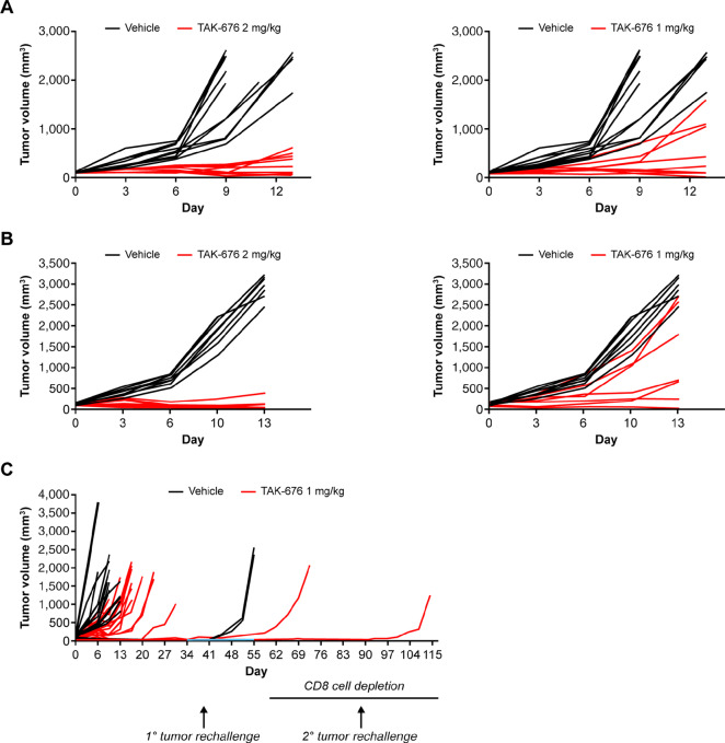
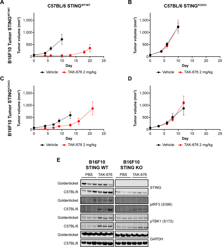
Oncology therapies targeting the immune system have improved patient outcomes across a wide range of tumor types, but resistance due to an inadequate T-cell response in a suppressive tumor microenvironment (TME) remains a significant problem. New therapies that activate an innate immune response and relieve this suppression may be beneficial to overcome this hurdle. TAK-676 is a synthetic novel stimulator of interferon genes (STING) agonist designed for intravenous administration. Here we demonstrate that TAK-676 dose-dependently triggers activation of the STING signaling pathway and activation of type I interferons. Furthermore, we show that TAK-676 is a highly potent modulator of both the innate and adaptive immune system and that it promotes the activation of dendritic cells, natural killer cells, and T cells in preclinical models. In syngeneic murine tumor models in vivo, TAK-676 induces dose-dependent cytokine responses and increases the activation and proliferation of immune cells within the TME and tumor-associated lymphoid tissue. We also demonstrate that TAK-676 dosing results in significant STING-dependent antitumor activity, including complete regressions and durable memory T-cell immunity. We show that TAK-676 is well tolerated, exhibits dose-proportional pharmacokinetics in plasma, and exhibits higher exposure in tumor. The intravenous administration of TAK-676 provides potential treatment benefit in a broad range of tumor types. Further study of TAK-676 in first-in-human phase I trials is ongoing.

Significance: TAK-676 is a novel systemic STING agonist demonstrating robust activation of innate and adaptive immune activity resulting in durable antitumor responses within multiple syngeneic tumor models. Clinical investigation of TAK-676 is ongoing.

Trial registration: ClinicalTrials.gov NCT04879849 NCT04420884 NCT04541108.


Conflict of interest statement

V.A. Appleman reports personal fees from Takeda Development Centers of America, Inc during the conduct of the study; personal fees from Takeda and other from Takeda outside the submitted work. V. Kolev reports other from Takeda during the conduct of the study. M. Mochizuki reports patent application WO2018/100558. S. Haridas reports personal fees from Takeda during the conduct of the study; personal fees from Takeda outside the submitted work. T.D. Thelen reports other from Takeda outside the submitted work. A.O. Abu-Yousif reports other from Takeda during the conduct of the study. M. Okaniwa reports patent application WO2018/100558. No other disclosures were reported.

Figures

FIGURE 1
FIGURE 1
Structure of STING agonist TAK-676 (A) and in vitro STING activation by TAK-676 in HEK293T, THP1-Dual and RAW-Lucia ISG cells (B). All data shown are representative of at least three independent experiments. WT, wild type.
FIGURE 2
FIGURE 2
TAK-676 dose-dependently activates the STING-TBK1-IRF3 pathway in THP1-Dual human AML cells (A) and CT26.WT cells (B). This activation is lost in STING-deficient CT26.WT cells following treatment with DMXAA (50 μg/mL, 1 hour) or TAK-676 (6.6 μmol/L, 2 hours) in C. Results are shown for two distinct STING KO clones and two distinct WT clones. All data shown are representative of at least three independent experiments. DMSO, dimethyl sulfoxide; GAPDH, glyceraldehyde 3-phosphate dehydrogenase; p, phosphorylated.
FIGURE 3
FIGURE 3
In vitro immune cell activation following treatment with TAK-676 in mouse BM-derived dendritic cells from 2 donors (A and B). C, Concentration-dependent activation in human MoDCs from 5 donors. NK-cell activation (D) and T-cell activation (E and F) in human whole blood from 5 donors following TAK-676 treatment for 24 hours. Conc., concentration; DMSO, dimethyl sulfoxide.
FIGURE 4
FIGURE 4
Antitumor effect of TAK-676 in: A, BALB/c mice bearing A20 syngeneic tumors; B, BALB/c mice bearing CT26.WT syngeneic tumors; C, BALB/c mice bearing CT26.WT syngeneic tumors following CR and subsequent rechallenge. Blue line denotes the period of rechallenge without CD8+ T-cell depletion. All data shown are representative of at least three independent experiments.
FIGURE 5
FIGURE 5
A–D, Mean tumor volume over time in WT (A and C) or STING-deficient C57BL/6J-Tmem173gt/J (Goldenticket; B and D) mice bearing STING WT (A and B) or deficient B16F10 (C and D) syngeneic tumors dosed with vehicle or TAK-676, and STING pathway activation (E) in STING WT or deficient B16F10 tumors implanted in WT C57BL/6 mice or STING-deficient Goldenticket mice.
FIGURE 6
FIGURE 6
Cytokine responses in the plasma and tumor of A20 tumor-bearing mice (left, center, all data shown are representative of at least three independent experiments) and the impact of STING deficiency on those responses in B16F10 tumor-bearing mice (right) following exposure to a single intravenous dose of vehicle or TAK-676: IFNα (A); IFNγ (B); and IP-10 (C). Δ Indicates that some samples in this group had values below the lower limit of quantitation of the assay or had values extrapolated beyond the standard range. *, P ≤ 0.05 relative to vehicle control; **, P ≤ 0.01 relative to vehicle control; ***, P < 0.00001 relative to vehicle control; IP-10, IFNγ-induced protein 10.
FIGURE 7
FIGURE 7
Effects of TAK-676 on immune cell populations within the tumor microenvironment in C57BL/6 mice bearing B16F10 syngeneic tumors (left) and BALB/c mice bearing CT26 syngeneic tumors (right). All data shown are representative of at least three independent experiments. A, Frequency of CD45+ cells in B16F10 tumors. B, Frequency of CD45+ cells in CT26 tumors. C, Frequency of CD8+ T cells in B16F10 tumors. D, Frequency of CD8+ T cells in CT26 tumors. E, Frequency of IFNγ+ CD8+ T cells in B16F10 tumors. F, MFI of Ki67 on CD8+ T cells in CT26 tumors. G, Frequency of CD8+ CD69+ T cells in B16F10 tumors. H, MFI of CD25 on CD8+ T cells in CT26 tumors. *, P ≤ 0.05; **, P ≤ 0.01. MHC, major histocompatibility complex.
FIGURE 8
FIGURE 8
Effects of TAK-676 on immune cell populations within the tumor-draining lymph nodes from mice bearing B16F10 tumors (left and middle) and CT26 tumors (right). All data shown are representative of at least three independent experiments. A, Frequency of total viable cells. B, Frequency of CD11+ MHCI+ cells. C, Frequency of CD8 CD80+ DCs. D, Frequency of CD8 CD86+ DCs. E, Frequency of IFNγ+ CD8+ T cells. F, Frequency of Ki67+ CD8+ T cells. G, Frequency of CD69+ CD8+ T cells. H, Frequency of CD11c+ MHCII+ cells. I, MFI of CD86 on DCs. J, MFI of CD80 on DCs. K, Frequency of CD69+ NK cells. **, P ≤ 0.01.

References

    1. Chyuan IT, Chu CL, Hsu PN. Targeting the tumor microenvironment for improving therapeutic effectiveness in cancer immunotherapy: focusing on immune checkpoint inhibitors and combination therapies. Cancers 2021;13:1188. - PMC - PubMed
    1. Nowicki TS, Hu-Lieskovan S, Ribas A. Mechanisms of resistance to PD-1 and PD-L1 blockade. Cancer J 2018;24:47–53. - PMC - PubMed
    1. Walsh RJ, Soo RA. Resistance to immune checkpoint inhibitors in non-small cell lung cancer: biomarkers and therapeutic strategies. Ther Adv Med Oncol 2020;12:1758835920937902. - PMC - PubMed
    1. Sun L, Wu J, Du F, Chen X, Chen ZJ. Cyclic GMP-AMP synthase is a cytosolic DNA sensor that activates the type I interferon pathway. Science 2013;339:786–91. - PMC - PubMed
    1. Ishikawa H, Ma Z, Barber GN. STING regulates intracellular DNA-mediated, type I interferon-dependent innate immunity. Nature 2009;461:788–92. - PMC - PubMed

Publication types

Associated data