. 2023 Feb 25;11(1):12-26.
eCollection 2023.

Rate of castration-induced prostate stroma regression is reduced in a mouse model of benign prostatic hyperplasia

Affiliations

Rate of castration-induced prostate stroma regression is reduced in a mouse model of benign prostatic hyperplasia

Renyuan Zhang et al. Am J Clin Exp Urol. .

Abstract

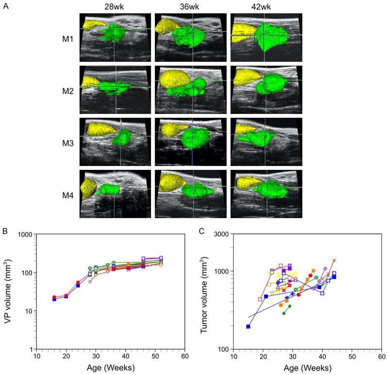
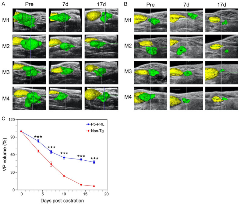
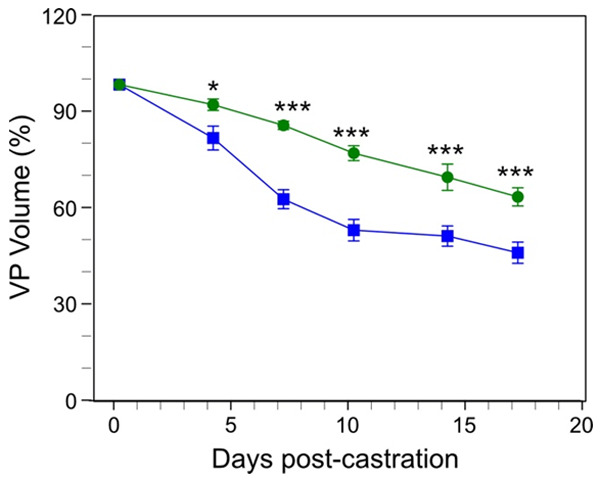
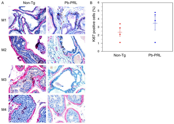
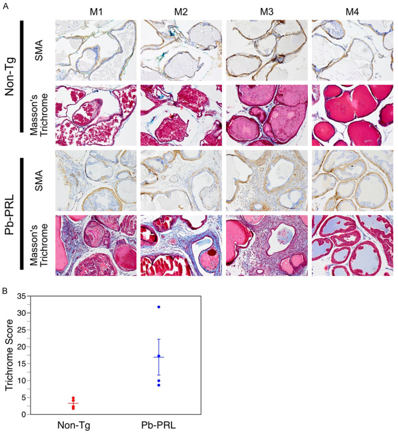
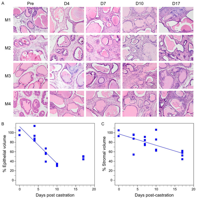
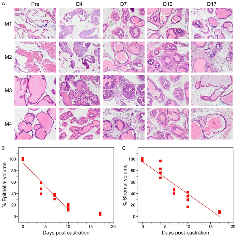
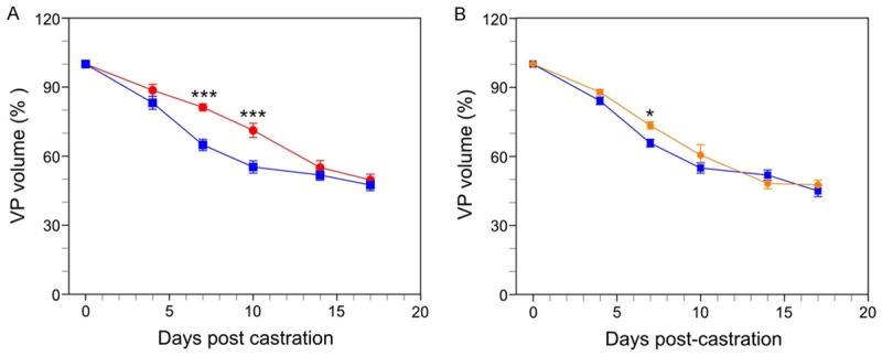
Benign prostatic hyperplasia (BPH) is a non-neoplastic proliferative disease producing lower urinary tract symptoms related to the resulting enlarged prostate. BPH is pathologically characterized by hyperplastic growth in both epithelial and stromal compartments. Androgen signaling is essential for prostate function and androgen blockade is the second-line medical therapy to relieve symptoms of BPH. Here we examined the prostates of probasin promoter-driven prolactin (Pb-PRL) transgenic mice, a robust model of BPH that spontaneously develops prostate enlargement, to investigate prostate regression in response to surgical castration. Serial ultrasound imaging demonstrated very uniform self-limited growth of Pb-PRL prostate volume that is consistent with the benign, limited cellular proliferation characteristic of BPH and that contrasts with the highly variable, exponential growth of murine prostate cancer models. Castration elicited only a partial reduction in prostate volume, relative to castration-induced regression of the normal prostate gland. The anti-androgen finasteride induced a diminished reduction of Pb-PRL prostate volume versus castration. The limited extent of Pb-PRL mouse prostate volume regression correlated with the initial volume of the stromal compartment, suggesting a differential sensitivity of the epithelial and stromal compartments to androgen withdrawal. Indeed, two-dimensional morphometric analyses revealed a distinctly reduced rate of regression for the stromal compartment in Pb-PRL mice. The myofibroblast component of the Pb-PRL prostate stroma appeared normal, but the stromal compartment contained more fibroblasts and extracellular collagen deposition. Like normal prostate, the rate of regression of the Pb-PRL prostate was partially dependent on TGFß and TNF signaling, but unlike the normal prostate, the extent of castration-induced regression was not affected by TGFß or TNF blockade. Our studies show that androgen deprivation can effectively reduce the overall volume of hyperplastic prostate, but the stromal compartment is relatively resistant, suggesting additional therapies might be required to offer an effective treatment for the clinical manifestations of BPH.

Keywords: LUTS; castration; high-frequency ultrasound imaging; hypertrophy; prostate; stromal.


Conflict of interest statement

None.

Figures

Figure 1
Figure 1
Growth of the ventral prostate lobe in Pb-PRL transgenic mice: sigmoidal growth curve kinetics and limited inter-animal variability. Prostate volumes of Pb-PRL and PbCre4 x PTENflox/flox mice were monitored during aging by serial HFUS imaging. A. Illustration of 3D-reconstruction of ventral prostate for four Pb-PRL mice acquired 28, 36 and 42 weeks after birth. Visualization of the bladder (yellow) and the ventral prostate (green) was performed by pseudo-coloring. B. Kinetics of ventral prostate growth in Pb-PRL transgenic mice. Volumes from the 3D-reconstructions are plotted for each animal at the indicated age. Each color/symbol combination represents an individual mouse (n = 44). C. Kinetics of prostate tumor growth in prostate-specific PTEN-deficient mice. Volumes from the 3D-reconstructions are plotted for each animal at the indicated age. Each color represents an individual mouse (n = 26).
Figure 2
Figure 2
Castration-induced ventral prostate regression reduced in BPH model mice versus normal mice. Pb-PRL transgenic and non-transgenic (Non-Tg) littermate mice were surgically castrated and monitored by serial HFUS imaging. A. 3D-reconstructions of ultrasound images corresponding to pre-castration (Pre) as well as 7- and 17-days following castration of Pb-PRL mice. Visualization of the bladder (yellow) and the ventral prostate (green) was performed by pseudo-coloring. B. Similar to A, for non-Tg littermate mice. C. Ventral prostate regression following castration of Pb-PRL mice (blue) and non-Tg littermate mice (red). Volumes of the 3D reconstructions are plotted at the indicated times following castration. Data points represent mean volume relative to starting volume, ± SEM (n = 4 (non-Tg), n = 12 (Pb-PRL). ***P<.001; vs non-Tg. Non-Tg and Pb-PRL mice were compared using two-way ANOVA with Tukey’s HSD test.
Figure 3
Figure 3
Comparison of ventral prostate regression induced by finasteride versus castration in the Pb-PRL mouse BPH model. Pb-PRL mice were surgically castrated (blue line, data from Figure 2), or left intact and treated daily with finasteride (green line). The ventral prostate (VP) of each mouse was monitored by serial HFUS imaging. Segmentation and 3D-reconstruction was performed on the resulting images to determine the volume of the ventral prostate. Data points represent mean volume relative to starting volume, ± SEM (n = 4). *P<.05; ***P<.001. Finasteride treated Pb-PRL mice and castrated Pb-PRL mice were compared using two-way ANOVA with Tukey’s HSD test.
Figure 4
Figure 4
Immunohistochemical characterization of prostate epithelium of Pb-PRL transgenic and non-transgenic mice. Non-transgenic (non-Tg) and Pb-PRL transgenic (Pb-PRL) mice were sacrificed, and the prostates were fixed, sectioned, and IHC-stained with anti-Ki-67 and anti-α-SMA, with hematoxylin as a counterstain. The epithelial glands are surrounded by α-SMA stained myofibroblasts. Ki-67-positive epithelial cells were counted in five high-power fields from each mouse prostate. A. Representative sections are shown for each mouse. Dual-staining for Ki-67 (brown) and α-SMA (red) in prostates from the indicated mouse strains. Magnification 400×. B. Quantification of Ki-67 positive cells in the epithelium of the prostates. Mean (± SEM) and individual values for each of four mice from each group is shown. Student’s unpaired t-test was used to compare non-Tg to Pb-PRL, P-value = 0.276.
Figure 5
Figure 5
Increased stroma in prostates of Pb-PRL mice. Non-transgenic (Non-Tg) and Pb-PRL transgenic (Pb-PRL) mice were sacrificed, and prostate tissue sections processed and stained. A. Images of immunostaining for α-SMA (smooth muscle actin, brown) and staining using Masson’s trichrome. Masson’s trichrome staining indicates collagen deposition (blue). Red staining indicates smooth muscle cells surrounding the glands (and non-specific red staining of the prostatic secretions intraluminally). Magnification 200×. B. Plots showing the mean (± SEM) trichome score in the stromal compartment for each of four mice of the two mouse strains. Note: Trichrome score is determined as described in Material and Methods section. Student’s unpaired t-test was used to compare non-Tg to Pb-PRL, P-value = 0.044.
Figure 6
Figure 6
Castration induced regression in epithelial and stromal compartments in Pb-PRL transgenic mice. Mature Pb-PRL mice were surgically castrated and monitored by serial HFUS imaging to determine prostate VP volume for compartment area normalization (see methods). Prostates from four mice were examined at each of the five indicated time points following castration. A. Representative H&E stained sections correspond to the indicated times post-castration. Magnification 200×. B. Plot of normalized epithelial compartment volume percentage during ventral prostate regression. C. Plot of normalized stromal compartment volume percentage during ventral prostate regression. B, C. Each filled square represents an individual animal. Lines represent data fitted by least squares linear regression. Note that the epithelial compartment volumes determined at day 17 post-castration are not fitted.
Figure 7
Figure 7
Castration induced regression in epithelial and stromal compartments in normal, non-transgenic mice. Mature littermate, non-transgenic mice were surgically castrated and monitored by serial HFUS imaging to determine prostate VP volume for compartment area normalization (see methods). Prostates from four mice were examined at each of the five indicated time points following castration. A. Representative H&E stained sections correspond to the indicated times post-castration. Magnification 200×. B. Plot of normalized epithelial compartment volume percentage during ventral prostate regression. C. Plot of normalized stromal compartment volume percentage during ventral prostate regression. B, C. Each filled square represents an individual animal. Lines represent data fitted by least squares linear regression. Note that the epithelial compartment volumes determined at day 17 post-castration are not fitted.
Figure 8
Figure 8
Blockade of cytokine signaling in Pb-PRL mice. Pb-PRL mice received vehicle (blue, data replicated from Figure 1C), TGFß/p38 signaling inhibitors (red) or TNF signaling blockade (orange) and were monitored after castration by serial HFUS imaging. A. Effect of TGF-β signaling blockade (red line) on castration-induced regression of VP. B. Effect of TNF signaling blockade (orange line) on castration-induced regression of VP. Data points represent mean volume relative to starting volume, ± SEM (n = 4). *P<.05; ***P<.001. TGF-β or TNF-α inhibitor treated castrated mice were compared to Pb-PRL Tg castrated mice (blue line, data from Figure 2) using two-way ANOVA with Tukey’s HSD test.

References

    1. Berry SJ, Coffey DS, Walsh PC, Ewing LL. The development of human benign prostatic hyperplasia with age. J Urol. 1984;132:474–479. - PubMed
    1. Lerner LB, McVary KT, Barry MJ, Bixler BR, Dahm P, Das AK, Gandhi MC, Kaplan SA, Kohler TS, Martin L, Parsons JK, Roehrborn CG, Stoffel JT, Welliver C, Wilt TJ. Management of lower urinary tract symptoms attributed to benign prostatic hyperplasia: AUA GUIDELINE PART I-initial work-up and medical management. J Urol. 2021;206:806–817. - PubMed
    1. Lerner LB, McVary KT, Barry MJ, Bixler BR, Dahm P, Das AK, Gandhi MC, Kaplan SA, Kohler TS, Martin L, Parsons JK, Roehrborn CG, Stoffel JT, Welliver C, Wilt TJ. Management of lower urinary tract symptoms attributed to benign prostatic hyperplasia: AUA GUIDELINE PART II-surgical evaluation and treatment. J Urol. 2021;206:818–826. - PubMed
    1. Lepor H. Alpha blockers for the treatment of benign prostatic hyperplasia. Rev Urol. 2007;9:181–190. - PMC - PubMed
    1. Cabot AT. II. The question of castration for enlarged prostate. Ann Surg. 1896;24:265–309. - PMC - PubMed

LinkOut - more resources