Review
. 2023 Sep;20(7):2924-2941.
doi: 10.1111/iwj.14158. Epub 2023 Mar 16.

Acellular fish skin for wound healing

Affiliations
Review

Acellular fish skin for wound healing

Ali Esmaeili et al. Int Wound J. 2023 Sep.

Abstract

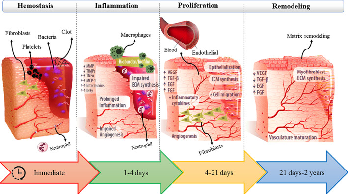
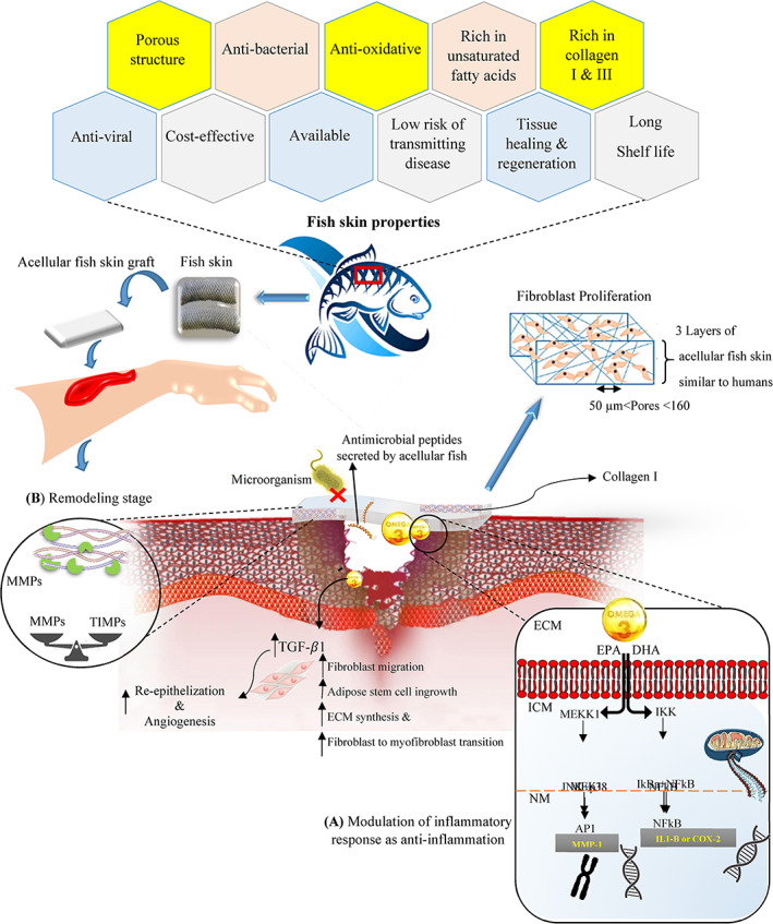
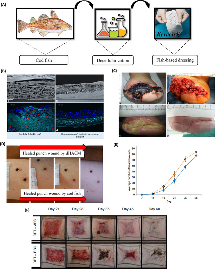
Fish skin grafting as a new skin substitute is currently being used in clinical applications. Acceleration of the wound healing, lack of disease transmission, and low cost of the production process can introduce fish skin as a potential alternative to other grafts. An appropriate decellularization process allows the design of 3D acellular scaffolds for skin regeneration without damaging the morphology and extracellular matrix content. Therefore, the role of decellularization processes is very important to maintain the properties of fish skin. In this review article, recent studies on various decellularization processes as well as biological, physical, and mechanical properties of fish skin and its applications with therapeutic effects in wound healing were investigated.

Keywords: biological and physical & mechanical properties; decellularization; fish skin; in-vivo studies; wound healing.


Conflict of interest statement

The authors declare that there is no conflict of interest.

Figures

FIGURE 1
FIGURE 1
Illustration of the process of skin wound healing.
FIGURE 2
FIGURE 2
(A) The decellularization method. (B) H&E and Trichrome Masson staining of (i, ii) native skin; (iii, iv) decellularized skin. (C) SEM images of decellularized (I) and recellularized fish skin (ii). (D) Animal model for evaluating skin regeneration by acellular fish skin and Plasma Rich in Growth Factor PRGF gel (E) Macroscopic evaluation of woumd (i) on the first day, (ii) treated wound in the 28th day with acellular fish skin and (iii) treated wound with acellular fish skin and PRGF gel in 28th. (F) Wound closure for all samples on the 28th day after grafting (G) H&E images of (i) native skin, (ii) treated with acellular fish skin, and (iii) treated with acellular fish skin and PRGF gel in the 28th.,
FIGURE 3
FIGURE 3
(A) design of tilapia skin acellular dermal matrix (TS‐ADM), (B) histological assays by H&E (I and II) and DAPI (III and IV) staining of TS‐ADM (Scale bar:100 μm), (C) scanning electron microscopy of TS‐ADM and DC‐ADM, (D) wound healing process on a rat model with TS‐ADM within 21 days, (E) wound closure on days 7, 14 and 21 (rat model), (F) wound healing process on porcine model with TS‐ADM on days 7, 14, 21, 28, 35, 42 and 180 postoperation, (G) wound closure on days 7, 14 and 21 (porcine model), (H) Schematic diagram: the partially degraded TS‐ADM promotes extracellular matrix deposition, angiogenesis, and reepithelialization by forming a microenvironment conducive to the expression of TGF‐β1, α‐SMA and CD31.
FIGURE 4
FIGURE 4
Acellular fish skin properties and the effect of Omega‐3 polyunsaturated fatty acids on signalling activity for inflammation stage of wound healing as well as mediating tissue remodelling. (A) Mechanism of action for Omega‐3 polyunsaturated fatty acids such eicosatetraenoic acid and docosahexaenoic acid (AP1, activator protein 1; COX‐2, cyclooxygenase‐2; DHA, docosahexaenoic acid; ECM, extracellular matrix; EPA, eicosatetraenoic acid; ICM, intracellular matrix; IkBa, NFkB inhibitor a; IKK, IkB kinase; IL1‐B, interleukin 1‐B; JNK, c‐Jun N‐terminal kinase; MEKK1, MAPK kinase 1; MEK1, MAPK kinases 1; MMP1, Matrix metallopeptidase 1; NM, nuclear matrix). (B) The enzymatic activities of MMPs by acting as an attachment site for breakdown of scaffold rather than developing host tissue and ECM components; so, it causes the balance between MMPs and tissue inhibitors of metalloproteinases (TIMPs) and finally the new ECM is formed and the inflammation time reduces.
FIGURE 5
FIGURE 5
(A) Kerecis® product representative of acellular Atlantic cod fish skin, (B) SEM of (a) Acellular dermal matrix derived from fish skin and (b) decellularized amnion matrix. Stem cells were cultured for 12 days in an acellular dermal matrix derived from fish skin (c) and amnion (d). The stem cells stained blue have migrated into the fish skin matrix, while the stem cells on the amniotic matrix have settled on the surface of the matrix, (C) Healed wound (wound area 29 cm2) following a total treatment duration of 33 week using acellular Cod fish skin, (D) Followed up to the healing of punch wounds that wounds on the bottom side of figure was treated with Cod fish skin graft, while the top wound was treated with dehydrated human amnion/chorion membrane or dHACM, (E) Clinical use of antibiotics before and after the application of AFS in a cohort of chronic wound patients, (F) Progression of porcine burn wound closure when treated with AFS with and without skin grafting. (AFS, acellular cod fish skin; DPT, deep partial thickness; FBC, fetal bovine collagen; SEM, Scanning electron microscope).

References

    1. Yao Y, Zhang A, Yuan C, Chen X, Liu Y. Recent trends on burn wound care: hydrogel dressings and scaffolds. Biomater Sci. 2021;9(13):4523‐4540. - PubMed
    1. Markiewicz‐Gospodarek A, Kozioł M, Tobiasz M, Baj J, Radzikowska‐Büchner E, Przekora A. Burn wound healing: clinical complications, medical care, treatment, and dressing types: the current state of knowledge for clinical practice. Int J Environ Res Public Health. 2022;19(3):1338. - PMC - PubMed
    1. Kaddoura I, Abu‐Sittah G, Ibrahim A, Karamanoukian R, Papazian N. Burn injury: review of pathophysiology and therapeutic modalities in major burns. Ann Burns Fire Disasters. 2017;30:95‐102. - PMC - PubMed
    1. Cartotto R. Topical antimicrobial agents for pediatric burns. Burn Trauma. 2017;5:33. doi: 10.1186/s41038-017-0096-6 - DOI - PMC - PubMed
    1. Khadjibayev AM, Fayazov AD, Djabriyev DA, Kamilov UR. Surgical treatment of deep burns. Ann Burns Fire Disasters. 2008;21:150‐152. - PMC - PubMed