. 2023 May 1;44(17):1495-1510.
doi: 10.1093/eurheartj/ehad067.

Cardiac sarcoidosis: phenotypes, diagnosis, treatment, and prognosis

Affiliations

Cardiac sarcoidosis: phenotypes, diagnosis, treatment, and prognosis

Jukka Lehtonen et al. Eur Heart J. .

Abstract

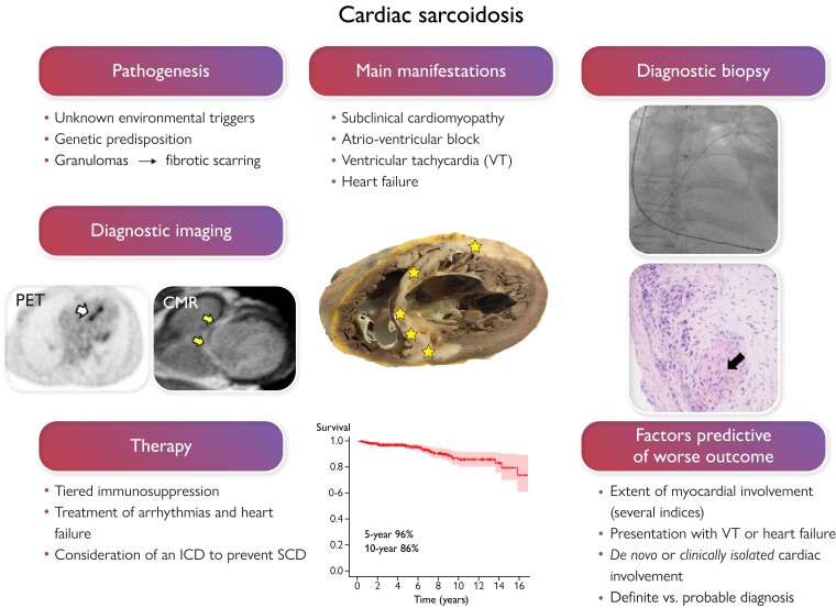
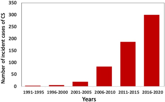
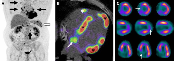
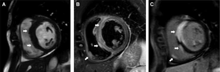
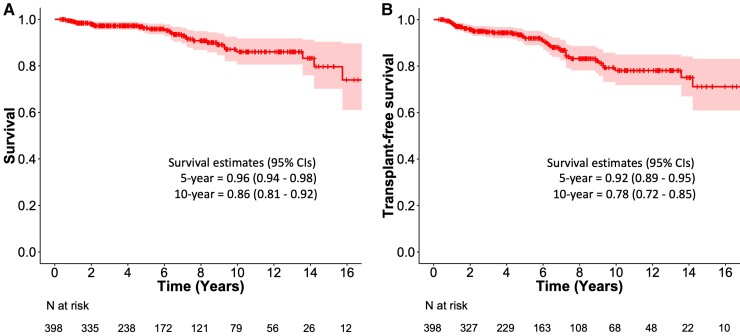
Cardiac sarcoidosis (CS) results from epithelioid cell granulomas infiltrating the myocardium and predisposing to conduction disturbances, ventricular tachyarrhythmias, and heart failure. Manifest CS, however, constitutes only the top of an iceberg as advanced imaging uncovers cardiac involvement 4 to 5 times more commonly than what is clinically detectable. Definite diagnosis of CS requires myocardial biopsy and histopathology, but a sufficient diagnostic likelihood can be achieved by combining extracardiac histology of sarcoidosis with clinical manifestations and findings on cardiac imaging. CS can appear as the first or only organ manifestation of sarcoidosis or on top of pre-existing extracardiac disease. Due to the lack of controlled trials, the care of CS is based on observational evidence of low quality. Currently, the treatment involves corticosteroid-based, tiered immunosuppression to control myocardial inflammation with medical and device-based therapy for symptomatic atrioventricular block, ventricular tachyarrhythmias, and heart failure. Recent outcome data indicate 90% to 96% 5-year survival in manifest CS with the 10-year figures ranging from 80% to 90%. Major progress in the care of CS awaits the key to its molecular-genetic pathogenesis and large-scale controlled clinical trials.

Keywords: Cardiac sarcoidosis; Heart failure; Implantable cardioverter-defibrillator; Inflammatory heart disease; Pacemaker.


Conflict of interest statement

Conflict of interest Lecture and/or advisory board fees from Boehringer Ingelheim, Bristol-Myers Squibb, MSD, Takeda, Bayer, Amgen, Roche, and Aiforia technologies oy (M.I.M.). Lecture fee from Pfizer and research collaboration with GE healthcare (V.U.).

Figures

Graphical abstract
Graphical abstract
In cardiac sarcoidosis (CS), inflammatory granulomas invade the heart leading to injury and fibrosis (yellow stars in the section of a sarcoidotic heart in the middle of the graph). CS is often subclinical, but, when clinically manifest, presents commonly with slow or fast arrhythmias or heart failure. On the left of the figure, positron emission tomography (PET) exposes focal septal uptake of 18-F fluorodeoxyglucose suggesting active inflammation (white arrow), and contrast-enhanced cardiac magnetic resonance (CMR) shows septal late gadolinium enhancement (arrows) indicating replacement fibrosis. Both constitute major diagnostic criteria for CS and entitle probable CS diagnosis if accompanied by confirmed extracardiac histology of sarcoidosis. Yet, the only way to definite diagnosis, demonstrated on the right, is myocardial biopsy showing non-necrotic granulomas (black arrow). The therapy of CS is based on immunosuppression and management of heart block, ventricular arrhythmias, and heart failure. The risk of sudden cardiac death (SCD) needs assessment and consideration of an implantable cardioverter-defibrillator (ICD). With current therapy, expected 5-year survival is well above 90% as shown by the Kaplan–Meier graph of a 398-patient Finnish CS cohort.
Figure 1
Figure 1
Incident cases of clinically manifest cardiac sarcoidosis (CS) in adults (>18 years) diagnosed in Finnish hospitals from 1991 through 2020. Curiously, 3 vs. 300 new cases were detected over the first and last 5-year periods, respectively. The population of Finland is 5.5 million with 4.5 million adults. The figure is based on Kandolin et al. and unpublished data.
Figure 2
Figure 2
18F-fluorodeoxyglucose (18F-FDG) positron emission tomography/computed tomography (PET/CT) scans of a patient with tri-fascicular block and depressed left ventricular (LV) function; cardiac sarcoidosis was verified by endomyocardial biopsy. (A) whole-body PET with 18F-FDG positive lymph nodes (arrows) and splenic radiotracer accumulation (hollow arrow). (B) 4-chamber PET/CT image showing 18F-FDG uptake on LV septum, apex, and basal lateral wall (asterisks), on right ventricular free wall (arrow), and on interatrial septum (arrow). (C) Single-photon emission computed tomography 99mTc-tetrofosmin scans showing perfusion defects (white arrows) on LV septum and apex overlapping areas of 18F-FDG uptake on PET (mismatch pattern). From top to bottom, the rows represent short-axis, vertical, and horizontal views of the heart.
Figure 3
Figure 3
Magnetic resonance images of a patient with cardiac sarcoidosis, 3rd degree atrioventricular block, and normal left and right ventricular ejection fraction. The arrows highlight key findings. (A) short-axis cardiac cine image showing thickened ventricular septum. (B) T2-weighted image showing septal and local right ventricular edema indicating active inflammation. (C) late gadolinium enhancement image showing transmural septal, papillary muscle, and local right ventricular free wall involvement.
Figure 4
Figure 4
Magnetic resonance images of a patient with 3rd degree atrioventricular block and depressed left ventricular function; cardiac sarcoidosis was verified by endomyocardial biopsy. (A) apical 4-chamber cine image showing basal septal thinning (arrow) and thickened mid-septum. (B and C) apical 4- and 2-chamber images, respectively, of late gadolinium enhancement showing patchy left ventricular involvement.
Figure 5
Figure 5
Magnetic resonance images of a patient with right bundle branch block, ventricular tachycardia, and depressed left and right ventricular function; cardiac sarcoidosis was verified by endomyocardial biopsy. The arrows point at key findings. (A) Short-axis cardiac cine image showing thickened left ventricular myocardium and inferior right ventricular wall. (B) A ‘hook sign’ pattern of cardiac sarcoidosis characterized by late gadolinium enhancement in the septum continuing to ventricular insertion points and right ventricular free wall.
Figure 6
Figure 6
Flowchart for cardiac imaging and biopsies at Helsinki University Hospital for suspected cardiac sarcoidosis (CS) after exclusion of ischemic heart disease and in the absence of histologically verified extracardiac sarcoidosis. If cardiac magnetic resonance (CMR) shows late gadolinium enhancement (LGE), imaging-guided endomyocardial biopsy (EMB) is performed first. If either CMR or EMB is negative, whole-body positron emission tomography (PET) is done. PET being positive, either EMB or extracardiac biopsy (ECB) is performed depending on PET and CMR findings and patient’s preferences. cFDG and ecFDG indicate cardiac and extracardiac uptake of fluorodeoxyglucose, respectively; histo, histology of sarcoidosis.
Figure 7
Figure 7
Histopathology of cardiac sarcoidosis. Sharply demarcated (‘geographical’) inflammatory lesions in a gross photo of an explanted heart (A) and in low-magnification hematoxylin-eosin (HE) staining (B). In panels (C–G), stars mark small coronary artery branches with vascular wall granulomas and circles encompass non-caseating granulomas. In panel D, CD3 immunostaining highlights T cells, and in panel E, CD4 antibody stains T cells intensely and macrophages and giant cells less strongly. Macrophages and giant cells can also be highlighted with CD68 (F) and PD-L1 (G) antibody staining. Scalebars are shown in each panel.
Figure 8
Figure 8
Kaplan–Meier curves of overall (A) and transplant-free (B) survival in a cohort of 398 patients with clinically manifest cardiac sarcoidosis diagnosed in Finland from 1988 through 2017 and followed for a median of 5.0 years. The figures are based on the data reported by Nordenswan et al. and summarized in Table 5. The survival graphs reflect the care of cardiac sarcoidosis based on the following principles: requirement of diagnostic histology and pursuit of definite diagnosis, consistent use of corticosteroids with azathioprine and infliximab as the main additional immunomodulators, clinical follow-up with selective instead of routine repeats of positron emission tomography, frequent use of implantable cardioverter-defibrillators, and no sarcoidosis-specific restrictions to heart transplantation.

References

    1. Drent M, Crouser ED, Grunewald J. Challenges of Sarcoidosis and Its Management. N Engl J Med 2021;385:1018–1032. - PubMed
    1. Kandolin R, Lehtonen J, Airaksinen J, Vihinen T, Miettinen H, Ylitalo K, et al. . Cardiac sarcoidosis: epidemiology, characteristics, and outcome over 25 years in a nationwide study. Circulation 2015;131:624–632. - PubMed
    1. Judson M, Costabel U, Drent Met al. . The WASOG Sarcoidosis Organ Assessment Instrument: an update of a previous clinical tool. Sarcoidosis Vasc Diffuse Lung Dis 2014;31:19–27. - PubMed
    1. Birnie DH, Sauer WH, Bogun F, Cooper JM, Culver DA, Duvernoy CS, et al. . HRS expert consensus statement on the diagnosis and management of arrhythmias associated with cardiac sarcoidosis. Heart Rhythm 2014;11:1305–1323. - PubMed
    1. Terasaki F, Azuma A, Anzai T, Ishizaka N, Ishida Y, Isobe M, et al. . Japanese Circulation Society Joint Working Group. JCS 2016 Guideline on Diagnosis and Treatment of Cardiac Sarcoidosis. Circ J 2019;83:2329–2388. - PubMed

Publication types