. 2023 May;299(5):104609.
doi: 10.1016/j.jbc.2023.104609. Epub 2023 Mar 15.

Mechanism and linkage specificities of the dual retaining β-Kdo glycosyltransferase modules of KpsC from bacterial capsule biosynthesis

Affiliations

Mechanism and linkage specificities of the dual retaining β-Kdo glycosyltransferase modules of KpsC from bacterial capsule biosynthesis

Liam Doyle et al. J Biol Chem. 2023 May.

Abstract

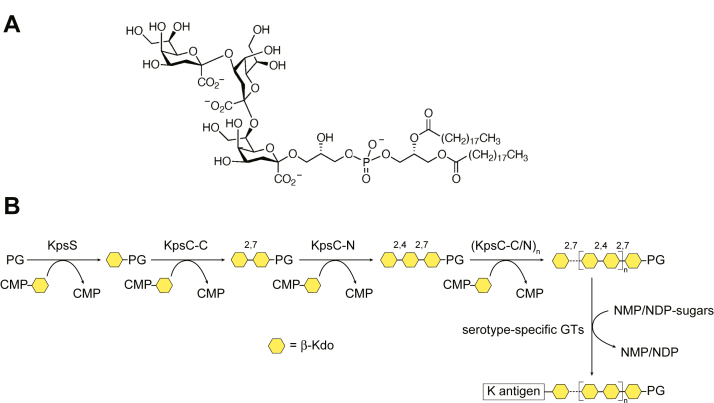
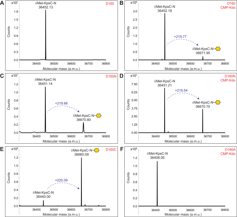
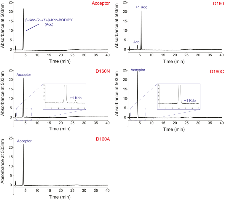
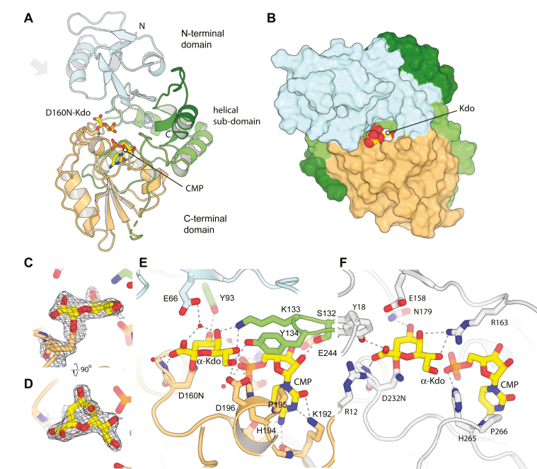
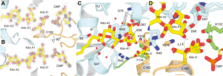
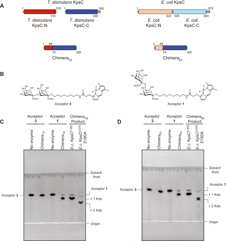
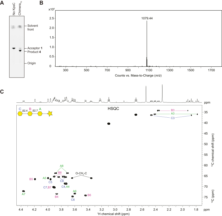
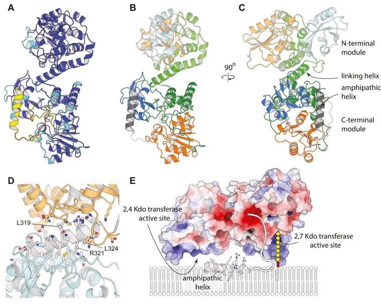
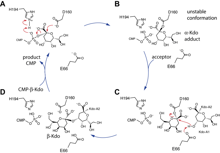
KpsC is a dual-module glycosyltransferase (GT) essential for "group 2" capsular polysaccharide biosynthesis in Escherichia coli and other Gram-negative pathogens. Capsules are vital virulence determinants in high-profile pathogens, making KpsC a viable target for intervention with small-molecule therapeutic inhibitors. Inhibitor development can be facilitated by understanding the mechanism of the target enzyme. Two separate GT modules in KpsC transfer 3-deoxy-β-d-manno-oct-2-ulosonic acid (β-Kdo) from cytidine-5'-monophospho-β-Kdo donor to a glycolipid acceptor. The N-terminal and C-terminal modules add alternating Kdo residues with β-(2→4) and β-(2→7) linkages, respectively, generating a conserved oligosaccharide core that is further glycosylated to produce diverse capsule structures. KpsC is a retaining GT, which retains the donor anomeric carbon stereochemistry. Retaining GTs typically use an SNi (substitution nucleophilic internal return) mechanism, but recent studies with WbbB, a retaining β-Kdo GT distantly related to KpsC, strongly suggest that this enzyme uses an alternative double-displacement mechanism. Based on the formation of covalent adducts with Kdo identified here by mass spectrometry and X-ray crystallography, we determined that catalytically important active site residues are conserved in WbbB and KpsC, suggesting a shared double-displacement mechanism. Additional crystal structures and biochemical experiments revealed the acceptor binding mode of the β-(2→4)-Kdo transferase module and demonstrated that acceptor recognition (and therefore linkage specificity) is conferred solely by the N-terminal α/β domain of each GT module. Finally, an Alphafold model provided insight into organization of the modules and a C-terminal membrane-anchoring region. Altogether, we identified key structural and mechanistic elements providing a foundation for targeting KpsC.

Keywords: 3-deoxy-β-D-manno-oct-2-ulosonic acid; Escherichia coli; KpsC; biosynthesis; capsular polysaccharide; cell surface; enzyme mechanism; enzyme structure; glycolipid; glycosyltransferase.


Conflict of interest statement

Conflict of interest The authors declare that they have no conflicts of interest with the contents of this article.

Figures

Figure 1
Figure 1
Structure and biosynthesis of the KpsC reaction product.Panel A shows the structure of the phosphatidylglycerol-(Kdo)3 glycolipid intermediate synthesized by the combined actions of KpsS and the two GT modules in KpsC. The overall reaction scheme for the synthesis of an Escherichia coli group 2 capsular polysaccharide is depicted in Panel B, KpsS and KpsC are β-Kdo GTs, which transfer Kdo residues from CMP-β-Kdo donor to create a phosphatidylglycerol (PG)-based acceptor. KpsS adds the initial Kdo residue. This is followed by sequential addition of Kdo residues by the alternating actions of the KpsC-C and KpsC-N GT modules, to create a glycolipid acceptor containing 5 to 9 Kdo residues. The conserved KpsC reaction product is then elongated by a serotype-specific GTs to create a CPS belonging to a given K-antigen serotype. GT, glycosyltransferase.
Figure 2
Figure 2
LC-MS analysis of KpsC-NEcand its active site variants to investigate the formation of covalent Kdo adducts. KpsC-NEc proteins were purified from Escherichia coli BL21 and analyzed by LC-MS before or after incubation with a CMP-Kdo generating reaction mixture. Kdo adducts are denoted by the yellow hexagons. Note that the N-terminal methionine residue is absent in in all cases. The formation of a Kdo adduct should result in a 220.18 Da mass increase to the protein. A, the spectrum of the WT protein shows a single species corresponding to the apoprotein. B, the spectrum of the WT protein following incubation with a CMP-Kdo generating reaction shows formation of a second species containing a modification with Kdo. C, the D160N protein appears to have formed Kdo adducts in vivo as two species differing by 219.66 Da were observed without incubation with CMP-Kdo. D, following incubation with CMP-Kdo, the D160N protein appears to have an increase in the amount of Kdo-modified protein compared to panel C. E, the D160C protein appears to be almost completely modified with Kdo in vivo as only small amounts of unmodified protein remain. F, the D160A protein remains unmodified even after incubation with CMP-Kdo.
Figure 3
Figure 3
UV-HPLC analysis of the in vitro β-Kdo GT activity of KpsC-NEcand its site-directed mutants. The top left panel shows the A503nm profile for acceptor 1 in the absence of added enzyme, while the top right panel shows the addition of a single Kdo residue to acceptor 1 by the WT KpsC-NEc enzyme (labeled D160). The identity of the D160 variant is indicated in each panel in red. The D160N and D160C variants of KpsC-NEc retain minimal activity, while the D160A variant shows no detectable activity in these conditions. Reactions were incubated at 30 °C for 30 min and performed in triplicate. GT, glycosyltransferase.
Figure 4
Figure 4
Structure of KpsC-N D160N Kdo adduct.A, shows the overall structure of KpsC-N in cartoon representation, with CMP and Kdo represented in yellow sticks. B, shows a surface view, from the approximate perspective of the gray arrow in panel (A). C and D, show orthogonal views of the electron density map around the D160N–Kdo adduct, contoured at 1.0 σ. Details of interactions mediated by the D160N–Kdo adduct are highlighted in (E), while (F) presents an equivalent view of the structure of WbbBGT99 D232N–Kdo adduct complex.
Figure 5
Figure 5
Structure of KpsC D160C ternary complex.A and B, show orthogonal views of the electron density map around the ligands in the D160C–Kdo ternary complex. The transparent surface is contoured at 1 σ, and the mesh is contoured at 4 σ. Note that most heteroatoms have clear, distinct peaks at 4 σ. Panel (C) shows details of interactions mediated by the D160C–Kdo acceptor disaccharide and (D) highlights the organization of the acceptor Kdo-A1 group and the Kdo–C160 adduct.
Figure 6
Figure 6
Functional characterization of KpsC chimeras.A, generation of KpsC chimeras from characterized KpsC-N and KpsC-C enzymes from Thermosulfurimonas dismutans and Escherichia coli. B, enzyme acceptors containing β-Kdo disaccharides with either (2→4)- (acceptor 3) or (2→7)-linkages (acceptor 1). C, TLC analysis of the reaction product synthesized by T. dismutans KpsC chimera (ChimeraTd) and E. coli KpsC enzymes with established specificity. D, equivalent TLC analysis of the E. coli KpsC chimera (ChimeraEc).
Figure 7
Figure 7
Structural characterization of product 4 synthesized by ChimeraTd. Panels (A) and (B) show TLC separation and the charge-deconvoluted ESI mass-spectrum of reaction product 4, respectively. The calculated monoisotopic mass of 4 is 1079.43. Panel (C) shows a selected region from the 1H,13C HSQC spectrum of product 4. Kdo residues are designated by letters, and the signals were assigned by comparison to previously characterized synthetic Kdo trisaccharide products (5, 9). Downfield shifts of the substituted carbon atoms in Kdo residues (due to a positive α-glycosylation effect) defined the positions of substitution.
Figure 8
Figure 8
AlphaFold model of full-length E. coli KpsC.A, KpsC colored by pLDDT; (blue: pLDDT > 90, cyan: pLDDT > 70, yellow: pLDDT > 50, orange: pLDDT <50). B and C, orthogonal views of KpsC, colored by module and domains within each module. The N-terminal α/β domain in blue, the helical inserts in green, and the C-terminal α/β domain in orange. The C-terminal β2,7-Kdo transferase is shown in darker shades than the N-terminal β-(2→4)-Kdo module. The C-terminal amphipathic helix is shown in dark gray, and the linker joining it to the β-(2→7)-Kdo module is shown in white. Note that the amphipathic C-terminal helix is attached by a disordered tether and likely does not pack on the rest of the structure. D, details of the interaction between domains. N-terminal domain is in cyan, C-terminal domain in orange, and the linking helix in white. E, model of KpsC interactions with the membrane. A cartoon representation of the substrate models how the substrate might reach the active sites.
Figure 9
Figure 9
The C-terminal amphipathic helix of dual-module KpsC enzymes influences the size distribution of in vitro oligo-Kdo product. The panels show the HPLC profiles of reaction products from a time course experiment with Escherichia coli (A) and Actinobacillus pleuropneumoniae. B, KpsC enzymes incubated with CMP-Kdo and acceptor 1. The reactions were performed using full-length proteins (blue) or a truncated derivatives (KpsCΔhelix), which contain both GT modules but have the predicted C-terminal amphipathic helices removed (red). Reactions were incubated at 30 °C and performed in triplicate with similar results. Numbers indicate the number of Kdo resides transferred to acceptor 1. The small peak of unreacted acceptor that remained unchanged over the time course is due to contaminating α-Kdo-(2→7)-β-Kdo-BODIPY, which is not a substrate for KpsC. GT, glycosyltransferase.
Figure 10
Figure 10
Proposed mechanism of KpsC-N. A, the anomeric carbon of the donor Kdo is attacked by Asp160; His94 acts as a general acid, promoting departure of the phosphate group. B, the immediate product of the reaction is an unstably bound adduct. C, the adduct rearranges into a more stable configuration. After binding the acceptor, O4 of Kdo-A1 is activated by Glu66 and attacks the anomeric carbon of the Kdo adduct. D, the resulting product can then be released, allowing binding of the next CMP-Kdo donor.

Comment in

References

    1. Whitfield C., Wear S.S., Sande C. Assembly of bacterial capsular polysaccharides and exopolysaccharides. Annu. Rev. Microbiol. 2020;74:1–23. - PubMed
    1. Sande C., Whitfield C. Capsules and extracellular polysaccharides in Escherichia coli and Salmonella. Ecosal Plus. 2021;9 - PMC - PubMed
    1. Geno K.A. Pneumococcal capsules and their types: past, present, and future. Clin. Microbiol. Rev. 2015;28:871–899. - PMC - PubMed
    1. Willis L.M., Stupak J., Richards M.R., Lowary T.L., Li J., Whitfield C. Conserved glycolipid termini in capsular polysaccharides synthesized by ATP-binding cassette transporter-dependent pathways in Gram-negative pathogens. Proc. Natl. Acad. Sci. U. S. A. 2013;110:7868–7873. - PMC - PubMed
    1. Doyle L., Ovchinnikova O.G., Myler K., Mallette E., Huang B.S., Lowary T.L., et al. Biosynthesis of a conserved glycolipid anchor for Gram-negative bacterial capsules. Nat. Chem. Biol. 2019;15:632–640. - PubMed

Publication types

MeSH terms

LinkOut - more resources