. 2023 Aug 1;94(3):239-248.
doi: 10.1016/j.biopsych.2023.01.009. Epub 2023 Jan 19.

A Ventromedial Prefrontal-to-Lateral Entorhinal Cortex Pathway Modulates the Gain of Behavioral Responding During Threat

Affiliations

A Ventromedial Prefrontal-to-Lateral Entorhinal Cortex Pathway Modulates the Gain of Behavioral Responding During Threat

Erin Hisey et al. Biol Psychiatry. .

Abstract

Background: The ability to correctly associate cues and contexts with threat is critical for survival, and the inability to do so can result in threat-related disorders such as posttraumatic stress disorder. The prefrontal cortex (PFC) and hippocampus are well known to play critical roles in cued and contextual threat memory processing. However, the circuits that mediate prefrontal-hippocampal modulation of context discrimination during cued threat processing are less understood. Here, we demonstrate the role of a previously unexplored projection from the ventromedial region of PFC (vmPFC) to the lateral entorhinal cortex (LEC) in modulating the gain of behavior in response to contextual information during threat retrieval and encoding.

Methods: We used optogenetics followed by in vivo calcium imaging in male C57/B6J mice to manipulate and monitor vmPFC-LEC activity in response to threat-associated cues in different contexts. We then investigated the inputs to, and outputs from, vmPFC-LEC cells using Rabies tracing and channelrhodopsin-assisted electrophysiology.

Results: vmPFC-LEC cells flexibly and bidirectionally shaped behavior during threat expression, shaping sensitivity to contextual information to increase or decrease the gain of behavioral output in response to a threatening or neutral context, respectively.

Conclusions: Glutamatergic vmPFC-LEC cells are key players in behavioral gain control in response to contextual information during threat processing and may provide a future target for intervention in threat-based disorders.

Keywords: Gain modulation; Lateral entorhinal; Prefrontal; Threat encoding; Threat retrieval.


Conflict of interest statement

Financial disclosures: K.J.R. has performed scientific consultation for Bioxcel, Bionomics, Acer, Takeda, and Jazz Pharma; serves on Scientific Advisory Boards for Sage and the Brain Research Foundation, and he has received sponsored research support from Takeda, Brainsway and Alto Neuroscience. He receives research funding from the NIH. E.H., A.P., Y.G., K.H., and S.H.S all report no biomedical financial interests or potential conflicts of interest.

Figures

Figure 1:
Figure 1:. vmPFC-LEC activation in same context as conditioning increases threat expression.
A) Schematic of viral injections and fiberoptic placement. B) Image showing fiberoptic placement in vmPFC and ChR2 expression throughout vmPFC. Scale bar, 1 millimeter. C) Magnified image of ChR2 expression of vmPFC-LEC cells and fibers in vmPFC. Scale bar, 50 microns. D) Schematic for A-A-A threat conditioning, first threat retrieval (TR1) and second threat retrieval (TR2) paradigm. E) Percent freezing during 30 second bins of baseline and tone presentation during threat conditioning in context A in GFP (black, n = 9) and ChR2 (pink, n =11) animals. F) Percent freezing during 30 second bins of baseline and tone presentation during TR1 in context A. 30 second 20 Hz ChR2 pulses were delivered at the onset of each 30 second tone. Shaded box compares average percent freezing during the first 5 tones between GFP and ChR2 injected animals. G) Percent freezing during 30 second bins of baseline and tone presentation during TR 2 in context A.
Figure 2:
Figure 2:. vmPFC-LEC activation in different context than conditioning decreases threat expression.
A) Schematic of viral injections and fiberoptic placement. B) Schematic for A-B-B threat conditioning, first threat retrieval (TR1) and second threat retrieval (TR2) paradigm. C) Percent freezing during 30 second bins of baseline and tone presentation during threat conditioning in context A in GFP (black, n = 12) and ChR2 (green, n =8) animals. D) Percent freezing during 30 second bins of baseline and tone presentation during TR1 in context B. 30 second 20 Hz ChR2 pulses were delivered at the onset of each 30 second tone. Shaded box compares average percent freezing during the first 5 tones between GFP and ChR2 injected animals. E) Percent freezing during 30 second bins of baseline and tone presentation during TR2 in context B.
Figure 3:
Figure 3:. vmPFC-LEC cells bidirectionally modulate context recall.
A) Schematic of contextual threat conditioning (context TC) and retrieval (context TR) paradigm with ChR2 stimulation in context C. B) Percent freezing during contextual threat conditioning in GFP (black, n =12) and ChR2 (blue, n=8) animals. Continuous 20 Hz laser stimulation was applied from minutes 2 through 8 of conditioning. C) Percent freezing during context recall. D) Percent freezing during the first minute of context recall. E) Schematic of contextual threat conditioning paradigm with Halo stimulation. F) Percent freezing during contextual threat conditioning in GFP (black, n =8) and Halo (orange, n=7) animals. Continuous 20 Hz laser stimulation was applied from minutes 2 through 8 of conditioning. G) Percent freezing during context recall. H) Percent freezing during the first minute of context recall.
Figure 4:
Figure 4:. vmPFC-LEC cells receive dense cortical inputs and more sparse subcortical inputs and send glutamatergic projections to L2/3 and 5 in LEC.
A) Schematic of viral injection. B) Percentage of inputs received by vmPFC-LEC from claustrum (CLA), anterior insula (AI), prelimbic/infralimbic cortex (PL/IL), orbitofrontal cortex (ORB), cingulate cortex (Cg), motor cortices (M1/2), basolateral amygdala (BLA), and thalamic regions (Thal) in n = 6 mice. C) Starter cells (white with white arrows) in IL among TVA-oG cells (green) and Rabies labeled cells (magenta). Monosynaptic inputs from Rabies labeled cells (magenta) in D) PL, E) ORB, F) CLA, and G) BLA. All scale bars are 50 um. H) Glutaminase staining (red) in GFP+ vmPFC-LEC cells. I) ChR2 light-evoked EPSCs (at −70 Mv: black: baseline with TTX and 4-AP; purple: with TTX and 4-AP plus glutamatergic blockers NBQX and APV; at 0 Mv: grey: with TTX and 4-AP plus glutamatergic blockers). J) Summary plot of light-evoked EPSC amplitude during ChR2 stim with and without glutamate blockers from cells in L2/3 and L5. White diamonds, layer 5, black diamonds, layer 2/3.
Figure 5:
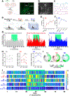
Figure 5:. vmPFC-LEC cells show increased activity and selectivity for tone during threat conditioning and threat expression.
A) Schematic of viral injections and GRIN lens placement. Inset, representative image of lens placement and GCaMP expression. Scale bar, 500 microns. B) Max. projection of vmPFC-LEC cells from representative animal. Scale bar, 50 microns. C) Delta F/F traces from 5 individual vmPFC-LEC cells. D) Schematic for behavioral battery performed during imaging. E) Percent freezing during baseline and tone presentation in threat conditioning in context A. F) Percent freezing during baseline and tone presentation during threat retrieval in context B. G) Normalized PSTH across all cells during tone habituation (TH) (n = 272 cells), threat conditioning (TC) (n = 313 cells), and threat retrieval (TR) (n = 298 cells), aligned to 30 seconds before tone. Normalized to average number of cumulative events in 30-second pretone interval. Tone indicated by green shading. H) Average number of events in each mouse (n= 5 mice) during cumulative pretone, tone, and posttone intervals for TH, TC, and TR. I) Selectivity [(number of events during tone – number of events during 30 s before tone) / (number of events during tone + number of events during 30 s before tone)] for 30 s tone interval versus 30 s pretone interval. Greater than zero indicates selective for tone (green), less than zero indicates selective for pretone (grey), equal to zero indicates non-selective (white). J) Heatmap of cumulative activity 30 s before, during and after tone presentation during TH, (tones 1–5), TC, and TR, for all 363 cells imaged. 1 s bins. Green box indicates tone.
Figure 6:
Figure 6:. vmPFC-LEC subpopulations show different tone selectivity in threat-related and neutral contexts.
A) Selectivity map for cells selective for tone during TC (inner ring) and their selectivity for tone during TR (outer ring). B) Selectivity map for cells selective for tone during TR (inner ring) and their selectivity for tone during TC (outer ring). C) Selectivity map for cells only active in context A (top) or in context B (bottom). D) Heatmap of cumulative activity during TC and TR tones from 30 s before the beginning to 30 s after the end of tone presentation in cells selective for tone in both TC and TR (purple section of selectivity plot in Fig. 7a,b). Green box indicates tone. E) As in D, but for cells only selective for tone in TC (red section of selectivity plot in Fig. 7a). F) As in D, but for cells only selective for tone in TR (blue section of selectivity plot in Fig. 7b).

Comment in

References

    1. Bowers ME & Ressler KJ An Overview of Translationally Informed Treatments for Posttraumatic Stress Disorder: Animal Models of Pavlovian Fear Conditioning to Human Clinical Trials. Biol Psychiatry 78, E15–E27 (2015). - PMC - PubMed
    1. Fenster RJ, Lebois LAM, Ressler KJ & Suh J Brain circuit dysfunction in post-traumatic stress disorder: from mouse to man. Nat Rev Neurosci 19, 535–551 (2018). - PMC - PubMed
    1. Vidal-Gonzalez I, Vidal-Gonzalez B, Rauch SL & Quirk GJ Microstimulation reveals opposing influences of prelimbic and infralimbic cortex on the expression of conditioned fear. Learning & Memory 13, 728–733 (2006). - PMC - PubMed
    1. Sierra-Mercado D, Corcoran KA, Lebrón-Milad K & Quirk GJ Inactivation of the ventromedial prefrontal cortex reduces expression of conditioned fear and impairs subsequent recall of extinction. European Journal of Neuroscience 24, 1751–1758 (2006). - PubMed
    1. Do-Monte FH, Manzano-Nieves G, Quinones-Laracuente K, Ramos-Medina L & Quirk GJ Revisiting the Role of Infralimbic Cortex in Fear Extinction with Optogenetics. Journal of Neuroscience 35, 3607–3615 (2015). - PMC - PubMed

Publication types

LinkOut - more resources