Review
. 2022 Dec 29;8(2):e10476.
doi: 10.1002/btm2.10476. eCollection 2023 Mar.

Microsampling tools for collecting, processing, and storing blood at the point-of-care

Affiliations
Review

Microsampling tools for collecting, processing, and storing blood at the point-of-care

Keith R Baillargeon et al. Bioeng Transl Med. .

Abstract

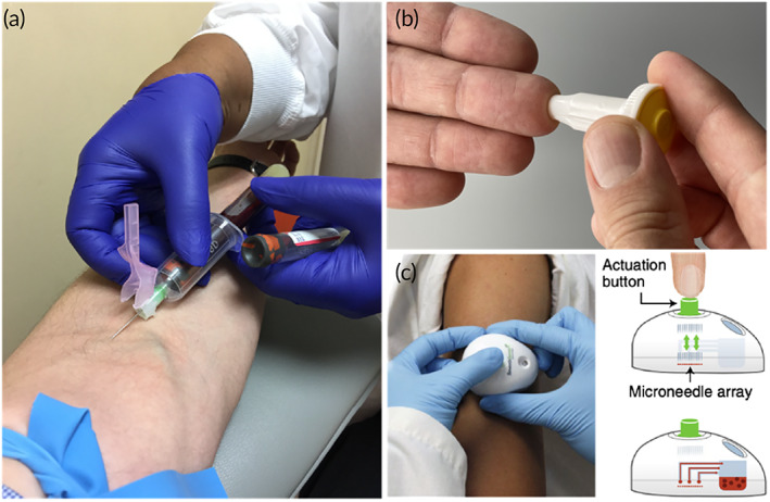
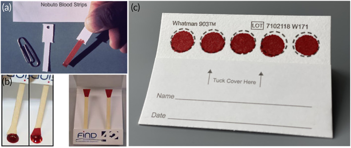
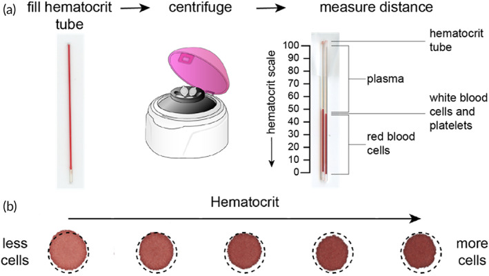
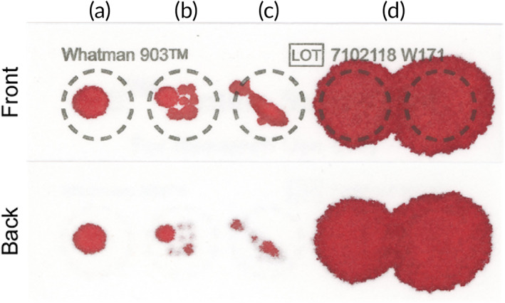
In the wake of the COVID-19 global pandemic, self-administered microsampling tools have reemerged as an effective means to maintain routine healthcare assessments without inundating hospitals or clinics. Finger-stick collection of blood is easily performed at home, in the workplace, or at the point-of-care, obviating the need for a trained phlebotomist. While the initial collection of blood is facile, the diagnostic or clinical utility of the sample is dependent on how the sample is processed and stored prior to transport to an analytical laboratory. The past decade has seen incredible innovation for the development of new materials and technologies to collect low-volume samples of blood with excellent precision that operate independently of the hematocrit effect. The final application of that blood (i.e., the test to be performed) ultimately dictates the collection and storage approach as certain materials or chemical reagents can render a sample diagnostically useless. Consequently, there is not a single microsampling tool that is capable of addressing every clinical need at this time. In this review, we highlight technologies designed for patient-centric microsampling blood at the point-of-care and discuss their utility for quantitative sampling as a function of collection material and technique. In addition to surveying methods for collecting and storing whole blood, we emphasize the need for direct separation of the cellular and liquid components of blood to produce cell-free plasma to expand clinical utility. Integrating advanced functionality while maintaining simple user operation presents a viable means of revolutionizing self-administered microsampling, establishing new avenues for innovation in materials science, and expanding access to healthcare.

Keywords: dried blood spot; microsampling; plasma separation; point‐of‐care; quality; volumetric.


Conflict of interest statement

Keith R. Baillargeon and Charles R. Mace are co‐inventors on patent applications for technologies related to blood and plasma microsampling devices.

Figures

FIGURE 1
FIGURE 1
Techniques for generating blood samples. (a) Venipuncture collection (≥10 ml) performed by a trained phlebotomist (Matthew Lammers, CC BY‐SA 4.0, via Wikimedia Commons). (b) Self‐administered finger stick (≥10 μl) collection. (c) Capillary collection device, TAP (≥100 μl), by YourBio Health (Reprinted by permission from Springer Nature: Nature Biomedical Engineering, Ref , Copyright 2018).
FIGURE 2
FIGURE 2
Whole blood collection technologies. (a) NOBUTO blood strips (Reprinted with permission from Ref 29). (b) Triangle paper dipsticks (Reprinted with permission from Ref 31). (c) Standard dried blood spot card (Whatman 903 protein saver). Each device is limited by unmetered sample absorption and the hematocrit effect.
FIGURE 3
FIGURE 3
The hematocrit effect. (a) Blood samples are collected into a hematocrit microcapillary tube, centrifuged, and measured to report the ratio of RBC volume to total volume of blood by distance (Database Center for Life Science, CC BY 3.0, via Wikimedia Commons). (b) The hematocrit effect can be directly visualized by the reduced diameter of dried blood spot as a function of increased hematocrit value. Higher hematocrit samples contain a higher ratio of cells to plasma, which reduces wicking radius and leads to sampling bias from subpunch analysis (Adapted with permission from Ref , Copyright 2021 American Chemical Society).
FIGURE 4
FIGURE 4
User‐errors associated with dried blood spot (DBS) sampling. Examples include (a) application of insufficient volume, (b) uneven sample application with multiple drops, (c) incomplete penetration through the thickness of the filter paper, and (d) coalescence of blood spots due to overfilling. Improper sampling can result in cards being discarded.
FIGURE 5
FIGURE 5
Volumetric dried blood spot (DBS) technologies. Each device operates independent of the hematocrit effect by restricting the area for sample absorption. (a) Volumetric absorptive paper disk (VAPD, Reprinted with permission from Ref 59) and (b) HemaSpot HF by Spot‐On Sciences (Ref , CC BY 4.0, via MDPI) precut the collection zones to restrict sample volume. (c) Patterned DBS (pDBS) cards use hydrophobic wax barriers to restrict sample flow and distribution (Adapted with permission from Ref , Copyright 2021 American Chemical Society).
FIGURE 6
FIGURE 6
Alternative volumetric dried blood spot (DBS) technologies. These devices present novel approaches for minimizing the hematocrit effect. (a) Mitras volumetric absorptive microsampling device (VAMs) by Neoteryx restricts sample volume to the absorbent polymeric tip but can be affected by touching surfaces after collection or oversaturating (Dove Medical Press Limited, CC BY 3.0, Ref 66). (b) HemaPen by Trajan Scientific and Medical (Reprinted from Ref , Copyright 2019, with permission from Elsevier) and (c) HemaXis DB device by DBS System SA operate independent of the hematocrit by incorporating fixed‐volume capillary tubes, which dispense a discrete volume of blood onto filter paper (Republished with permission of Royal Society of Chemistry, from Ref ; permission conveyed through Copyright Clearance Center, Inc.). (d) The Capitainer device by KTH protects against overfilling by inclusion of PVA valves that redirect excess sample prior to directing a discrete volume of sample to filter paper (Reprinted with permission from Ref , Copyright 2019 American Chemical Society). (e) In contrast, 3D blood spheroids utilize hydrophobic treated paper to eliminate chromatographic effects (Reprinted with permission from Ref , Copyright 2018 American Chemical Society).
FIGURE 7
FIGURE 7
Hand‐powered centrifuges. (a) Eggbeater adapted with tubing containing blood (Reprinted with permission from Ref 88). (b) Salad spinner outfitted with a series of combs to hold microcapillary tubes containing blood (Reprinted with permission from Ref 89). (c) Low‐cost paperfuge with capillary tubes contained within straw segments (Reprinted by permission from Springer Nature: Ref , Copyright 2017.). (d) Unmodified fidget‐spinner toy with individually sealed capillary tubes on each arm (Reprinted with permission from Ref . Copyright 2019 American Chemical Society). Each device requires considerable energy and time input by the user.
FIGURE 8
FIGURE 8
Passive separation techniques and liquid plasma output. Each device incorporates at least one layer of plasma separation membrane for filtering cells by size exclusion. The first device (a) collects diluted liquid plasma directly into a capillary tube (Reprinted from Ref , Copyright 2018, with permission from Elsevier). In contrast, the following devices require a pipette to provide a source of vacuum for recovering plasma. Devices (b) (Reprinted with permission from Ref , Copyright 2013 American Chemical Society) and (c) (Reprinted with permission from Ref 96) operate by size exclusion and sedimentation. While device (d) operates by size exclusion and immunocapture (Reprinted with permission from Ref 97).
FIGURE 9
FIGURE 9
Passive separation techniques and dried plasma output stored in a porous matrix. Two vertically stacked devices enhanced separation efficiency by (a) treating the plasma separation membrane (PSM) layer (Reprinted from Ref , Copyright 2011, with permission from Elsevier) and (b) inclusion of a prefilter material (Reprinted with permission from Ref , Copyright 2020 American Chemical Society). Plasma separation cards comprise a basic structure of PSM layered with filter paper. The (c) Noviplex UNO produces a single plasma sample in a precut disk (Reprinted with permission from Ref , Copyright 2013 American Chemical Society). (d) The volume‐defined dried plasma spot (DPS) meters plasma using a PVA valve to remove excess plasma before filling an absorbent paper strip (Reprinted with permission from Ref , Copyright 2019 American Chemical Society). (e) The Cobas PSC by Roche produces a single sample of plasma in a shield‐shaped fleece located beneath the PSM. (f) The patterned DPS (pDPS) card incorporates a prefilter material and filter paper impregnated with hydrophobic barriers to control plasma flow and distribution for improved purity (Ref 108).
FIGURE 10
FIGURE 10
Lateral flow separation devices. (a) The blood collection device comprises a spreading mesh and rectangular strip of glass fiber to produce a gradient of plasma (Reprinted with permission from Ref 110). A lateral flow test with upstream plasma separation unit (b) comprising three layers of stacked plasma separation membrane (PSM) with unique chemical treatments for increased hydrophilicity (Reprinted with permission from Ref , Copyright 2021 American Chemical Society). (c) Microfluidic paper analytical device (μPAD) comprising overlapped glass fiber separation membrane and filter paper patterned with hydrophobic barriers for detection of plasma proteins (Reprinted with permission from Ref , Copyright 2012 American Chemical Society).
FIGURE 11
FIGURE 11
Plasma separation membrane (PSM)‐free separation technologies. (a) Agglutination‐based approach in a single layer of filter paper with four plasma output zones (Reprinted with permission from Ref 125). (b) A microfluidic device utilizing constriction‐expansion inertial cell sorting and liquid plasma collection (Reprinted with permission from Ref , Copyright 2016 American Chemical Society). (c) Porous superabsorbent polymer (PSAP) beads for separation and storage of plasma by size exclusion (Reprinted with permission from Ref 133).

Similar articles

Cited by

References

    1. Basu D, Kulkarni R. Overview of blood components and their preparation. Indian J Anaesth. 2014;58:529‐537. - PMC - PubMed
    1. Wians HF. Clinical laboratory tests: which, why, and what do the results mean? Lab Med. 2009;40:105‐113.
    1. World Health Organization . Best practices in phlebotomy. WHO Guidelines on Drawing Blood: Best Practices in Phlebotomy. World Health Organization; 2010. https://www.ncbi.nlm.nih.gov/books/NBK138665/. Accessed September 2021 - PubMed
    1. World Health Organization . Annex 3, Collection, storage and shipment of specimens for laboratory diagnosis and interpretation of results. Surveillance Guidelines for Measles, Rubella and Congenital Rubella Syndrome in the WHO European Region. World Health Organization; 2012. https://www.ncbi.nlm.nih.gov/books/NBK143256/. Accessed June 2021 - PubMed
    1. Lalongo C, Bernardini S. Phlebotomy, a bridge between laboratory and patient. Biochem Med. 2016;26:17‐33. - PMC - PubMed

LinkOut - more resources