Review
. 2023 Sep;18(9):1931-1939.
doi: 10.4103/1673-5374.367834.

Mechanisms and treatment strategies of demyelinating and dysmyelinating Charcot-Marie-Tooth disease

Affiliations
Review

Mechanisms and treatment strategies of demyelinating and dysmyelinating Charcot-Marie-Tooth disease

Nadège Hertzog et al. Neural Regen Res. 2023 Sep.

Abstract

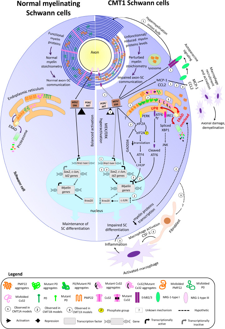
Schwann cells, the myelinating glia of the peripheral nervous system, wrap axons multiple times to build their myelin sheath. Myelin is of paramount importance for axonal integrity and fast axon potential propagation. However, myelin is lacking or dysfunctional in several neuropathies including demyelinating and dysmyelinating Charcot-Marie-Tooth disease. Charcot-Marie-Tooth disease represents the most prevalent inherited neuropathy in humans and is classified either as axonal, demyelinating or dysmyelinating, or as intermediate. The demyelinating or dysmyelinating forms of Charcot-Marie-Tooth disease constitute the majority of the disease cases and are most frequently due to mutations in the three following myelin genes: peripheral myelin protein 22, myelin protein zero and gap junction beta 1 (coding for Connexin 32) causing Charcot-Marie-Tooth disease type 1A, Charcot-Marie-Tooth disease type 1B, and X-linked Charcot-Marie-Tooth disease type 1, respectively. The resulting perturbation of myelin structure and function leads to axonal demyelination or dysmyelination and causes severe disabilities in affected patients. No treatment to cure or slow down the disease progression is currently available on the market, however, scientific discoveries led to a better understanding of the pathomechanisms of the disease and to potential treatment strategies. In this review, we describe the features and molecular mechanisms of the three main demyelinating or dysmyelinating forms of Charcot-Marie-Tooth disease, the rodent models used in research, and the emerging therapeutic approaches to cure or counteract the progression of the disease.

Keywords: Charcot-Marie-Tooth disease; Schwann cells; demyelination and dysmyelination; emerging treatments; endoplasmic reticulum stress; gene therapy; myelin; repair; rodent models; unfolded protein response.


Conflict of interest statement

None

Figures

Figure 1
Figure 1
Overview of myelin structure, molecular pathways, and gene transcription in normal myelinating SCs (left) and in demyelinating/dysmyelinating CMT SCs (right). In normal conditions, myelin proteins are functional and present in the proper stoichiometry, allowing myelin compaction and optimal axon-SC communication. Axon-derived NRG1-type III can bind the ErbB2/3 receptor on SCs and represses Nrg1-type I expression via MEK/ERK signaling. Myelin protein gene transcription is maintained by Krox20, while transcription of negative regulators of myelination (c-Jun, Id2, and Sox2) is silenced, and the activity of PI3K/AKT and MEK/ERK signaling pathways is balanced, thereby maintaining SCs in their differentiation state. In the ER, the misfolded myelin proteins are normally degraded via the ERAD and proteasome pathway. In the case of CMT, the presence of non-functional/dysfunctional myelin proteins and/or the reduced or increased levels of functional myelin proteins perturbate the myelin stoichiometry and impair axon-SC communication. This results in axon dysmyelination, hypomyelination, and demyelination. In CMT1A (1), the perturbed axon-glia communication decreases PI3K/AKT signaling activity, which results in MEK/ERK hyperactivation. The imbalanced activity of PI3K/AKT and MEK/ERK signaling pathways activates the transcription of SC immature and repair markers, leading to impaired SC differentiation and myelination. The absence of axon-SC contact prevents NRG1-type III binding to ErbB2/3 receptors, leading to Nrg1-type I expression. SC-derived NRG1-type I is secreted in an autoparacine way and activates ErbB2/3 receptors, which signal through MEK/ERK and further trigger the expression of negative regulators of myelination. The chronic NRG1-type I expression also leads to hypermyelination and onion bulb formation. In CMT1B (2) and CMT1X (3), misfolded mutant P0 and Cx32 aggregate and accumulate in the ER. Furthermore, some mutant misfolded forms of P0 and Cx32 can trap the wild-type forms of P0 and Cx32 in the ER. Consequently, the levels of functional P0 and Cx32 are reduced in the myelin sheath. PMP22 aggregates are also observed in the ER, cytoplasm, and lysosomes in CMT1A. The accumulation of protein aggregates overloads the ERAD capacity, leading to ER stress and activation of the three UPR arms. The PERK-mediated CHOP activation and the cleaved form of ATF6 reduce myelin gene transcription. GADD34 is then activated by CHOP and dephosphorylates eIF2A. The dephosphorylated form of eIF2A restores normal translational levels and activates MER/ERK. How eIF2A induces MEK/ERK activation is still unknown. In parallel, the IRE1 arm of the UPR may upregulate c-Jun levels via c-Jun N-terminal kinase, leading to Krox20 downregulation and reduced myelin gene transcription. The decreased levels of myelin proteins may then reduce ERAD overloading and ER stress. In CMT1A, CMT1B, and CMT1X, MEK/ERK-mediated MCP-1/CCL2 expression in SCs recruits and activates macrophages, leading to axonal damage and demyelination. In CMT1X, endoneurial fibroblasts form cell-cell contact with macrophages and secrete the macrophage activator CSF-1. The resulting inflammation further impairs SC differentiation. How CMT1X SCs induce CSF-1 expression in fibroblasts is still unknown. ATF4: Activating transcription factor 4; ATF6: activating transcription factor 6; CHOP: CCAAT/enhancer-binding protein homologous gene; c-Jun: gene coding for Jun proto-oncogene; CMT1A: Charcot-Marie-Tooth disease type 1A; CMT1B: Charcot-Marie-Tooth disease type 1B; CMT1X: X-linked Charcot-Marie-Tooth disease type 1; CSF-1: colony-stimulating factor 1; Cx32: connexin 32; eIF2A: eukaryotic translation initiation factor 2A; ERAD: endoplasmic reticulum-associated degradation; ErbB2/3: heterodimer of erb-b2 receptor tyrosine kinases 2 and 3; GADD34: DNA damage inducible protein 34; Id2: gene coding for inhibitor of DNA binding 2; IRE1: inositol-requiring enzyme 1; JNK: c-Jun N-terminal kinase; Krox20: also called Egr2, early growth response 2; MCP-1/CCL2: monocyte chemoattractant protein 1/C-C motif chemokine ligand 2; MEK/ERK: extracellular signal-regulated kinase/mitogen-activated protein kinase; Nrg1-type I: gene coding for neuregulin 1 type I; NRG1-type I: neuregulin 1 type I; NRG1-type III: neuregulin 1 type III; P0: myelin protein zero; PERK: protein kinase RNA-like endoplasmic reticulum kinase; PI3K/AKT: phosphoinositide-3 kinase/v-Akt murine thymoma viral oncogene homolog 1; PMP22: peripheral myelin protein 22; Sox2: gene coding for SRY-box transcription factor 2; UPR: unfolded protein response; XBP1: X-box-binding protein 1.

References

    1. Amici SA, Dunn WA, Jr, Murphy AJ, Adams NC, Gale NW, Valenzuela DM, Yancopoulos GD, Notterpek L. Peripheral myelin protein 22 is in complex with alpha6beta4 integrin, and its absence alters the Schwann cell basal lamina. J Neurosci. 2006;26:1179–1189. - PMC - PubMed
    1. Arthur-Farraj P, Coleman MP. Lessons from injury:how nerve injury studies reveal basic biological mechanisms and therapeutic opportunities for peripheral nerve diseases. Neurotherapeutics. 2021;18:2200–2221. - PMC - PubMed
    1. Arthur-Farraj PJ, Latouche M, Wilton DK, Quintes S, Chabrol E, Banerjee A, Woodhoo A, Jenkins B, Rahman M, Turmaine M, Wicher GK, Mitter R, Greensmith L, Behrens A, Raivich G, Mirsky R, Jessen KR. c-Jun reprograms Schwann cells of injured nerves to generate a repair cell essential for regeneration. Neuron. 2012;75:633–647. - PMC - PubMed
    1. Attarian S, Vallat JM, Magy L, Funalot B, Gonnaud PM, Lacour A, Péréon Y, Dubourg O, Pouget J, Micallef J, Franques J, Lefebvre MN, Ghorab K, Al-Moussawi M, Tiffreau V, Preudhomme M, Magot A, Leclair-Visonneau L, Stojkovic T, Bossi L, et al. An exploratory randomised double-blind and placebo-controlled phase 2 study of a combination of baclofen, naltrexone and sorbitol (PXT3003) in patients with Charcot-Marie-Tooth disease type 1A. Orphanet J Rare Dis. 2014;9:199. - PMC - PubMed
    1. Bai Y, Ianokova E, Pu Q, Ghandour K, Levinson R, Martin JJ, Ceuterick-de Groote C, Mazanec R, Seeman P, Shy ME, Li J. Effect of an R69C mutation in the myelin protein zero gene on myelination and ion channel subtypes. Arch Neurol. 2006;63:1787–1794. - PubMed