. 2023 Sep;18(9):1990-1998.
doi: 10.4103/1673-5374.366497.

Low-temperature 3D-printed collagen/chitosan scaffolds loaded with exosomes derived from neural stem cells pretreated with insulin growth factor-1 enhance neural regeneration after traumatic brain injury

Affiliations

Low-temperature 3D-printed collagen/chitosan scaffolds loaded with exosomes derived from neural stem cells pretreated with insulin growth factor-1 enhance neural regeneration after traumatic brain injury

Xiao-Yin Liu et al. Neural Regen Res. 2023 Sep.

Abstract

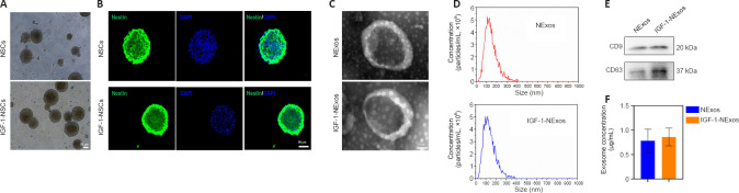
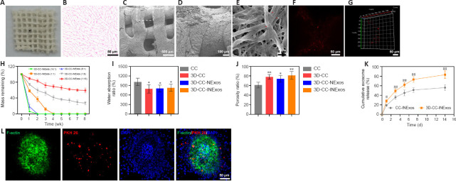
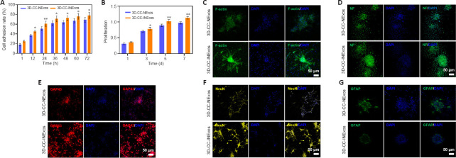
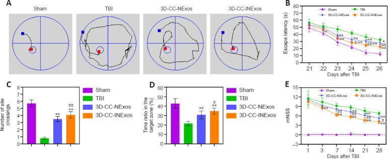
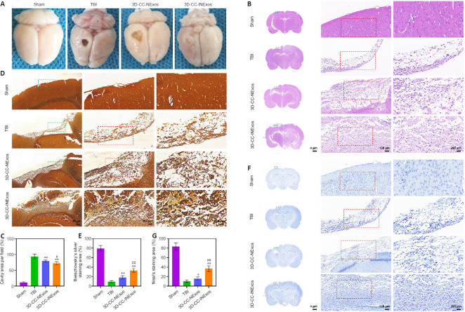
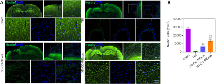
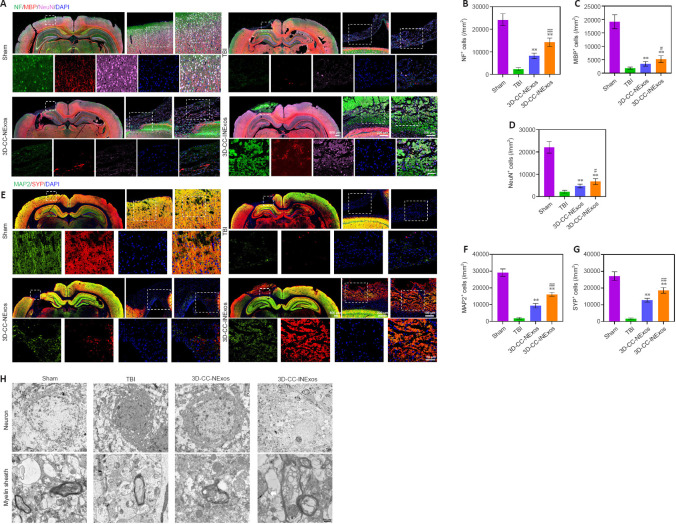
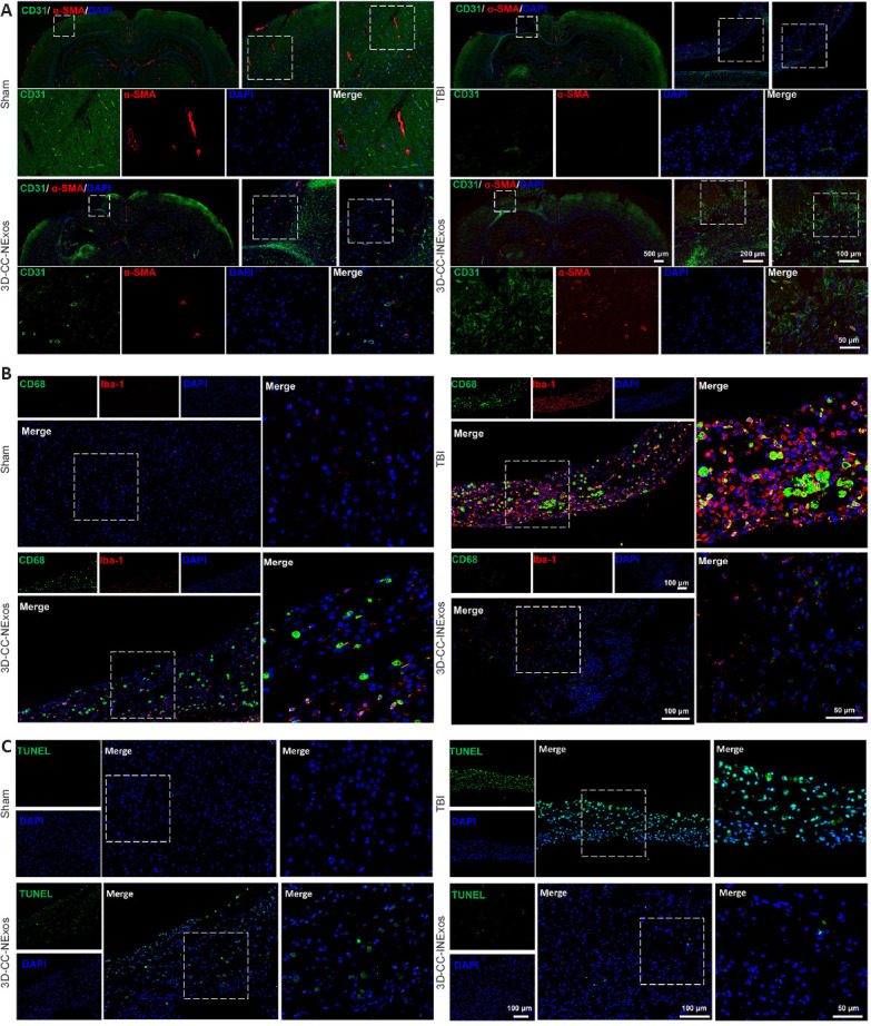
There are various clinical treatments for traumatic brain injury, including surgery, drug therapy, and rehabilitation therapy; however, the therapeutic effects are limited. Scaffolds combined with exosomes represent a promising but challenging method for improving the repair of traumatic brain injury. In this study, we determined the ability of a novel 3D-printed collagen/chitosan scaffold loaded with exosomes derived from neural stem cells pretreated with insulin-like growth factor-1 (3D-CC-INExos) to improve traumatic brain injury repair and functional recovery after traumatic brain injury in rats. Composite scaffolds comprising collagen, chitosan, and exosomes derived from neural stem cells pretreated with insulin-like growth factor-1 (INExos) continuously released exosomes for 2 weeks. Transplantation of 3D-CC-INExos scaffolds significantly improved motor and cognitive functions in a rat traumatic brain injury model, as assessed by the Morris water maze test and modified neurological severity scores. In addition, immunofluorescence staining and transmission electron microscopy showed that 3D-CC-INExos implantation significantly improved the recovery of damaged nerve tissue in the injured area. In conclusion, this study suggests that transplanted 3D-CC-INExos scaffolds might provide a potential strategy for the treatment of traumatic brain injury and lay a solid foundation for clinical translation.

Keywords: 3D printing; angiogenesis; chitosan; collagen; exosomes; functional recovery; insulin-like growth factor-1; neural regeneration; neural stem cells; traumatic brain injury.


Conflict of interest statement

None

Figures

Figure 1
Figure 1
Identification of NSCs and characteristics of exosomes from NSCs. (A) Morphology of NSCs with or without IGF-1 pretreatment under a light microscope. Scale bar: 50 μm. (B) Immunofluorescence staining images of nestin (green) in NSCs with or without IGF-1 pretreatment. Scale bar: 50 μm. (C) Morphology of exosomal structures observed by transmission electron microscopy. Scale bar: 50 nm. (D) Particle size distribution of exosomes measured by nanoparticle tracking analysis. (E) Exosomal markers detected by western blot analyses. (F) Exosome concentrations of NExos and INExos. DAPI: 4′,6-Diamidino-2-phenylindole; IGF-1: insulin-like growth factor 1; NSCs: neural stem cells. NExos: Exosomes extracted from supernatant of NSCs cultured in ordinary complete medium; IGF-1-NExos (INExos): exosomes extracted from supernatant of NSCs cultured in ordinary complete medium supplemented with IGF-1.
Figure 2
Figure 2
Characteristics of 3D-CC-INExos scaffolds. (A) Shape of 3D-CC-INExos scaffolds. (B) 3D-CC-INExos scaffolds shown by hematoxylin and eosin staining. Scale bar: 50 μm. (C, D) Structure of 3D-CC-INExos scaffolds by SEM micrographs (D is enlarged image of white box in C). Scale bars: 500 μm in C, 100 μm in D. (E) SEM micrographs showing exosomes (red arrows) adhered to 3D-CC-INExos scaffolds. Scale bar: 1 μm. (F, G) Immunofluorescence staining images revealed distribution of exosomes in scaffolds. Scale bars: 50 μm. (H) Degradation time curves of scaffolds with different ratios. (I, J) Water absorption (I) and porosity ratios (J) of scaffolds. (K) Cumulative release curves of exosomes from scaffolds within 14 days. (L) IF images showing exosomes labeled with PKH26 (red) released from scaffolds phagocytosed by NSCs (F-actin, green). Scale bars: 50 μm. *P < 0.05, **P < 0.01, vs. CC; #P < 0.05, ##P < 0.01, vs. CC-INExos (two-sample t-tests or one-way analyses of variance followed by Student-Newman-Keuls tests; experiment repeated five times). Four kinds of scaffolds were used: collagen/chitosan (CC), 3D-printed collagen/chitosan (3D-CC), 3D-printed collagen/chitosan/NExos (3D-CC-NExos), and 3D-printed collagen/chitosan/INExos scaffolds (3D-CC-INExos). NExos: exosomes extracted from supernatant of NSCs cultured in ordinary complete medium; IGF-1-NExos (INExos): exosomes extracted from supernatant of NSCs cultured in ordinary complete medium supplemented with IGF-1. DAPI: 4′,6-Diamidino-2-phenylindole; NSCs: neural stem cells; IGF-1: insulin-like growth factor 1; SEM: scanning electron microscopy.
Figure 3
Figure 3
Good biocompatibility of 3D-CC-INExos scaffolds and effects on neural differentiation of NSCs. (A) Cell adhesion rate of NSCs cocultured with scaffolds. (B) Proliferative capacity of NSCs cocultured with scaffolds. (C–G) Typical images of F-actin (green) (C), NF (green) (D), GAP43 (red) (E), NeuN (yellow) (F), and GFAP (green) (G) after coculture of NSCs with 3D-CC-NExos and 3D-CC-INExos. Scale bars: 50 μm. Data are presented as mean ± SD. *P < 0.05, **P < 0.01, vs. 3D-CC-NExos (two-sample t-tests or one-way analyses of variance followed by Student-Newman-Keuls tests; experiments repeated five times). 3D-CC-NExos: 3D-printed collagen/chitosan/NExos; 3D-CC-INExos: 3D-printed collagen/chitosan/INExos scaffolds. NExos: Exosomes extracted from supernatant of NSCs cultured in ordinary complete medium; IGF-1-NExos (INExos): exosomes extracted from supernatant of NSCs cultured in ordinary complete medium supplemented with IGF-1. DAPI: 4′,6-Diamidino-2-phenylindole; GAP43: growth associated protein-43; GFAP: glial fibrillary acidic protein; IGF-1: insulin-like growth factor 1; NeuN: neuronal nuclear protein; NF: neurofilament.
Figure 4
Figure 4
Implantation of 3D-CC-INExos scaffolds improves cognitive function in rats after TBI. (A) Typical swimming path in spatial-learning phase 26 days after TBI. (B) Escape latency analyzed in the spatial-learning stage. (C) Number of site crossings in the spatial-memory stage. (D) Proportion of time in the target zone in the spatial-memory stage. (E) mNSS at 1, 3, 7, 14, 21, and 28 days after TBI. Data are presented as mean ± SD. *P < 0.05, **P < 0.01, vs. TBI; #P < 0.05, ##P < 0.01, vs. 3D-CC-NExos (two-sample t-tests or one-way analyses of variance followed by Student-Newman-Keuls tests; experiments repeated five times). 3D-CC-NExos: 3D-printed collagen/chitosan/NExos; 3D-CC-INExos: 3D-printed collagen/chitosan/INExos scaffolds. NExos: Exosomes extracted from supernatant of NSCs cultured in ordinary complete medium; IGF-1-NExos (INExos): exosomes extracted from supernatant of NSCs cultured in ordinary complete medium supplemented with IGF-1. TBI: Traumatic brain injury.
Figure 5
Figure 5
Tissue regeneration and reparative ability of scaffolds in vivo 2 months after TBI. (A) General images of damaged area in the TBI model. (B) Representative hematoxylin and eosin (HE) staining images of damaged area in sham, TBI, 3D-CC-NExos, and 3D-CC-INExos groups. Middle image is enlarged image of green box in left-hand image; right-hand image is enlarged image of red box in middle image. (C) Quantitative statistical analysis of HE staining. (D) Representative Bielschowsky’s silver staining images of damaged area in sham, TBI, 3D-CC-NExos, and 3D-CC-INExos groups. Middle image is enlarged image of green box in left-hand image; right-hand image is enlarged image of red box in middle image. (E) Quantitative statistical analysis of Bielschowsky’s silver staining. (F) Representative Nissl staining images of damaged area in sham, TBI, 3D-CC-NExos, and 3D-CC-INExos groups. (G) Quantitative statistical analysis of Nissl staining. Middle image is enlarged image of green box in left-hand image; right-hand image is enlarged image of red box in middle image. Data are presented as mean ± SD. *P < 0.05, **P < 0.01, vs. TBI; #P < 0.05, ##P < 0.01, vs. 3D-CC-NExos (two-sample t-tests or one-way analyses of variance followed by Student-Newman-Keuls tests; experiments repeated five times). 3D-CC-NExos: 3D-printed collagen/chitosan/NExos; 3D-CC-INExos: 3D-printed collagen/chitosan/INExos scaffolds. NExos: Exosomes extracted from supernatant of NSCs cultured in ordinary complete medium; IGF-1-NExos (INExos): exosomes extracted from supernatant of NSCs cultured in ordinary complete medium supplemented with IGF-1. TBI: traumatic brain injury.
Figure 6
Figure 6
IF analysis of NSC recruitment in damaged area 2 months post-injury. (A) Typical IF images of nestin (green) in the lesion area in sham, TBI, 3D-CC-NExos, and 3D-CC-INExos groups. Middle image is enlarged image of white box in left-hand image; right-hand image is enlarged image of white box in middle image; image below is enlarged image of white box in right-hand image. (B) Quantitative analysis of nestin-positive cells in lesion area in each group. Data are presented as mean ± SD. **P < 0.01 vs. TBI; ##P < 0.01 vs. 3D-CC-NExos (two-sample t-tests or one-way analyses of variance followed by Student-Newman-Keuls tests; experiments repeated five times). 3D-CC-NExos: 3D-printed collagen/chitosan/NExos; 3D-CC-INExos: 3D-printed collagen/chitosan/INExos scaffolds. NExos: Exosomes extracted from supernatant of NSCs cultured in ordinary complete medium; IGF-1-NExos (INExos): exosomes extracted from supernatant of NSCs cultured in ordinary complete medium supplemented with IGF-1. IF: Immunofluorescence staining; TBI: traumatic brain injury.
Figure 7
Figure 7
Regeneration of nerve fibers, myelin sheaths, and axons 2 months post-injury. (A) Typical IF images of NF (green), MBP (red), and NeuN (pink) in lesion area in sham, TBI, 3D-CC-NExos, and 3D-CC-INExos groups. Middle image is enlarged image of white box in left-hand image; right-hand image is enlarged image of white box in middle image; image below is enlarged image of white box in right-hand image. (B–D) Quantification of NF- (B), MBP- (C), and NeuN- (D) positive cells in lesion area in each group. (E) Typical IF images of MAP2 (green) and SYP (red) in lesion area in sham, TBI, 3D-CC-NExos, and 3D-CC-INExos groups. Middle image is enlarged image of white box in left-hand image; right-hand image is enlarged image of white box in middle image; image below is enlarged image of white box in right-hand image. (F–G) Quantification of MAP2- (F) and SYP-positive (G) cells in lesion area in each group. (H) Typical TEM images of lesion area in each group. Scale bars: 500 nm in H. Data are presented as mean ± SD. **P < 0.01, vs. TBI; #P < 0.05, ##P < 0.01, vs. 3D-CC-NExos (two-sample t-tests or one-way analyses of variance followed by Student-Newman-Keuls tests; experiments repeated five times). 3D-CC-NExos: 3D-printed collagen/chitosan/NExos; 3D-CC-INExos: 3D-printed collagen/chitosan/INExos scaffolds. NExos: Exosomes extracted from supernatant of NSCs cultured in ordinary complete medium; IGF-1-NExos (INExos): exosomes extracted from supernatant of NSCs cultured in ordinary complete medium supplemented with IGF-1. DAPI: 4′,6-Diamidino-2-phenylindole; IF: Immunofluorescence staining; MAP2: microtubule-associated protein 2; MBP: myelin basic protein; NeuN: neuronal nuclear protein; NF: neurofilament; SYP: synaptophysin; TBI: traumatic brain injury.
Figure 8
Figure 8
Angiogenesis, inflammation, and apoptosis in the injured area 2 months post-injury. (A) Typical IF images of angiogenesis markers CD31 (green) and α-SMA (red) in lesion area in sham, TBI, 3D-CC-NExos, and 3D-CC-INExos groups. Middle image is enlarged image of white box in left-hand image; right-hand image is enlarged image of white box in middle image; image below is enlarged image of white box in right-hand image. (B) Typical immunofluorescence staining images of inflammatory markers CD68 (green) and Iba-1 (red) in lesion area in sham, TBI, 3D-CC-NExos, and 3D-CC-INExos groups. Right-hand image is enlarged image of white box in left-hand image. (C) Typical images of TUNEL staining (green) for apoptosis in lesion area in sham, TBI, 3D-CC-NExos, and 3D-CC-INExos groups. Right-hand image is enlarged image of white box in left-hand image. 3D-CC-NExos: 3D-printed collagen/chitosan/NExos; 3D-CC-INExos: 3D-printed collagen/chitosan/INExos scaffolds. NExos: Exosomes extracted from supernatant of NSCs cultured in ordinary complete medium; IGF-1-NExos (INExos): exosomes extracted from supernatant of NSCs cultured in ordinary complete medium supplemented with IGF-1. α-SMA: Alpha smooth muscle actin; Iba1: ionized calcium binding adaptor molecule 1; TUNEL: terminal deoxynucleotidyl transferase dUTP nick-end labeling; DAPI: 4′,6-diamidino-2-phenylindole.
Figure 9
Figure 9
Biocompatibility of scaffolds in vivo. (A) Representative HE staining of the heart, lung, liver, spleen, and kidney in sham, TBI, 3D-CC-NExos, and 3D-CC-INExos groups 1 month after TBI and 2 months post-injury. Scale bar: 100 μm. (B) ALT, AST, BUN, and CR levels. Data are presented as mean ± SD. Experiments repeated five times. 3D-CC-NExos: 3D-printed collagen/chitosan/NExos; 3D-CC-INExos: 3D-printed collagen/chitosan/INExos scaffolds. NExos: Exosomes extracted from supernatant of NSCs cultured in ordinary complete medium; IGF-1-NExos (INExos): exosomes extracted from supernatant of NSCs cultured in ordinary complete medium supplemented with IGF-1. ALT: Alanine transaminase; AST: aspartate aminotransferase; BUN: blood urea nitrogen; CR: creatinine.

References

    1. Acosta SA, Tajiri N, Hoover J, Kaneko Y, Borlongan CV. Intravenous bone marrow stem cell grafts preferentially migrate to spleen and abrogate chronic inflammation in stroke. Stroke. 2015;46:2616–2627. - PMC - PubMed
    1. Ayer-le Lievre C, Stahlbom PA, Sara VR. Expression of IGF-I and -II mRNA in the brain and craniofacial region of the rat fetus. Development. 1991;111:105–115. - PubMed
    1. Bianchi VE, Locatelli V, Rizzi L. Neurotrophic and neuroregenerative effects of GH/IGF1. Int J Mol Sci. 2017;18:2441. - PMC - PubMed
    1. Chen Y, Lin J, Yan W. A prosperous application of hydrogels with extracellular vesicles release for traumatic brain injury. Front Neurol. 2022;13:908468. - PMC - PubMed
    1. Cui L, Saeed Y, Li H, Yang J. Regenerative medicine and traumatic brain injury:from stem cell to cell-free therapeutic strategies. Regen Med. 2022;17:37–53. - PubMed