Review
. 2023 Mar 28;17(6):5163-5186.
doi: 10.1021/acsnano.2c08913. Epub 2023 Mar 16.

Ex Situ Characterization of 1T/2H MoS2 and Their Carbon Composites for Energy Applications, a Review

Affiliations
Review

Ex Situ Characterization of 1T/2H MoS2 and Their Carbon Composites for Energy Applications, a Review

Alexandar D Marinov et al. ACS Nano. .

Abstract

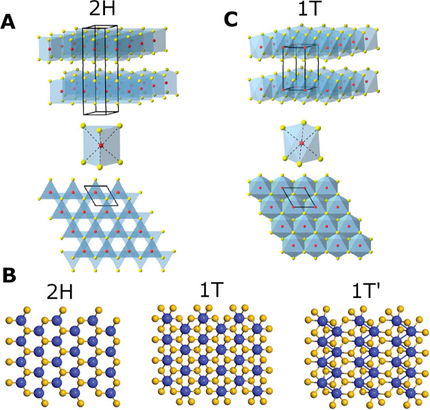
The growing interest in the development of next-generation net zero energy systems has led to the expansion of molybdenum disulfide (MoS2) research in this area. This activity has resulted in a wide range of manufacturing/synthesis methods, controllable morphologies, diverse carbonaceous composite structures, a multitude of applicable characterization techniques, and multiple energy applications for MoS2. To assess the literature trends, 37,347 MoS2 research articles from Web of Science were text scanned to classify articles according to energy application research and characterization techniques employed. Within the review, characterization techniques are grouped under the following categories: morphology, crystal structure, composition, and chemistry. The most common characterization techniques identified through text scanning are recommended as the base fingerprint for MoS2 samples. These include: scanning electron microscopy (SEM), X-ray diffraction (XRD), X-ray photoelectron spectroscopy (XPS), and Raman spectroscopy. Similarly, XPS and Raman spectroscopy are suggested for 2H or 1T MoS2 phase confirmation. We provide guidance on the collection and presentation of MoS2 characterization data. This includes how to effectively combine multiple characterization techniques, considering the sample area probed by each technique and their statistical significance, and the benefit of using reference samples. For ease of access for future experimental comparison, key numeric MoS2 characterization values are tabulated and major literature discrepancies or currently debated characterization disputes are highlighted.

Keywords: HER; LIB; MoS2; SIB; TEM; battery; characterization; energy application; supercapacitor; text scanning.


Conflict of interest statement

The authors declare no competing financial interest.

Figures

Figure 1
Figure 1
(A) Distribution of scan hits per relevant energy application category for 2011–2022 using Web of Science API (performed on 30/11/2022) and custom text classification. For detailed analysis methods, refer to the Supporting Information. Total count of 8,898 research articles presented. (B) Distribution of MoS2 production techniques used within the 100 manually surveyed energy application-oriented papers. (C) Production routes for 1T MoS2 reported by 29 out of the 100 manually surveyed energy application-oriented papers. Variance for all bar charts included in Table SI 5.
Scheme 1
Scheme 1. Life Cycle of MoS2 Samples in Research for Energy Applications
Comparison of the similarities and differences in production routes, including top-down or bottom-up methods. Indication of the general similarity between sub-categories of production routes, highlighting the various stages at which characterization techniques are typically applied, followed by electrode fabrication and different energy applications. The direction of the arrows indicates the flow of processing steps. Solid arrows represent synthesis or material processing steps, and dashed arrows represent characterization steps. Color coding: morphology–red, crystal structure–purple, composition–green, chemistry–blue, and other–grey. Abbreviations: Scanning electron microscopy (SEM), transmission electron microscopy (TEM), atomic force microscopy (AFM), gas adsorption (Gas Ads.), X-ray diffraction (XRD), selected area electron diffraction (SAED), X-ray photoelectron spectroscopy (XPS), energy dispersive spectroscopy (EDS), thermogravimetric analysis (TGA), differential scanning calorimetry (DSC), photoluminescence (PL), and ultraviolet-visible spectroscopy (UV-vis).
Figure 2
Figure 2
Structural representation of the polymorphs of MoS2. Red/blue atoms indicate Mo, and yellow atoms depict S. (A) Alignment of atoms in two adjacent layers of 2H MoS2, trigonal prismatic coordination of Mo atom, and top view (c-axis direction) of 2H nanosheet plane. (B) Top view of c-axis plane of the 2H, 1T, and 1T’ MoS2. (C) Alignment of two adjacent layers of 1T MoS2, octahedral coordination of Mo atom, and top view of 1T nanosheet plane. (A, C) Adapted from ref (24). Copyright 2013 American Chemical Society. (B) Adapted with permission from ref (126). Copyright 2017 Royal Society of Chemistry.
Figure 3
Figure 3
(A) Histogram of scan hits per relevant characterization category for 2011–2022 using Web of Science API (performed on 30/11/2022) and custom text classification. For detailed analysis methods, refer to the Supporting Information. Total count of 11,227 research articles presented, which can contribute to each of the characterization sub-categories. General color grouping of the techniques is based on the key information each technique provides regarding the material. (B) Schematic of different sample forms to which various characterization techniques can be applied. Abbreviations: Scanning electron microscopy (SEM), transmission electron microscopy (TEM), high-resolution TEM (HRTEM), high angle annular dark field STEM (HAADF-STEM), atomic force microscopy (AFM), gas adsorption (Gas Ads.), X-ray diffraction (XRD), selected area electron diffraction (SAED), X-ray photoelectron spectroscopy (XPS), energy dispersive spectroscopy (EDS), thermogravimetric analysis (TGA), differential scanning calorimetry (DSC), photoluminescence (PL), ultraviolet–visible spectroscopy (UV–vis), and Fourier transform infrared (FTIR).
Figure 4
Figure 4
Microscopy techniques. SEM: (A) MoO3 on carbon filter paper. Adapted from ref (12). Copyright 2014 American Chemical Society. (B) MoS2/graphene composite (1:1). Adapted from ref (146). Copyright 2011 American Chemical Society. (C) MoS2/carbon nanoflowers. Adapted from ref (54). Copyright 2014 American Chemical Society. (D) MoS2 spheres. Adapted with permission from ref (38). Copyright 2018 Elsevier. (E) Perpendicular 1T MoS2 nanosheets on 2H MoS2 substrate. Adapted from (18). Copyright 2016 American Chemical Society. (F) MoS2 on a 3D graphene network. Adapted with permission from (74). Copyright 2013 Wiley. TEM: (G) MoS2 on a 3D graphene network. Adapted with permission from ref (74). Copyright 2013 Wiley. HRTEM: (H) heterogeneous structure of 1T/2H MoS2 nanosheet. Insets: structures of 2H and 1T MoS2. (I, J) Enlarged segment from H indicating the atomic arrangement of 2H and 1T MoS2 respectively. Insets: FFT of image. Adapted from ref (26). Copyright 2016 American Chemical Society. HAADF-STEM: (K) heterogeneous MoS2 produced from ball-milling. Adapted with permission from (110). Copyright 2016 Royal Society of Chemistry. For (H–K), blue and yellow atoms represent Mo and S, respectively. (L, M) Intensity distributions along lines in (I) and (J), respectively. Adapted from ref (26). Copyright 2016 American Chemical Society.
Figure 5
Figure 5
Morphology techniques. AFM: (A) and (B) of quantum dots exfoliated from Li electrochemically intercalated in MoS2 with current densities of 1.0 A g–1 and 0.001 A g–1, respectively. Scale bars are 200 nm. Adapted from ref (9). Copyright 2018 American Chemical Society. CS-AFM: (C, D) micrographs of 2H MoS2 from CVD and 1T MoS2 from lithiation and exfoliation of the 2H MoS2. (E, F) Conductivity maps of (C) and (D), respectively. Inset in (F): current–voltage sweeps of 2H and 1T MoS2. Adapted with permission from ref (24). Copyright 2013 American Chemical Society. DLS: (G) size distribution of liquid-exfoliated MoS2. Inset: exfoliated MoS2 solution. Adapted with permission from (29). Copyright 2018 Elsevier. N2 adsorption: (H) adsorption/desorption isotherms and (I) pore diameter distribution for 2H MoS2 and 1T MoS2/carbon composite. Inset in (H): table of BET and BJH values. Adapted with permission from ref (39). Copyright 2019 Wiley.
Figure 6
Figure 6
Crystal structure techniques. XRD: (A) Diffraction patterns of top-down precursor MoS2 ball-milled for 2, 5, and 10 h. Adapted with permission from ref (110). Copyright 2016 Royal Society of Chemistry. (B) Schematic representation of a single flake of MoS2 viewed from the direction of the different Miller indices plane groups. (C) Statistical analysis of the MoS2 XRD peaks from literature. Note, the 2θ positions are not exact. Approximations and averages were used where literature was vague. Additionally, the intensity is not representative of an XRD pattern but the number of literature sources reporting the different peaks. Only peaks with more than five mentions were indexed. XRD: (D, E) diffraction patterns of intercalation of species in the MoS2 lattice using sodium naphthalenide. Schematic displays the varying degree of interlayer space expansion due to different intercalants. Adapted with permission from ref (118). Copyright 2014 Nature. SAED: patterns of commercial MoS2 powder in Na cells discharged to (F) 60, (G) 80, (H) 160, and (I) 256 mAh g–1. Adapted from ref (75). Copyright 2014 American Chemical Society. (J, K) SAED: of 1T MoS2/graphene electrodes in a LIB after the (J) third lithiation (discharge) and after the (K) third delithiation (charge), respectively. Adapted with permission from ref (37). Copyright 2019 Elsevier.
Figure 7
Figure 7
Composition techniques. XPS: (A) survey scan of MoS2 grown vertically on graphene sheets. Adapted from ref (56). Copyright 2016 American Chemical Society. (B, C) Fitting of the Mo 3d and S 2p peaks with models to quantify the transition from 2H top-down precursor crystals to 1T nanodots. Adapted with permission from ref (25). Copyright 2018 Wiley. (D) Mo 3d region scans of 2H and 1T’ MoS2 crystals. Adapted with permission from ref (20). Copyright 2018 Nature. XPS statistics: (E) bar chart of the reported energy down-shift of the Mo 3d peaks for 2H to 1T phase shift in literature. TGA: (F) 2H and 1T MoS2/C composites. Adapted with permission from ref (39). Copyright 2019 Wiley. EDS: mapping of MoS2/carbon corn stalk composites for elements; (G) Mo, (H) S, and (I) C. (J) The original SEM image. Adapted from ref (36). Copyright 2018 American Chemical Society.
Figure 8
Figure 8
Chemistry techniques. (A) Illustration of the Raman phonon interactions on MoS2 atoms. Adapted with permission from (161). Copyright 2014 Nature. Raman: spectra of (B) top-down precursor MoS2 measured with different excitation wavelengths. Adapted with permission from ref (109). Copyright 2015 IOP Science. (C) Frequencies of E2g1 and A1g varying with laser wavelength and layer number. Adapted with permission from ref (144). Copyright 2012 Wiley. (D) Typical MoS2 precursors used in bottom-up approaches (MoO3 and MoO2) and pure compounds expected to be formed during LIB discharge (Li2S and S), and (E) ex situ MoS2 electrode at various states during a discharge and recharge cycle in a LIB (black–pristine, red–discharged to 0.8 V, green–discharged to 0.8 V and charged to 3.0 V, blue–discharged to 0.05 V, magenta–discharged to 0.05 V and charged to 3.0 V). Adapted from ref (69). Copyright 2018 American Chemical Society. PL: spectra of (F) the distinction between 2H and 1T MoS2. Adapted with permission from ref (20). Copyright 2018 Nature. (G–J) Optical microscopy, AFM, J3 Raman mode, and PL of a masked MoS2 sample bombarded with Ar+ ions to cause controlled transition to 1T. The green boxes in (G) indicate the masking, which has subsequently been removed for (H–J) scans. Adapted from ref (131). Copyright 2017 American Chemical Society.

References

    1. Novoselov K. S.; Geim A. K.; Morozov S. V.; Jiang D.; Zhang Y.; Dubonos S. V.; Griogorieva I. V.; Firsov A. A. Electric Field Effect in Atomically Thin Carbon Films. Science 2004, 306, 666–669. 10.1126/science.1102896. - DOI - PubMed
    1. Chng E. L. K.; Sofer Z.; Pumera M. MoS2 exhibits stronger toxicity with increased exfoliation. Nanoscale 2014, 6, 14412–14418. 10.1039/C4NR04907A. - DOI - PubMed
    1. Moore C.; Movia D.; Smith R. J.; Hanlon D.; Lebre F.; Lavelle E. C.; Burne H. J.; Coleman J. N.; Volkov Y.; McIntyre J. Industrial grade 2D molybdenum disulphide (MoS2): An in vitro exploration of the impact on cellular uptake, cytotoxicity, and inflammation. 2D Materials 2017, 4, 025065.10.1088/2053-1583/aa673f. - DOI
    1. Arefi-Oskoui S.; Khataee A.; Ucun O. K.; Kobya M.; Hanci T. Ö.; Arslan-Alaton I. Toxicity evaluation of bulk and nanosheet MoS2 catalysts using battery bioassays. Chemosphere 2021, 268, 128822.10.1016/j.chemosphere.2020.128822. - DOI - PubMed
    1. Winer W. O. Molybdenum Disulpide As a Lubricant: a Review of Fundamental Knowledge. Wear 1967, 10, 422–452. 10.1016/0043-1648(67)90187-1. - DOI