. 2023 Mar 17;379(6637):1140-1149.
doi: 10.1126/science.abq5622. Epub 2023 Mar 16.

Mechanism of STMN2 cryptic splice-polyadenylation and its correction for TDP-43 proteinopathies

Affiliations

Mechanism of STMN2 cryptic splice-polyadenylation and its correction for TDP-43 proteinopathies

Michael W Baughn et al. Science. .

Abstract

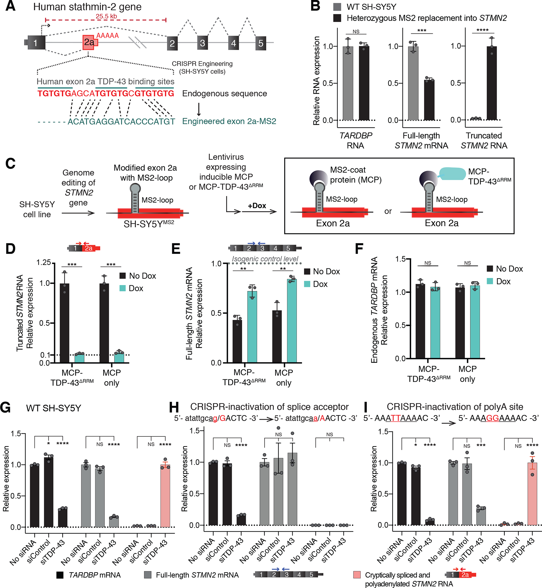
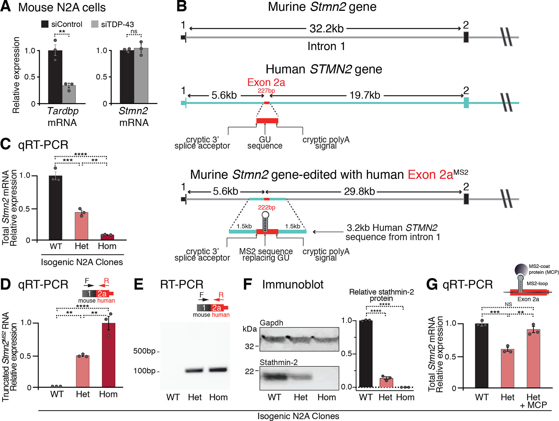
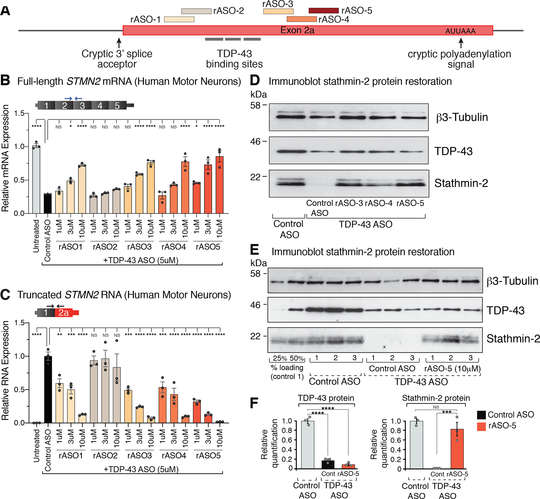
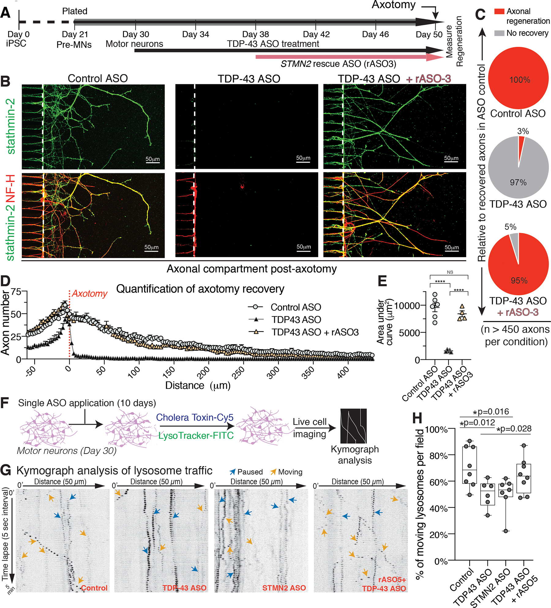
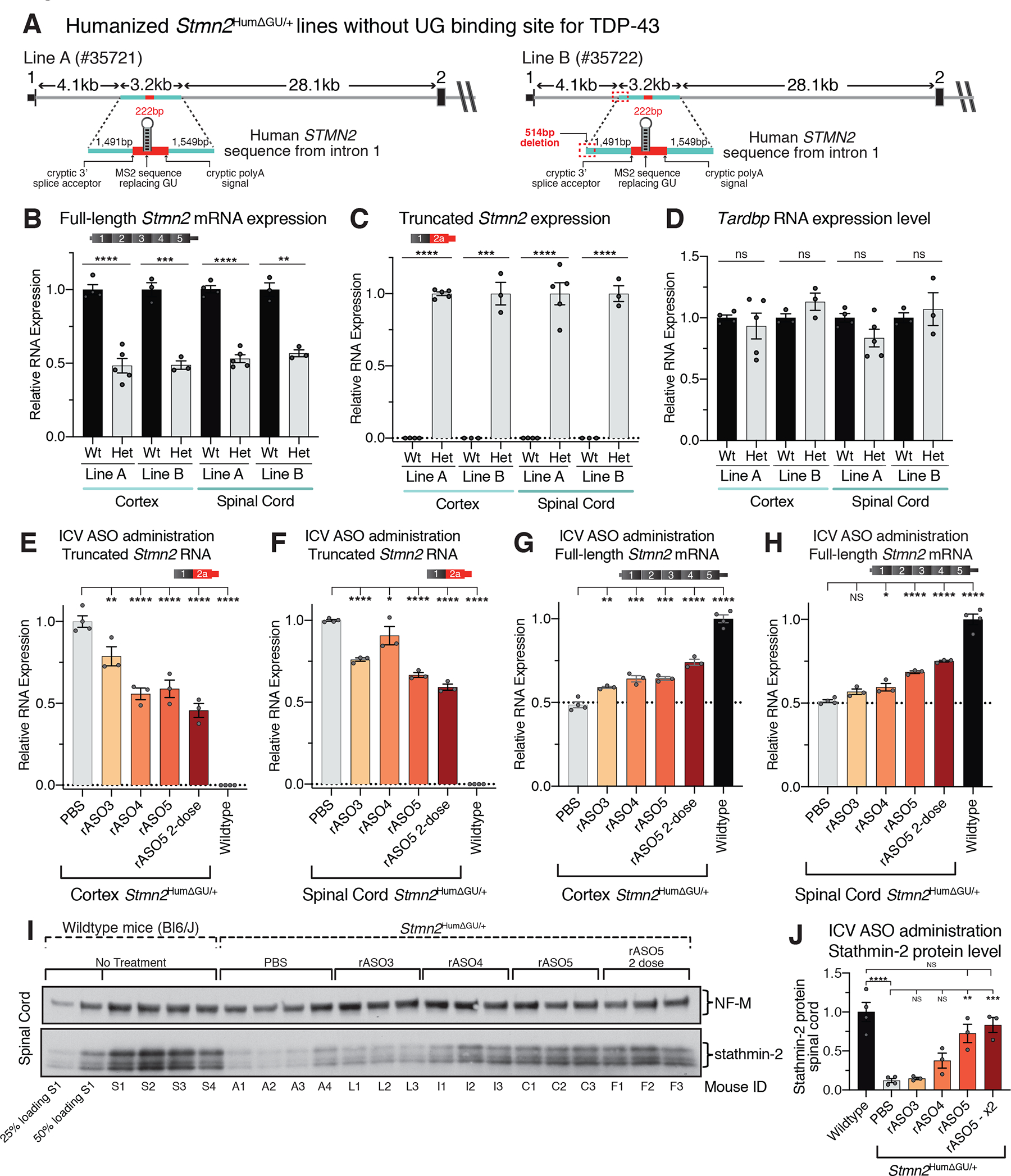
Loss of nuclear TDP-43 is a hallmark of neurodegeneration in TDP-43 proteinopathies, including amyotrophic lateral sclerosis (ALS) and frontotemporal dementia (FTD). TDP-43 mislocalization results in cryptic splicing and polyadenylation of pre-messenger RNAs (pre-mRNAs) encoding stathmin-2 (also known as SCG10), a protein that is required for axonal regeneration. We found that TDP-43 binding to a GU-rich region sterically blocked recognition of the cryptic 3' splice site in STMN2 pre-mRNA. Targeting dCasRx or antisense oligonucleotides (ASOs) suppressed cryptic splicing, which restored axonal regeneration and stathmin-2-dependent lysosome trafficking in TDP-43-deficient human motor neurons. In mice that were gene-edited to contain human STMN2 cryptic splice-polyadenylation sequences, ASO injection into cerebral spinal fluid successfully corrected Stmn2 pre-mRNA misprocessing and restored stathmin-2 expression levels independently of TDP-43 binding.


Conflict of interest statement

Competing interests:

A patent application has been filed describing composition and methods for restoring stathmin-2 expression; publication number US 2021-0252039. D.W.C. and Z.M. are inventors on the patent.

CFB, KL, PJN, FR, are employees of Ionis Pharmaceuticals.

DWC is a consultant for Ionis Pharmaceuticals.

Figures

Figure 1.
Figure 1.. Human GU-motif removal and MS2-directed tethering demonstrate TDP-43 binding locus, while cryptic site mutations identify TDP-43 dependent misprocessing requiring cryptic splice acceptor.
(A) Schematic of CRISPR-engineering strategy for conversion of the GU binding motif in exon 2a into an MS2 aptamer sequence in one STMN2 allele of diploid SH-SY5Y neuroblastoma cells. (B) qRT-PCR demonstrating that SH-SY5Y cells carrying heterozygous GU to MS2 edit misprocess STMN2 RNA, leading to 50% loss of stathmin-2 encoding mRNAs compared with wildtype cells and accumulation of truncated RNA. (C) Schematic depicting MS2:MCP directed strategy to direct MCP-tethered proteins to the normal TDP-43 binding locus. (D-F) qRT-PCR measurement of (D) truncated STMN2, (E) full-length STMN2 mRNA, or (F) endogenous TARDBP mRNA levels with and without induction of MCP-fusion protein expression in SH-SY5Y cells carrying heterozygous MS2 aptamer insertion. (G-I) qRT-PCR measured expression of TARDBP, full-length STMN2 mRNA and truncated STMN2 RNAs 96 hours after siRNA treatment with a control siRNA pool or a pool targeting TARDBP in (G) wildtype SH-SY5Y cells, (H) SH-SY5Y cells harboring homozygous mutation of the human exon 2a 3’ splice acceptor site and (I) SH-SY5Y cells harboring a homozygous mutation of the human exon 2a premature polyadenylation signal to the murine sequence AGGAAA. For all qPCR analysis individual data points are independently treated wells of cells. Error bars are SEM. Statistical significance was determined by 2-tailed Student’s T-test (B-F), or 1-way ANOVA with Dunnett correction (G-I). ****, p <0.0001; ***, p < 0.001; **, p < 0.01; *, p <0.05.
Figure 2.
Figure 2.. Humanization of murine Stmn2 gene in N2A cells demonstrates human-specific inhibition of altered STMN2 pre-mRNA processing via non-conserved TDP-43 binding sites.
(A) qRT-PCR demonstrating that depletion of TDP-43 in wildtype murine N2A cells does not affect Stmn2 expression levels. (B) Schematics of the human and mouse Stmn2 genomic regions before and after genome-editing to insert a 3kb human fragment of STMN2 intron 1 into murine N2A cells. (C) qRT-PCR demonstrating dose-dependent reduction of N2A Stmn2 mRNA level that correlates with the number of alleles carrying human STMN2 gene fragment. (D) qRT-PCR and (E) RT-PCR confirming expression of chimeric Stmn2 truncated RNA with murine exon 1 spliced to human exon 2a, in N2A clones carrying the human STMN2 gene fragment. (F) Immunoblotting confirming reduced expression of full-length stathmin-2 protein levels in N2A clones that carry humanized Stmn2 gene fragment. (G) qRT-PCR showing restoration of normal Stmn2 pre-mRNA processing in N2A cells upon doxycycline induction of MCP expression. For all qPCR analyses, each data point represents an independently treated well of N2A cells. Error bars are SEM. Statistical significance was determined by 2-tailed Student’s T-test (A), or 1-way ANOVA with Dunnett (F) or Tukey (C, D, G) correction. ****, p <0.0001; ***, p < 0.001; **, p < 0.01; *, p <0.05.
Figure 3.
Figure 3.. Dose dependent suppression of STMN2 cryptic splicing and polyadenylation by rASOs in iPSC-derived motor neurons with TDP-43 depletion.
(A) Schematic representation of the exon 2a region of human STMN2 gene with TDP-43 binding sites and selected rASOs that show splice-modifying activity. (B) STMN2 mRNA restoration analyzed by qRT-PCR after treatment with 5 representative rASOs in iPSC-derived motor neurons depleted of TDP-43. Expression of TFRC mRNA was used as endogenous control. (C) qRT-PCR analysis of truncated STMN2 RNA levels after treatment with 5 representative rASOs in iPSC-derived motor neurons depleted of TDP-43. Expression of TFRC mRNA was used as endogenous control. (D) Immunoblot showing TDP-43 and stathmin-2 protein levels in iPS motor neurons treated with control or TDP-43 suppressing ASOs, subsequently treated with control or splice-rescuing rASOs 3, 4, or 5 to restore stathmin-2. Beta3 Tubulin used as an endogenous control. (E) Immunoblot showing TDP-43 and stathmin-2 levels in motor neurons depleted of TDP-43 with a targeted ASO and subsequently treated with control or rescue ASO-5. Linearity of antibody detection with 25% and 50% control treated motor neuron lysate loading controls included at the far-left side of the blot. Immunoblot quantified in (F). Each lane and data point represents an independently differentiated and ASO treated neuronal culture. Error bars are SEM. Statistical significance was determined by 1-way ANOVA with Dunnett correction. ****, p <0.0001; ***, p < 0.001; **, p < 0.01; *, p <0.05.
Figure 4.
Figure 4.. Restoration of axonal regeneration capacity using rASOs that rescue stathmin-2 levels in iPSC-derived motor neurons with TDP-43 depletion.
(A) Timeline of iPSC-derived motor neuron maturation, ASO/rASO treatment and axotomy. (B) Immunofluorescence images of microgrooves (left of dotted line) and distal compartments (right), 36 hours post-axotomy. Axonal regeneration and growth cones are observed by immunofluorescence detection of stathmin-2 (green) and NF-H (red) in the terminals of motor neurons. (C-E) Quantification of axonal recovery for at least 450 axons per condition represented as the percentage of recovered axons relative to control ASO treated motor neurons in (C), the overall number of axons per micrometer from the axotomy site plotted in (D), and corresponding area-under the curve in (E). Statistical significance determined by one-way ANOVA with Tukey’s multiple comparison correction. (F) Schematic of ASO treatment and live motor neuron lysosomal tracking and analysis. (G) Representative kymographs of lysosomal transport in axons of ASO-treated motor neurons. (H) Quantification of moving tracked axonal lysosomes after ASO treatment, statistical significance determined by one-way ANOVA indicated, three independently differentiated chambers quantified per condition. Error bars are SEM. ****, p <0.0001; ***, p < 0.001; **, p < 0.01; *, p <0.05.
Figure 5.
Figure 5.. In vivo ASO-mediated restoration of Stmn2 pre-mRNA processing in a humanized mouse, engineered to constitutively misprocess Stmn2 RNAs from a partly humanized allele.
(A) Schematic showing the strategy to produce a Stmn2 human exon 2A knock-in mouse without a TDP-43 GU binding site to drive constitutive misprocessing of the humanized allele, and resulting mouse lines obtained after CRISPR editing. (B) qRT-PCR showing full-length murine Stmn2 mRNA levels were reduced by half in animals heterozygous for the humanized Stmn2HumΔGU/+ allele compared with wildtype littermate controls. (C) qRT-PCR showing that truncated chimeric RNAs consisting of a murine exon 1 fused to a modified human exon 2a were abundantly and specifically expressed in mice heterozygously carrying the modified humanized Stmn2HumΔGU/+ allele. (D) qRT-PCR showing normal murine Tardbp mRNA levels in both heterozygous Stmn2HumΔGU/+ mice and wildtype littermate controls. (E-F) qRT-PCR showing suppression of truncated chimeric RNA accumulation in both brain (E), and spinal cord (F), of mice dosed by ICV injection with rASOs. (G-H) qRT-PCR Sshowing rASO-mediated restoration of full-length murine Stmn2 mRNAs in both brain (G), and spinal cord (H), of mice after ICV injection of rASOs. (I) Immunoblot showing restoration of stathmin-2 protein in the spinal cords of heterozygous Stmn2HumΔGU/+ mice dosed by ICV injection with rASOs, compared with PBS injected animals or wildtype C57 BL/6J mice. 25% and 50% loading of lysates from wildtype mouse S1at the far left, NF-M shown as an endogenous loading control protein. (J) Quantification of relative stathmin-2 protein restoration. Each lane and data point represents an individual mouse. Error bars are SEM. Statistical significance was determined by 2-tailed Student’s T-test (B-D), or 1-way ANOVA with Dunnett (E-H) or Tukey (J) corrections. ****, p <0.0001; ***, p < 0.001; **, p < 0.01; *, p <0.05.

Comment in

References

    1. Nussbacher JK, Tabet R, Yeo GW, Lagier-Tourenne C, Disruption of RNA Metabolism in Neurological Diseases and Emerging Therapeutic Interventions. Neuron 102, 294–320 (2019). - PMC - PubMed
    1. Lagier-Tourenne C, Polymenidou M, Cleveland DW, TDP-43 and FUS/TLS: emerging roles in RNA processing and neurodegeneration. Hum Mol Genet 19, R46–64 (2010). - PMC - PubMed
    1. Nicolas A et al., Genome-wide Analyses Identify KIF5A as a Novel ALS Gene. Neuron 97, 1268–1283 e1266 (2018). - PMC - PubMed
    1. Weskamp K, Barmada SJ, RNA Degradation in Neurodegenerative Disease. Adv Neurobiol 20, 103–142 (2018). - PMC - PubMed
    1. Gitler AD, Shorter J, RNA-binding proteins with prion-like domains in ALS and FTLD-U. Prion 5, 179–187 (2011). - PMC - PubMed