. 2023 Apr 19;43(16):2973-2987.
doi: 10.1523/JNEUROSCI.1778-22.2023. Epub 2023 Mar 16.

Threat and Reward Imminence Processing in the Human Brain

Affiliations

Threat and Reward Imminence Processing in the Human Brain

Dinavahi V P S Murty et al. J Neurosci. .

Abstract

In the human brain, aversive and appetitive processing have been studied with controlled stimuli in rather static settings. In addition, the extent to which aversive-related and appetitive-related processing engage distinct or overlapping circuits remains poorly understood. Here, we sought to investigate the dynamics of aversive and appetitive processing while male and female participants engaged in comparable trials involving threat avoidance or reward seeking. A central goal was to characterize the temporal evolution of responses during periods of threat or reward imminence. For example, in the aversive domain, we predicted that the bed nucleus of the stria terminalis (BST), but not the amygdala, would exhibit anticipatory responses given the role of the former in anxious apprehension. We also predicted that the periaqueductal gray (PAG) would exhibit threat-proximity responses based on its involvement in proximal-threat processes, and that the ventral striatum would exhibit threat-imminence responses given its role in threat escape in rodents. Overall, we uncovered imminence-related temporally increasing ("ramping") responses in multiple brain regions, including the BST, PAG, and ventral striatum, subcortically, and dorsal anterior insula and anterior midcingulate, cortically. Whereas the ventral striatum generated anticipatory responses in the proximity of reward as expected, it also exhibited threat-related imminence responses. In fact, across multiple brain regions, we observed a main effect of arousal. In other words, we uncovered extensive temporally evolving, imminence-related processing in both the aversive and appetitive domain, suggesting that distributed brain circuits are dynamically engaged during the processing of biologically relevant information regardless of valence, findings further supported by network analysis.SIGNIFICANCE STATEMENT In the human brain, aversive and appetitive processing have been studied with controlled stimuli in rather static settings. Here, we sought to investigate the dynamics of aversive/appetitive processing while participants engaged in trials involving threat avoidance or reward seeking. A central goal was to characterize the temporal evolution of responses during periods of threat or reward imminence. We uncovered imminence-related temporally increasing ("ramping") responses in multiple brain regions, including the bed nucleus of the stria terminalis, periaqueductal gray, and ventral striatum, subcortically, and dorsal anterior insula and anterior midcingulate, cortically. Overall, we uncovered extensive temporally evolving, imminence-related processing in both the aversive and appetitive domain, suggesting that distributed brain circuits are dynamically engaged during the processing of biologically relevant information regardless of valence.

Keywords: anxiety; reward; threat.


Figures

Figure 1.
Figure 1.
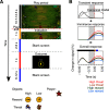
Experimental design. A, The temporal evolution is indicated vertically. At the outset of the trial, an icon appeared at the top of the screen and descended its length in ∼12 s. The participant controlled the turtle at the bottom, which could only move horizontally. During the indication period, the turtle icon either turned red (if caught by the threat) or green (if it caught the reward) or did not change color (if the subject escaped the threat or missed the reward). The inset shows the icons used as a function of experimental condition, and the icon controlled by the participant (“player”). B, Schematic responses illustrating our primary hypothesis: potential imminence-related responses. Here, responses assumed a brief initial transient response followed by imminence-related responses for different threat and reward levels. The final simulated response was obtained by summing transient and sustained hypothesized components. The gray zone indicates the temporal window considered for analysis at trial end. The dashed line in the top row represents a canonical hemodynamic response filter convolved with a boxcar function lasting for the duration of the stimulus, as was assumed in most prior studies, for comparison.
Figure 2.
Figure 2.
Skin conductance responses aligned to trial end (t = 0). The gray zone indicates the temporal window considered for analysis. A, Responses for the four experimental conditions. B, Temporal evolution of arousal and valence effects. The effect of arousal was statistically significant (red “A”), but no effect of valence was detected (black “V”). Colored bars above plots indicate trial timing: play period (black), indication period (yellow), and blank screen (blue; variable length).
Figure 3.
Figure 3.
Estimated fMRI responses aligned to trial end (t = 0) for regions of interest (all in left hemisphere). The gray zone indicates the temporal window considered for analysis. A, Responses for the four experimental conditions. B, Temporal evolution of arousal and valence differential effects. Red letters “A” (arousal) or “V” (valence) indicate that effects were detected at the FDR corrected significance level of 0.05; black letters indicate effects that were not detected. Colored bars above plots indicate trial timing: play period (black), indication period (yellow), and blank screen (blue; variable length).
Figure 4.
Figure 4.
Estimated fMRI responses aligned to trial end (t = 0) for regions of interest (left hemisphere). Same format as in Figure 3.
Figure 5.
Figure 5.
Estimated fMRI responses aligned to trial end (t = 0) for regions of interest (left hemisphere). Same format as in Figure 3.
Figure 6.
Figure 6.
Estimated fMRI responses aligned to trial end (t = 0) for regions of interest (left hemisphere). Same format as in Figure 3.
Figure 7.
Figure 7.
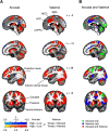
Voxelwise analysis. Effects of arousal and valence (A) were thresholded using a false discovery rate of 0.001. The last column (B) shows the same information as the arousal and valence maps, but color coded to indicate where effects overlap spatially. ACC, anterior cingulate cortex; MCC, midcingulate cortex; SMA, supplementary motor area; vmPFC, ventromedial prefrontal cortex.
Figure 8.
Figure 8.
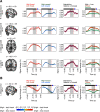
Estimated fMRI responses aligned to trial end (t = 0) for sets of voxels of the voxelwise analysis. Location of the voxels is indicated by black ellipses in the brain slices in B. Effects were illustrated by averaging 7 neighboring voxels (center voxel plus 6 adjacent ones). The gray zone indicates the temporal window considered for analysis. A, Responses for the four experimental conditions. B, Temporal evolution of arousal and valence differential effects. Colored bars above plots indicate trial timing: play period (black), indication period (yellow), and blank screen (blue; variable length). FEF, Frontal eye field; MFG, middle frontal gyrus; OFC, orbitofrontal cortex.
Figure 9.
Figure 9.
Valence by arousal interactions, voxelwise analysis. Effects are illustrated by averaging all voxels from clusters detected (cluster size indicated by k). Effect for high threat > low threat in all clusters. (A) clusters showing effect for high reward > low reward, (B) clusters showing effect for low reward > high reward. Colored bars above plots indicate trial timing: play period (black), indication period (yellow), and blank screen (blue; variable length).
Figure 10.
Figure 10.
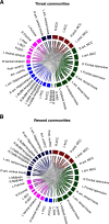
Functional networks. Regions of interest grouped into different communities (colors) based on imminence-related responses for high threat (A) and high reward (B). The hypothalamus ROI was excluded because of poor SNR. ACC, anterior cingulate cortex; ant., anterior; BLBM, basolateral/basomedial; CeMe, centromedial; L, left; MCC, midcingulate cortex; PAG, periaqueductal gray; PCC, posterior cingulate cortex; post., posterior; R, right.

Update of

References

    1. Adolphs R, Nummenmaa L, Todorov A, Haxby JV (2016) Data-driven approaches in the investigation of social perception. Phil Trans R Soc B 371:20150367. 10.1098/rstb.2015.0367 - DOI - PMC - PubMed
    1. Alvarez RP, Chen G, Bodurka J, Kaplan R, Grillon C (2011) Phasic and sustained fear in humans elicits distinct patterns of brain activity. Neuroimage 55:389–400. 10.1016/j.neuroimage.2010.11.057 - DOI - PMC - PubMed
    1. Ambadar Z, Schooler JW, Cohn JF (2005) Deciphering the enigmatic face: the importance of facial dynamics in interpreting subtle facial expressions. Psychol Sci 16:403–410. 10.1111/j.0956-7976.2005.01548.x - DOI - PubMed
    1. Amir A, Lee S-C, Headley DB, Herzallah MM, Pare D (2015) Amygdala signaling during foraging in a hazardous environment. J Neurosci 35:12994–13005. 10.1523/JNEUROSCI.0407-15.2015 - DOI - PMC - PubMed
    1. Amir A, Kyriazi P, Lee S-C, Headley DB, Paré D (2019) Basolateral amygdala neurons are activated during threat expectation. J Neurophysiol 121:1761–1777. 10.1152/jn.00807.2018 - DOI - PMC - PubMed

Publication types

LinkOut - more resources