. 2023 Apr 24;51(7):3465-3484.
doi: 10.1093/nar/gkad165.

Precise homology-directed installation of large genomic edits in human cells with cleaving and nicking high-specificity Cas9 variants

Affiliations

Precise homology-directed installation of large genomic edits in human cells with cleaving and nicking high-specificity Cas9 variants

Qian Wang et al. Nucleic Acids Res. .

Abstract

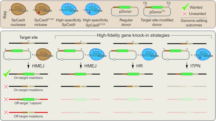
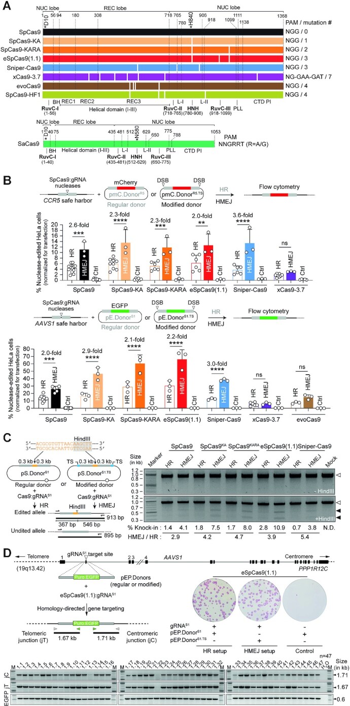
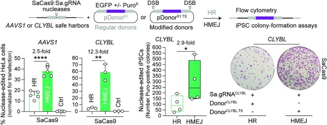
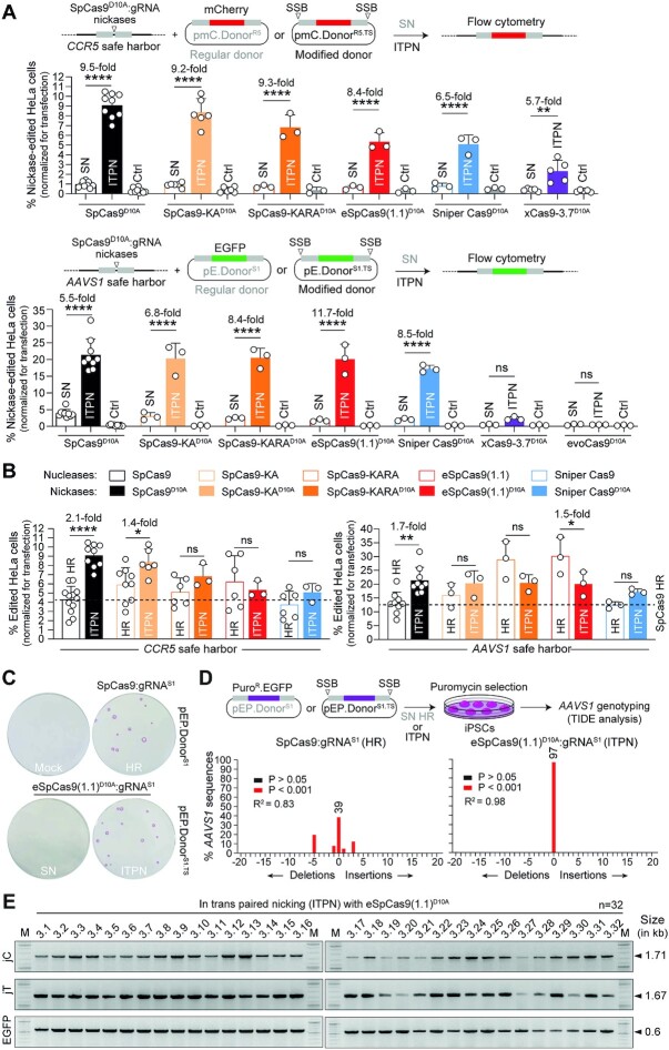
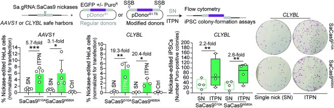
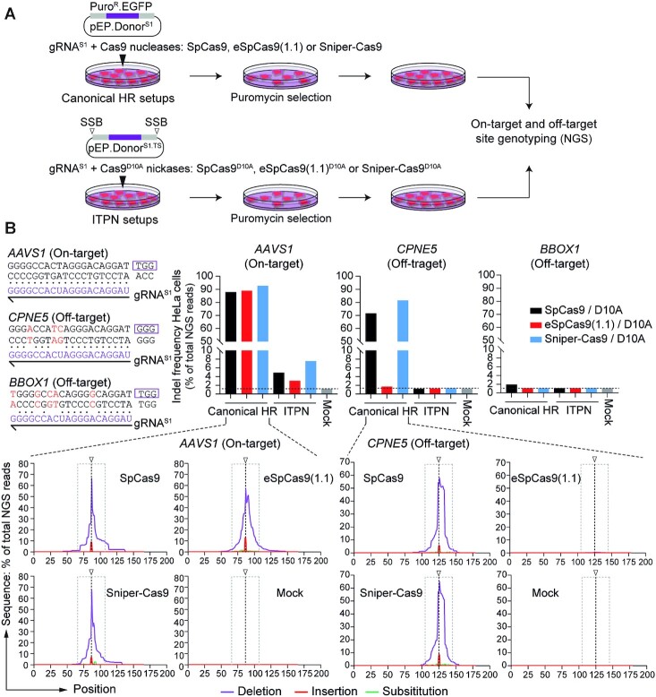
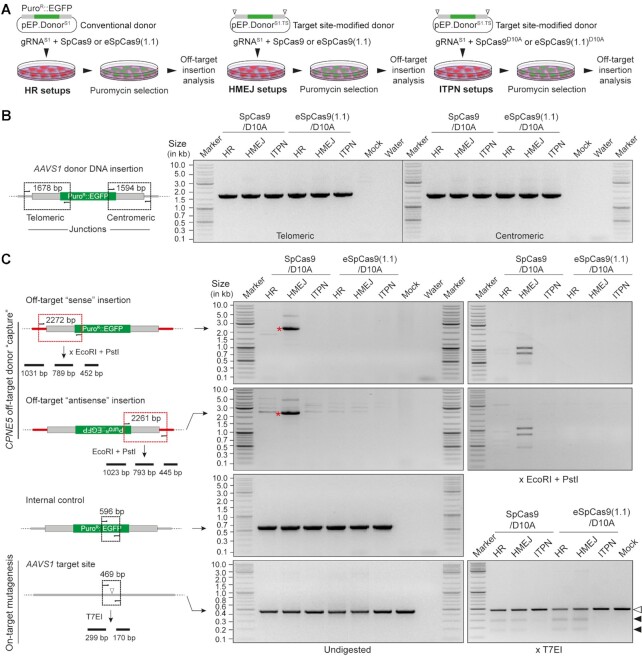
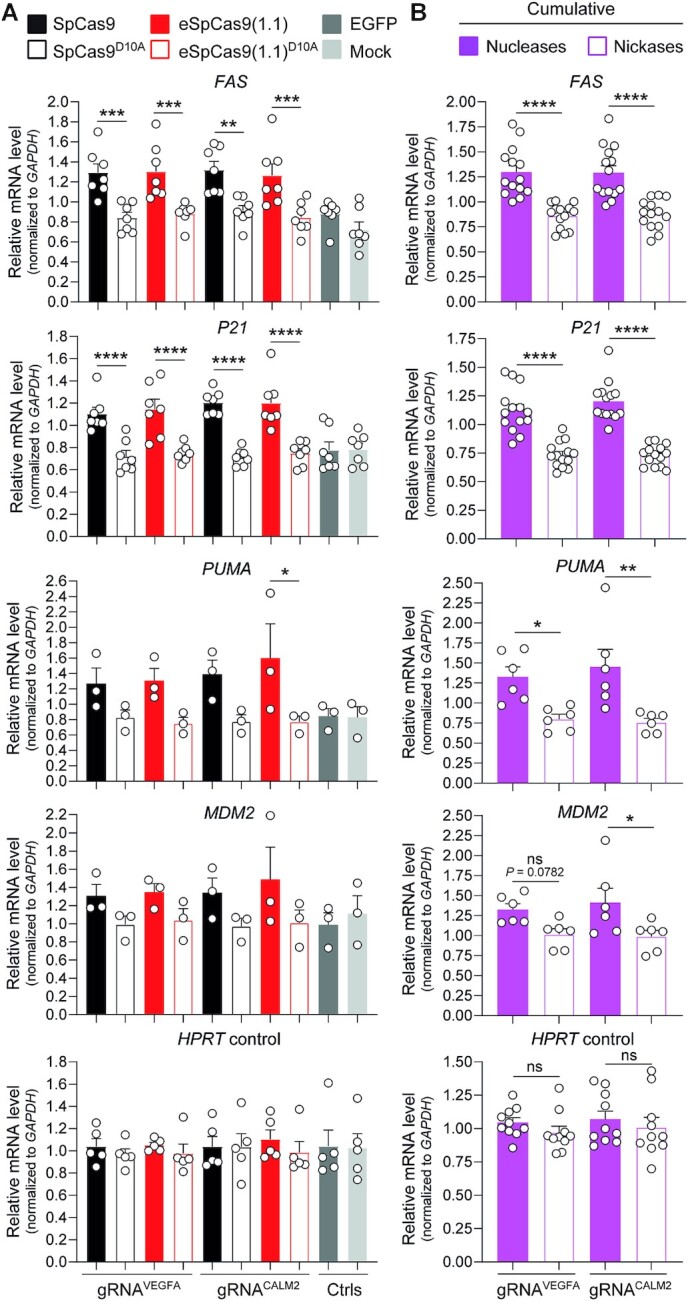
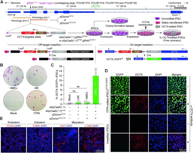
Homology-directed recombination (HDR) between donor constructs and acceptor genomic sequences cleaved by programmable nucleases, permits installing large genomic edits in mammalian cells in a precise fashion. Yet, next to precise gene knock-ins, programmable nucleases yield unintended genomic modifications resulting from non-homologous end-joining processes. Alternatively, in trans paired nicking (ITPN) involving tandem single-strand DNA breaks at target loci and exogenous donor constructs by CRISPR-Cas9 nickases, fosters seamless and scarless genome editing. In the present study, we identified high-specificity CRISPR-Cas9 nucleases capable of outperforming parental CRISPR-Cas9 nucleases in directing genome editing through homologous recombination (HR) and homology-mediated end joining (HMEJ) with donor constructs having regular and 'double-cut' designs, respectively. Additionally, we explored the ITPN principle by demonstrating its compatibility with orthogonal and high-specificity CRISPR-Cas9 nickases and, importantly, report that in human induced pluripotent stem cells (iPSCs), in contrast to high-specificity CRISPR-Cas9 nucleases, neither regular nor high-specificity CRISPR-Cas9 nickases activate P53 signaling, a DNA damage-sensing response linked to the emergence of gene-edited cells with tumor-associated mutations. Finally, experiments in human iPSCs revealed that differently from HR and HMEJ genome editing based on high-specificity CRISPR-Cas9 nucleases, ITPN involving high-specificity CRISPR-Cas9 nickases permits editing allelic sequences associated with essentiality and recurrence in the genome.


Figures

Graphical Abstract
Graphical Abstract
Genome editing strategies based on precision homology-directed gene targeting with high-specificity Cas9 variants minimize the complexity of bystander chromosomal effects, including off-target donor DNA insertions.
Figure 1.
Figure 1.
Testing DSB-dependent genome editing using regular versus high-specificity SpCas9 nucleases. (A) Diagrams of engineered Cas9 nucleases derived from S. pyogenes and S. aureus type II CRISPR systems. Protein domains and mutation positions are marked by dashed and white lines, respectively. HNH, histidine-asparagine-histidine nuclease domain; RuvC, RuvC-like nuclease domain composed of a tripartite assembly of RuvC-I, -II and -III. The HNH and RuvC domains in the nuclease lobe cut the target and non-target DNA strands, respectively. L-I and L-II, linker region I and II, respectively. BH, Arginine-rich bridge helix; CTD, C-terminal domain in which the PAM-interacting motif (PI) is integrated; NUC and REC, nuclease and recognition lobes, respectively; PLL, phosphate lock loop. Asterisks mark residues D10 and H840 crucial for RuvC and HNH catalytic activities, respectively. (B) Genome editing based on donors prone to canonical HR and HMEJ upon high-specificity SpCas9 delivery. Nuclease-dependent genome editing frequencies in HeLa cells transfected with the depicted reagents targeting CCR5 and AAVS1 were quantified by reporter-directed flow cytometry at 17 days post-transfection (top and bottom graphs, respectively). HeLa cells exposed to corresponding Cas9 nucleases and regular donor plasmids in the absence of locus-specific gRNAs served as negative controls. Data are plotted as mean ± SD of at least 3 independent biological replicates. Significant differences between the indicated datasets were determined by two-way ANOVA followed by Šidák's multiple comparisons tests; ****P < 0.0001, ***0.0001 < P< 0.001, **0.001 < P< 0.01; P> 0.05 was considered non-significant (ns). (C) Genotyping assay assessing HDR-mediated restriction site knock-ins. Regular pS.DonorS1 and modified pS.DonorS1.TS constructs, designed to introduce a HindIII recognition site at AAVS1 through HR and HMEJ processes, respectively, were transfected into HeLa cells together with plasmids expressing SpCas9 nucleases and gRNAS1. The HindIII polymorphism is detected by restriction-fragment length analysis (RFLA) of amplicons covering the target site (left panel). RFLA products diagnostic for unedited and edited AAVS1 alleles retrieved from HeLa cells exposed to the indicated reagents were measured through densitometry and are marked with open and closed arrowheads, respectively (right panel). (D) Genotyping assay assessing HDR-mediated transgene knock-ins. Regular pEP.DonorS1 and modified pEP.DonorS1.TS plasmids, tailored for inserting the live-cell selectable marker PuroR.EGFP at AAVS1 via HR and HMEJ processes, respectively, were transfected into iPSCs together with constructs expressing eSpCas9(1.1):gRNAS1 complexes. HDR-derived gene knock-ins were identified by junction PCR analysis of randomly selected iPSC clones engineered through pEP.DonorS1 and eSpCas9(1.1):gRNAS1 delivery. Puromycin-resistant iPSC colonies were identified by staining for the pluripotency marker alkaline phosphatase.
Figure 2.
Figure 2.
Genome editing combining plasmid donors with regular HR or modified HMEJ templates and orthogonal SaCas9 complexes. SaCas9-dependent genome editing at AAVS1 and CLYBL loci in HeLa cells using EGFP-encoding donors, and at CLYBL in iPSCs using PuroR.EGFP-encoding donors was determined by reporter-directed flow cytometry and colony-formation assays, respectively. The latter assays detected the pluripotency marker alkaline phosphatase to identify puromycin-resistant iPSCs. Controls consisted of cells exposed to regular donor plasmids and SaCas9 nucleases with non-targeting gRNAs. Data are presented as mean ± SD of at least three independent biological replicates. Significant differences between the indicated datasets were calculated by two-tailed unpaired Student's t tests (left and middle graphs) and two-tailed paired ratio t test (right graph); ****P < 0.0001, **0.001 < P< 0.01, *0.01 < P< 0.05.
Figure 3.
Figure 3.
Testing SSB-dependent genome editing using regular versus high-specificity SpCas9D10A nickases. (A) Single nicking and in trans paired nicking genome editing based on high-specificity SpCas9D10A variants. Nickase-dependent genome editing frequencies in HeLa cells transfected with the depicted components targeting CCR5 and AAVS1 were measured by reporter-directed flow cytometry at 17 days post-transfection (top and bottom graphs, respectively). HeLa cells treated with corresponding Cas9D10A nickases and regular donor plasmids in the absence of locus-specific gRNAs served as negative controls. Results are plotted as mean ± SD of at least three independent biological replicates. Significant differences between the indicated datasets were assessed by two-way ANOVA followed by Šidák's multiple comparisons test; ****P < 0.0001, **0.001 < P< 0.01; P> 0.05 was considered non-significant (ns). (B) Comparing standard and in trans paired nicking genome editing strategies at CCR5 and AAVS1. Plotting of datasets presented in panel A corresponding to HeLa cells subjected to nucleases and regular donors or to nickases and target site-modified donors (canonical HR or ITPN strategies, respectively). Dashed lines mark the means of the DSB-dependent genome editing levels obtained with conventional SpCas9 and unmodified HR donor templates. Data are shown as mean ± SD of at least 3 independent biological replicates. Significant differences between the indicated datasets were calculated by two-way ANOVA followed by Šidák's multiple comparisons tests; ****P < 0.0001, **0.001 < P< 0.01, *0.01 < P< 0.05; P> 0.05 was considered non-significant (ns). (C) Testing standard and in trans paired nicking in iPSCs using high-specificity cleaving and nicking CRISPR complexes. iPSCs edited upon exposure to the indicated AAVS1-targeting reagents were selected in the presence of puromycin and the resulting colonies were stained for the pluripotency marker alkaline phosphatase. (D) Probing mutagenic loads in genome-edited iPSCs. iPSCs edited after exposure to the indicated AAVS1-targeting reagents were selected in the presence of puromycin and indel profiles at AAVS1 were examined through tracking of indels by decomposition (TIDE) analysis. (E) Establishing HDR-mediated transgene insertion in iPSCs edited through in trans paired nicking. Junction PCR analysis was performed on randomly picked iPSC clones engineered through pEP.DonorS1.TS and eSpCas9(1.1)D10A:gRNAS1 delivery.
Figure 4.
Figure 4.
Genome editing combining regular SN plasmid donors or modified ITPN donors and nicking orthogonal SaCas9 complexes. SaCas9D10A- or SaCas9N580A-dependent genome editing at AAVS1 and CLYBL loci in HeLa cells using EGFP-encoding donors, and at these loci in iPSCs using PuroR.EGFP-encoding donors, was assessed by reporter-directed flow cytometry and colony-formation assays, respectively. The latter assay detected the pluripotency marker alkaline phosphatase to identify puromycin-resistant iPSCs. Controls consisted of cells exposed to regular donor plasmids and nickases lacking locus-specific gRNAs. Data are shown as mean ± SD of at least three independent biological replicates. Significant differences between the indicated datasets were calculated by two-tailed unpaired Student's t tests (left and middle graphs) and two-tailed paired ratio t test (right graph); ***0.0001 < P< 0.001, **0.001 < P< 0.01, *0.01 < P< 0.05.
Figure 5.
Figure 5.
Assessing mutagenic loads in cells edited through canonical homologous recombination versus in trans paired nicking. (A) Experimental design. HeLa cells were exposed to regular and modified donors conferring puromycin resistance together with SpCas9 nucleases and SpCas9D10A nickases, respectively. SpCas9, eSpCas9(1.1) and Sniper-Cas9 nucleases, and their D10A nickase derivatives, were coupled to AAVS1-targeting gRNAS1. Indel frequencies at on-target and off-target sites was done by amplicon deep sequencing genotyping of puromycin-resistant cell populations. (B) Quantification of indels at on-target and off-target sites. CRISPR complex-derived indels at the AAVS1 target site and at two validated off-target sites (i.e. CPNE5 and BBOX1) were quantified by amplicon deep sequencing (∼100,000 paired-end reads per sample). Nucleotide mismatch positions between gRNAS1 spacer and off-target CPNE5 and BBOX1 sequences are highlighted in red. The types and distributions of indels detected within AAVS1, CPNE5 and BBOX1 in cells treated with regular and high-specificity nucleases are plotted. HeLa cells not exposed to CRISPR complexes provided for negative controls (Mock).
Figure 6.
Figure 6.
Assessing off-target chromosomal donor DNA insertions resulting from HR, HMEJ and ITPN using regular and high-specificity Cas9 enzymes. (A) Experimental design. HeLa cells were subjected to HR, HMEJ and ITPN procedures using the indicated combinations of donor DNA constructs and Cas9 proteins coupled to AAVS1-targeting gRNAS1. Genetically modified cells, selected through puromycin exposure, were screened for donor DNA ‘capture’ at the prevalent gRNAS1 off-target site CPNE5 by junction PCR analysis. (B) On-target donor DNA insertion analysis. Amplicons diagnostics for HDR-mediated AAVS1 knock-ins are illustrated and shown. (C) Off-target insertion and on-target mutagenesis analysis. Amplicons diagnostics for HDR-independent ‘capture’ of donor DNA sequences at CPNE5 in the ‘sense’ and ‘antisense’ orientations are illustrated and marked with asterisks. Specific donor DNA ‘capture’ at CPNE5 off-target alleles and mutagenesis at AAVS1 target alleles were probed via restriction enzyme (EcoRI and PstI) and T7 endonuclease I (T7EI) digestions, respectively. Solid arrowheads point to T7EI-digested products derived from indel-containing AAVS1 sequences. PCR amplifications of a 596-bp EGFP tract served as internal controls.
Figure 7.
Figure 7.
Cell survival assay for assessing P53 functionality in human iPSCs. (A) Schematics of post-transcriptional P53 activity control by DNA damage and Nutlins. In cells with normal amounts of P53, DNA damage activates ATM/ATR kinases that disrupt P53-MDM2 interaction through P53 phosphorylation. Free P53 escapes proteasomal degradation and upregulates the expression of downstream target genes (e.g. cyclin-dependent kinase inhibitor P21) inducing cell cycle arrest and apoptosis. Nutlins disrupt the P53-MDM2 interaction by instead occupying the P53 binding pocket in MDM2 mimicking a P53-dependent DNA damage response. Conversely, in cells with no or low amounts of P53, nutlins induce neither cell cycle arrest nor apoptosis (not drawn). (B and C) Realtime cell proliferation assay. The proliferation of human iPSCs incubated in the presence of Nutlin-3a (10 μM) or vehicle (DMSO) was quantified in a live-cell imaging system (IncuCyte) for 3 days. Data are shown as mean ± SD of 6 technical replicates. Significant differences between the indicated datasets were calculated by two-way ANOVA tests; ****P < 0.0001. (D and E) Cell survival assays. The survival of human iPSCs incubated in regular medium (Mock) or in medium supplemented with DMSO or Nutlin-3a (2 μM and 10 μM) was monitor for 3 days by using the MTS cell metabolic activity readout (panel D). The frequencies of apoptotic human iPSCs were determined with a combined annexin V/propidium iodide assay (panel E). Annexin V positive cells and annexin V/propidium iodine doubly positive cells measured by flow cytometry scored for early and late apoptosis, respectively. Prior to flow cytometry the cells were incubated in regular medium (Mock) and in medium supplemented with DMSO or with Nutlin-3a (10 μM) for different periods. Staurosporine applied at the indicated conditions served as an apoptosis-inducing control. (F) Assessing P53-dependent responses in human iPSCs exposed to Nutlin-3a. RT-qPCR analysis of transcripts for P53 and P53-responsive genes were conducted in human iPSCs incubated for 5 h in regular medium or in medium supplemented with Nutlin-3a (10 μM). RT-qPCR analysis of HPRT1 transcripts served to measure the expression of a P53-independent control gene (n = 3 independent biological replicates). Significances were calculated with two-way ANOVA followed by Šidák's test for multiple comparisons; ****P < 0.0001; P> 0.05 was considered non-significant (ns). (G) P53-dependent P21 protein detection assay. Western blot analysis of P21 expression in human iPSCs incubated in the presence of Nutlin-3a (10 μM) or vehicle (DMSO) for 12 h. Transformed P53-defective HEK293T cells exposed to the same experimental conditions served as control. Western blotting of the housekeeping GAPDH provided for loading controls.
Figure 8.
Figure 8.
Assessing activation of P53-dependent DNA damage responses in human iPSCs exposed to nucleases versus nickases. (A) Expression analysis of P53 activation-responsive genes. Constructs encoding the indicated Cas9 enzymes and gRNAs conferring high (gRNAVEGFA) or low (gRNACALM2) off-target activities (Supplementary Figure S9), were transfected into iPSCs. RT-qPCR measurements of FAS, P21, PUMA and MDM2 transcripts whose expression is upregulated upon P53 activation (minimum n = 3 independent biological replicates). Targeting HPRT1 transcripts served for RT-qPCR measurements of a housekeeping control gene (n = 5 independent biological replicates). Additional controls consisted of targeting FAS, P21, PUMA, MDM2 and HPRT1 transcripts in mock-transfected iPSCs and in iPSCs transfected with an EGFP-encoding plasmid. Significances were calculated with one-way ANOVA followed by Tukey's test for multiple comparisons; ****P < 0.0001, ***0.0001 < P< 0.001, **0.001 < P< 0.01, *0.01 < P< 0.05. (B) Cumulative comparison of cleaving versus nicking effects on P53-responsive gene modulation. Combined RT-qPCR datasets derived from iPSCs treated with nucleases SpCas9 and eSpCas9(1.1) or nickases SpCas9D10A and eSpCas9(1.1)D10A. Significances were calculated with two-way ANOVA followed by Šidák's test for multiple comparisons; ****P < 0.0001, **0.001 < P< 0.01, *0.01 < P< 0.05; P> 0.05 was considered non-significant (ns).
Figure 9.
Figure 9.
Testing DSB- versus SSB-dependent genome editing strategies at essential OCT4 alleles in human iPSCs using high-specificity CRISPR complexes. (A) Experimental setup for tracking OCT4 gene editing events. iPSCs exposed to the indicated reagents designed to elicit canonical HR, HMEJ or ITPN were traced by colony-formation assays upon puromycin selection and alkaline phosphatase staining and by a genetic assay reporting live-cell OCT4 gene targeting events upon Cre recombinase delivery. (B) Detection of stably transfected iPSC colonies. Picture of a representative colony-formation assay is shown. (C) Detection of OCT4 gene editing events. The frequencies of OCT4 edited cells (OCT4::EGFP+) in puromycin-resistant iPSC populations were determined by EGFP-directed flow cytometry following transduction with Cre-expressing lentivector particles (20 vector particles per cell). Data are presented as mean ± S.D. of independent biological replicates (n = 3). (D) Confocal microscopy analysis of iPSCs edited at OCT4 through ITPN. OCT4::EGFP-expressing iPSCs engineered through ITPN and Cre delivery (iPSCOCT4::EGFP) were analysed through immunofluorescence microscopy for detecting OCT4 and EGFP, respectively. Nuclei were stained with DAPI. The merge of the three fluorescence signals highlights the nuclear localization of the OCT4::EGFP fusion product. Unedited iPSCs served as negative controls. iPSC and iPSCOCT4::EGFP specimens not incubated with the OCT4-specific primary antibody served as staining controls. (E) Assessing the multi-lineage differentiation capacity of iPSCs edited at OCT4 through ITPN. iPSCsOCT4::EGFP generated by ITPN using high-specificity eSpCas9(1.1)D10A were induced to differentiate into cell lineages corresponding to the three embryonic germ layers, i.e. mesoderm, ectoderm and endoderm. Immunofluorescence microscopy detected the indicated embryonic germ layer-specific markers. Nuclei were stained with DAPI.

References

    1. Ernst M.P.T., Broeders M., Herrero-Hernandez .P., Oussoren E., van der Ploeg A.T., Pijnappel W. Ready for repair? Gene editing enters the Clinic for the treatment of Human disease. Mol. Ther. Methods Clin. Dev. 2020; 18:532–557. - PMC - PubMed
    1. Chen F., Ding X., Feng Y., Seebeck T., Jiang Y., Davis G.D.. Targeted activation of diverse CRISPR-cas systems for mammalian genome editing via proximal CRISPR targeting. Nat. Commun. 2017; 8:14958. - PMC - PubMed
    1. Ran F.A., Cong L., Yan W.X., Scott D.A., Gootenberg J.S., Kriz A.J., Zetsche B., Shalem O., Wu X., Makarova K.S.et al. .. In vivo genome editing using Staphylococcus aureus Cas9. Nature. 2015; 520:186–191. - PMC - PubMed
    1. Jinek M., Chylinski K., Fonfara I., Hauer M., Doudna J.A., Charpentier E.. A programmable dual-RNA-guided DNA endonuclease in adaptive bacterial immunity. Science. 2012; 337:816–821. - PMC - PubMed
    1. Sternberg S.H., LaFrance B., Kaplan M., Doudna J.A.. Conformational control of DNA target cleavage by CRISPR-Cas9. Nature. 2015; 527:110–113. - PMC - PubMed

Publication types