. 2023 May 1;133(9):e161929.
doi: 10.1172/JCI161929.

Aurora A kinase inhibition compromises its antitumor efficacy by elevating PD-L1 expression

Affiliations

Aurora A kinase inhibition compromises its antitumor efficacy by elevating PD-L1 expression

Xiaobo Wang et al. J Clin Invest. .

Abstract

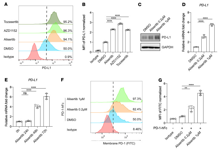
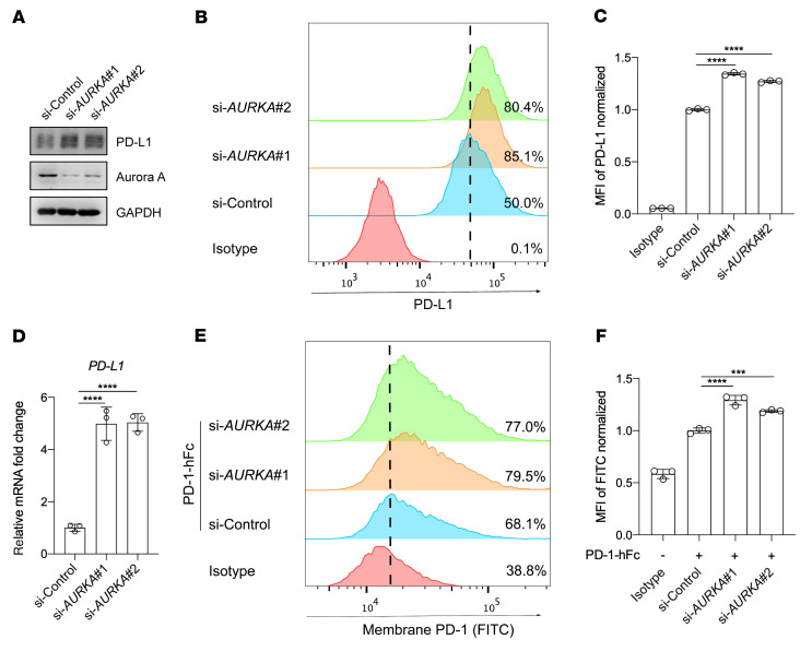
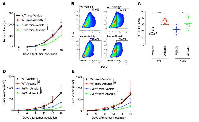
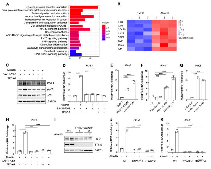
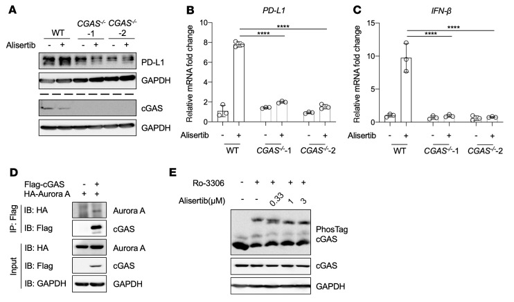
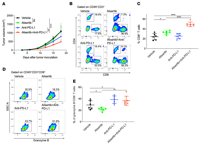
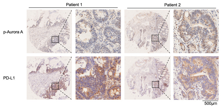
Aurora A plays a critical role in G2/M transition and mitosis, making it an attractive target for cancer treatment. Aurora A inhibitors showed remarkable antitumor effects in preclinical studies, but unsatisfactory outcomes in clinical trials have greatly limited their development. In this study, the Aurora A inhibitor alisertib upregulated programmed death ligand 1 (PD-L1) expression in a panel of tumor cells both in vitro and in vivo. Upregulation of the checkpoint protein PD-L1 reduced antitumor immunity in immune-competent mice, paradoxically inhibiting the antitumor effects of alisertib. Mechanistically, Aurora A directly bound to and phosphorylated cyclic GMP-AMP synthase (cGAS), suppressing PD-L1 expression in tumor cells. Aurora A inhibition by alisertib activated the cGAS/stimulator of IFN genes (STING)/NF-κB pathway and promoted PD-L1 expression. Combining alisertib with anti-PD-L1 antibody improved antitumor immunity and enhanced the antitumor effects of alisertib in immune-competent mice. Our results, which reveal the immunomodulatory functions of Aurora A inhibitors and provide a plausible explanation for the poor clinical outcomes with their use, offer a potential approach to improve the antitumor efficacy of these inhibitors.

Keywords: Cancer; Drug therapy; Oncology.


Figures

Figure 1
Figure 1. Aurora A kinase inhibitor elevates PD-L1 expression in tumor cells.
(A) Flow cytometric analysis of cell-surface PD-L1 expression in BxPC3 cells following treatment for 72 hours with 1 μmol/L alisertib, 0.5 μmol/L AZD1152, or 0.5 μmol/L tozasertib. (B) Normalized MFI for the data shown in A (n = 3). (C) Western blot showing PD-L1 expression in BxPC3 cells after treatment for 72 hours with the indicated concentrations of alisertib. (D and E) qRT-PCR analysis of PDL1 expression in BxPC3 cells after treatment with alisertib at the indicated concentrations (D) and for the indicated durations (E) (n = 3). (F) Flow cytometric analysis of cell-surface PD-1 binding of BxPC3 cells after treatment for 72 hours with the indicated concentrations of alisertib. (G) Normalized MFI for the data in F (n = 3). hFc, human Fc. Data indicate the mean ± SD. **P < 0.01 and ****P < 0.0001, by 1-way ANOVA.
Figure 2
Figure 2. Knockdown of AURKA elevates PD-L1 expression in tumor cells.
(A) Western blot analysis of the expression of indicated proteins following treatment with an AURKA siRNA or a control siRNA. (B) Flow cytometric analysis of surface expression of PD-L1 on BxPC3 cells after treatment with the indicated siRNA. (C) Normalized MFI for the data shown in B (n = 3). (D) qRT-PCR analysis of PDL1 expression in BxPC3 cells after treatment with the indicated siRNA (n = 3). (E) Flow cytometric analysis of cell-surface PD-1 binding of BxPC3 cells after treatment with the indicated siRNAs. (F) Normalized MFI for the data in E (n = 3). Data indicate the mean ± SD. ***P < 0.001 and ****P < 0.0001, by 1-way ANOVA.
Figure 3
Figure 3. Aurora A inhibition upregulates PD-L1 expression in vivo, which compromises the inhibitor’s antitumor efficacy.
(AC) (A) Effect of vehicle or alisertib treatment on CT26 growth in BALB/c WT or BALB/c nude mice (n = 6). (B) Excised tumor tissues were digested to a single-cell suspension, and PD-L1+ cells were evaluated by flow cytometry. SSC-A, side scatter area. (C) Cumulative data for the percentage of PD-L1+ cells in B (n = 6). (D) Effect of vehicle or alisertib treatment on tumor growth in WT or Pdl1–/– MC38 mouse tumor models (n = 6). (E) Effect of vehicle or alisertib treatment on MC38 growth in WT or Pdl1–/– C57BL/6 mice (n = 6). Data indicate the mean ± SD. A 2-way ANOVA was applied to compare time-dependent tumor growth. *P < 0.05 and ****P < 0.0001, by unpaired t test.
Figure 4
Figure 4. PD-L1 upregulation caused by Aurora A inhibition depends on STING/NF-κB activation.
(A and B) RNA-Seq analysis was performed on BxPC3 cells after treatment for 72 hours with DMSO or 1 μmol/L alisertib. (A) KEGG pathway analysis of genes differentially expressed between the DMSO- and alisertib-treated groups. The most substantially enriched pathways are shown. p.adjust, adjusted P value. (B) Heatmap of gene expression levels of the indicated cytokines or chemokines in DMSO- or alisertib-treated BxPC3 cells. (C and D) BxPC3 cells were pretreated for 6 hours with 10 μmol/L TPCA-1 or with 5 μmol/L BAY11-7082, followed by treatment for 72 hours with 1 μmol/L alisertib, and PD-L1 expression was assessed by Western blotting (C) and qRT-PCR (D). (E and F) qRT-PCR analysis of IFNB expression in BxPC3 cells after the indicated concentrations (E) and durations (F) of alisertib treatment (n = 3). (G) qRT-PCR analysis of IFNB expression in BxPC3 cells after treatment with the indicated siRNA (n = 3). (H) qRT-PCR analysis of IFNB expression (n = 3). (IK) WT BxPC3 cells or STING–/– BxPC3 cells were treated with 1 μmol/L alisertib for 72 hours. (I) Western blot analysis of PD-L1 and STING protein levels. qRT-PCR analysis of PDL1 (J) and IFNB (K) mRNA levels (n = 3). Data indicate the mean ± SD. ***P < 0.001 and ****P < 0.0001, by 1-way ANOVA (DH) and 2-way ANOVA (J and K).
Figure 5
Figure 5. Aurora A inhibition–induced PD-L1 expression is mediated by cGAS dephosphorylation.
(AC) WT BxPC3 cells or CGAS–/– BxPC3 cells were treated with 1 μmol/L alisertib for 72 hours. (A) Western blot analysis of PD-L1 and cGAS protein levels. PD-L1 and cGAS were detected separately in 2 gels using the same biological samples, and GAPDH in each gel served as the loading control. qRT-PCR analysis of PDL1 (B) and IFNB (C) mRNA levels (n = 3). (D) Co-immunoprecipitation of Aurora A and cGAS. HEK293T cells were transfected with the indicated vectors encoding HA–Aurora A and Flag-cGAS. Whole-cell lysates were immunoprecipitated with anti-Flag beads, and the interactions were analyzed by Western blotting. (E) BxPC3 cells were synchronized with 10 μmol/L Ro-3306 for 16 hours and released into mitosis in the presence of alisertib at the indicated concentrations. Phosphorylation of cGAS was analyzed by Phos-tag electrophoresis. Data indicate the mean ± SD. ****P < 0.0001, by 2-way ANOVA.
Figure 6
Figure 6. PD-L1 blockade therapy improves the antitumor activity of alisertib.
(AE) BALB/c mice were inoculated with CT26 cells and administered alisertib, anti–PD-L1 antibody alone, or their combination. (A) Tumor growth curves of CT26 in BALB/c mice. Tumor volumes were measured at the indicated time points (n = 6). (B and D) Flow cytometric analysis of tumor-infiltrating CD8+ T cells (B) and granzyme B+CD8+ T cells (D). Representative plots are shown. (C and E) Cumulative data for B and D (n = 6). Data indicate the mean ± SD. A 2-way ANOVA was applied to compare time-dependent tumor growth. *P < 0.05, **P < 0.01, and ****P < 0.0001, by unpaired t test.
Figure 7
Figure 7. Active Aurora A levels are negatively associated with PD-L1 expression in human tumor tissues.
Representative images of IHC staining for p–Aurora A and PD-L1 in human colon cancer tissues. Scale bar: 500 μm (enlarged insets).

References

    1. Barr AR, Gergely F. Aurora-A: the maker and breaker of spindle poles. J Cell Sci. 2007;120(pt 17):2987–2996. doi: 10.1242/jcs.013136. - DOI - PubMed
    1. Liewer S, Huddleston A. Alisertib: a review of pharmacokinetics, efficacy and toxicity in patients with hematologic malignancies and solid tumors. Expert Opin Investig Drugs. 2018;27(1):105–112. doi: 10.1080/13543784.2018.1417382. - DOI - PubMed
    1. Durlacher CT, et al. An update on the pharmacokinetics and pharmacodynamics of alisertib, a selective Aurora kinase A inhibitor. Clin Exp Pharmacol Physiol. 2016;43(6):585–601. doi: 10.1111/1440-1681.12571. - DOI - PubMed
    1. Bavetsias V, Linardopoulos S. Aurora kinase inhibitors: current status and outlook. Front Oncol. 2015;5:278. - PMC - PubMed
    1. O’Connor OA, et al. Randomized phase III study of alisertib or investigator’s choice (selected single agent) in patients with relapsed or refractory peripheral T-cell lymphoma. J Clin Oncol. 2019;37(8):613–623. doi: 10.1200/JCO.18.00899. - DOI - PMC - PubMed

Publication types