. 2023 Mar 16;13(1):4373.
doi: 10.1038/s41598-023-31474-2.

Probiotic disruption of quorum sensing reduces virulence and increases cefoxitin sensitivity in methicillin-resistant Staphylococcus aureus

Affiliations

Probiotic disruption of quorum sensing reduces virulence and increases cefoxitin sensitivity in methicillin-resistant Staphylococcus aureus

Monica Angela Cella et al. Sci Rep. .

Abstract

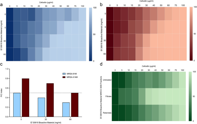
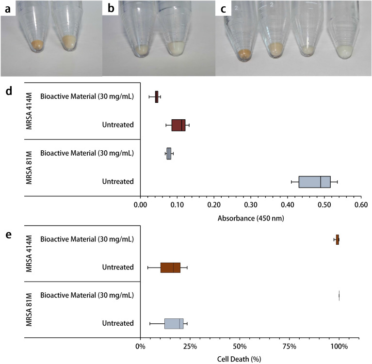
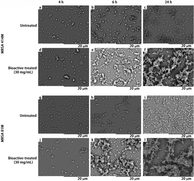
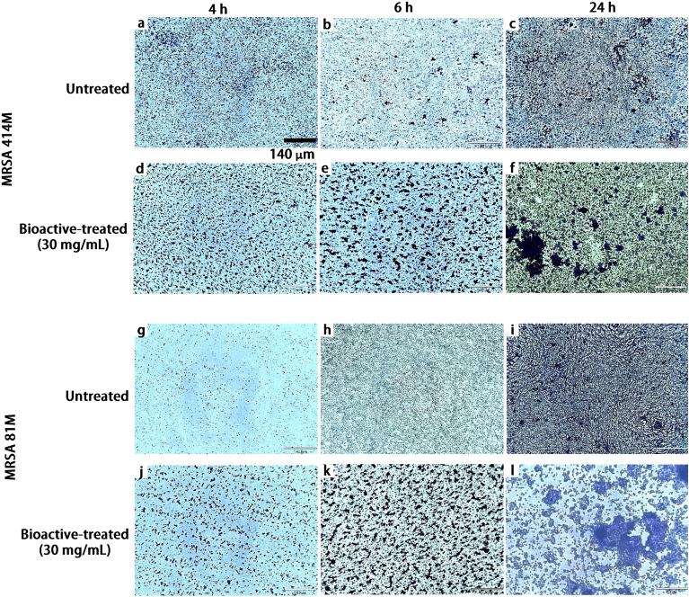
Therapies which target quorum sensing (QS) systems that regulate virulence in methicillin-resistant Staphylococcus aureus (MRSA) are a promising alternative to antibiotics. QS systems play a crucial in the regulation of MRSA antibiotic resistance, exotoxin production, antioxidant protection and immune cell evasion, and are therefore attractive therapeutic targets to reduce the virulence of a pathogen. In the present work the the effects of bioactive peptides isolated from two strains of lactic acid bacteria were tested against antibiotic resistance, carotenoid production, resistance to oxidative killing and biofilm structure in two clinical MRSA isolates. The results obtained from fractional-inhibitory concentration assays with bulk and semi-purified bioactive molecules showed a significant synergistic effect increasing cefoxitin mediated killing of MRSA. This was coupled to a six-fold decrease of the major membrane pigment staphyloxanthin, and a 99% increase in susceptibility to oxidative stress mediated killing. Real-time quantitative PCR analysis of the QS-genes agrA and luxS, showed differential expression between MRSA strains, and a significant downregulation of the hemolysin gene hla. Light microscopy and scanning electron microscopy revealed alteration in biofilm formation and clustering behavior. These results demonstrate that bioactive metabolites may be effectively applied in tandem with beta-lactam antibiotics to sensitize MRSA to cefoxitin. Moreover, these results shown that several key QS-controlled virulence mechanisms are diminished by probiotic metabolites.


Conflict of interest statement

The authors declare no competing interests.

Figures

Figure 1
Figure 1
Heat plots of MIC percent growth inhibition of the two representative clinical MRSA strains by combinational testing of the beta-lactam antibiotic cefoxitin (0–100 µg/mL) and Ef 30616 bioactive metabolites (0, 5, 30 and 60 mg/mL): (a) MIC heat plots of MRSA 81M and (b) MIC heat plots of MRSA 414M. (c) FIC index values for the combinatory effects of EfMSI1 bioactive metabolites and cefoxitin for MRSA strains 81M and 414M. (d) Heat plot of the percent growth inhibition of MRSA 81M for the combination testing of cefoxitin (0–100 µg/mL) and Ef 30616 bioactive metabolites (30 mg/mL) MWCO 3000 filtrate (i.e. < 3000 Da) and retentate (i.e. > 3000 Da) (n = 3). Averages of biological replicates are shown for heat maps and are presented as the means in terms of percent growth inhibition (ANOVA, p < 0.05 Dunnett’s multiple comparison test).
Figure 2
Figure 2
Heat plots of MIC percent growth inhibition of MRSA 81M in combination with cefoxitin (0–140 µg/mL) and SEC fraction four from (a) Ef 30616 or (b) Ll 11454 at 5, 15 and 30 mg/mL of equivalent bulk material. (c) FIC index values for Ef 30616 fractions one through four and (d) Ll 11454 fractions one through four. Averages of biological replicates are shown for heat maps and are presented as the means in terms of percent growth inhibition (ANOVA, p < 0.05 Dunnett’s multiple comparison test).
Figure 3
Figure 3
(a) Colonies of MRSA 81M growing on blood agar after treatment with 30 mg/mL Ef 30616 bioactive material (left) or no treatment (right) and (b) colonies of MRSA 414M growing on blood agar after treatment with 30 mg/ml Ef 30616 bioactive material (left) or no treatment (right). Arrows indicate SCVs lacking carotenoid pigmentation and reduced hemolysis. (c) Percentage of SCV to wild-type colonies for MRSA 81M (n = 3) and MRSA 414M (n = 3) following 24 h incubation at 37 +/− 1 °C with and without Ef 30616 bioactive material (30 mg/mL) (U = 0; p < 0.05; Mann–Whitney). Central black bars indicate the mean, and the upper and lower whiskers how the maximum and minimum, respectively.
Figure 4
Figure 4
MRSA cell pellets lack carotenoid pigmentation following 24 h incubation with Ef 30616 bioactive material (30 mg/mL) at 37 °C ± 1 °C: (a) untreated MRSA 81M (left) and bioactive-treated MRSA 81M (right) pellets, (b) untreated MRSA 414M (left) and bioactive-treated MRSA 414M (right) pellets, and (c) comparison of all four pellets (from left to right) untreated MRSA 81M, bioactive-treated MRSA 81M, untreated MRSA 414M, and bioactive-treated MRSA 414M. (d) Carotenoid absorbance measurement at 450 nm (A450) from each of the two treatments described for both strains (n = 3) (U = 0; p < 0.05; Mann–Whitney). (e) Percent decrease of Staphylococcal survival to 1.5% v/v hydrogen peroxide for MRSA 81M (n = 3) and MRSA 414M (n = 3) following 1 h incubation at 37 °C ± 1 °C in PBS solution with and without Ef 30616 bioactive treatment (30 mg/mL) (W = 0; p < 0.05; Wilcoxon signed-rank). Central black bars indicate median, and the upper and lower whisker caps show the maximum and minimum, respectively, for box plots (d) and (e).
Figure 5
Figure 5
Differential expression of MRSA virulence-related genes in relation to untreated controls of MRSA strains 81M and 414M following incubation with Ef 30616 bioactive material (30 mg/mL) at 37 °C ± 1 °C; cultures were incubated to the late exponential phase and stationary phase. The 16s rRNA was used as a housekeeping gene. Data are shown as averages and bars indicate the standard deviation (n = 3). Statistically significant differences between strains indicated by asterisks (ANOVA; p < 0.05).
Figure 6
Figure 6
Scanning electron microscopy indicating the progression of biofilm formation of both untreated and bioactive-treated MRSA 81M and 414M cultures incubated over a 24 h period at 37 °C ± 1 °C and imaged at the following intervals: untreated MRSA 414M images at (a) 4 h, (b) 6 h and (c) 24 h, Ef 30616 bioactive-treated MRSA 414M at (d) 4 h, (e) 6 h and (f) 24 h, untreated MRSA 81M at (g) 4 h, (h) 6 h and (i) 24 h, and Ef 30616 bioactive-treated MRSA 81 M at (j) 4 h, (k) 6 h and (l) 24 h. Magnification = 4000×. Scale bar = 20 µm.
Figure 7
Figure 7
Light microscopy images of different growth stages of both untreated and Ef 30616 bioactive-treated MRSA 81M and 414M cultures incubated over a 24 h period at 37 °C ± 1 °C and imaged at the following intervals: untreated MRSA 414M at (a) 4 h, (b) 6 h and (c) 24 h, bioactive-treated MRSA 414M at (d) 4 h, (e) 6 h and (f) 24 h, untreated MRSA 81M at (g) 4 h, (h) 6 h and (i) 24 h, and Ef 30616 bioactive-treated MRSA 81M at (j) 4 h, (k) 6 h and (l) 24 h. Objective = 10×. Scale bar = 140 µm.

References

    1. Podolsky SH. The evolving response to antibiotic resistance (1945–2018) Palgrave Commun. 2018;4:124. doi: 10.1057/s41599-018-0181-x. - DOI
    1. Turner NA, et al. Methicillin-resistant Staphylococcus aureus: An overview of basic and clinical research. Nat. Rev. Microbiol. 2019;17:203–218. doi: 10.1038/s41579-018-0147-4. - DOI - PMC - PubMed
    1. Tyers M, Wright GD. Drug combinations: A strategy to extend the life of antibiotics in the 21st century. Nat. Rev. Microbiol. 2019;17:141–155. doi: 10.1038/s41579-018-0141-x. - DOI - PubMed
    1. Holmes AH, et al. Understanding the mechanisms and drivers of antimicrobial resistance. Lancet. 2016;387:176–187. doi: 10.1016/S0140-6736(15)00473-0. - DOI - PubMed
    1. Wright GD. Antibiotic adjuvants: Rescuing antibiotics from resistance. Trends Microbiol. 2016;24:862–871. doi: 10.1016/j.tim.2016.06.009. - DOI - PubMed

Publication types