Review
. 2023 Mar;17(1):85-98.
doi: 10.1007/s12105-023-01525-1. Epub 2023 Mar 16.

Cystic Lesions of the Jaws: The Top 10 Differential Diagnoses to Ponder

Affiliations
Review

Cystic Lesions of the Jaws: The Top 10 Differential Diagnoses to Ponder

Anne C McLean et al. Head Neck Pathol. 2023 Mar.

Abstract

Background: Cystic lesions of the gnathic bones present challenges in differential diagnosis. This category includes a smorgasbord of odontogenic and non-odontogenic entities that may be reactive or neoplastic in nature. While most cystic jaw lesions are benign, variability in biologic behavior makes distinction between these entities absolutely crucial.

Methods: Review.

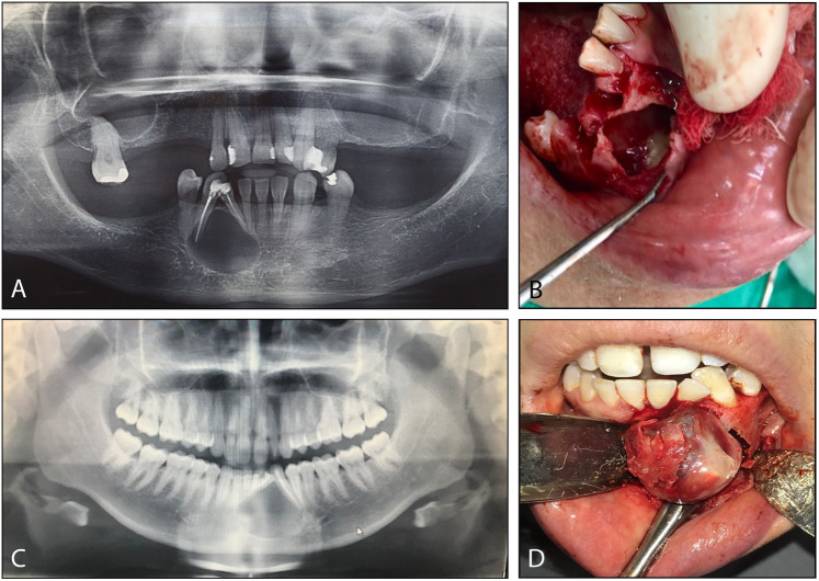
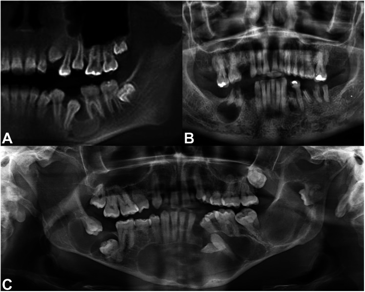
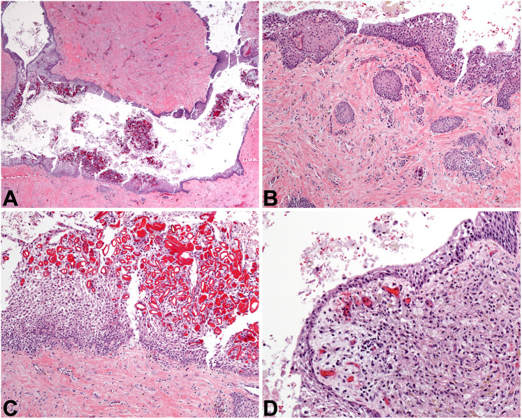
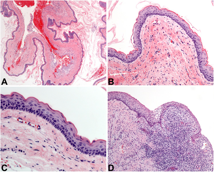
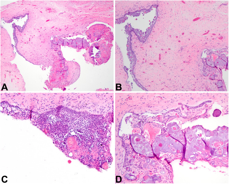
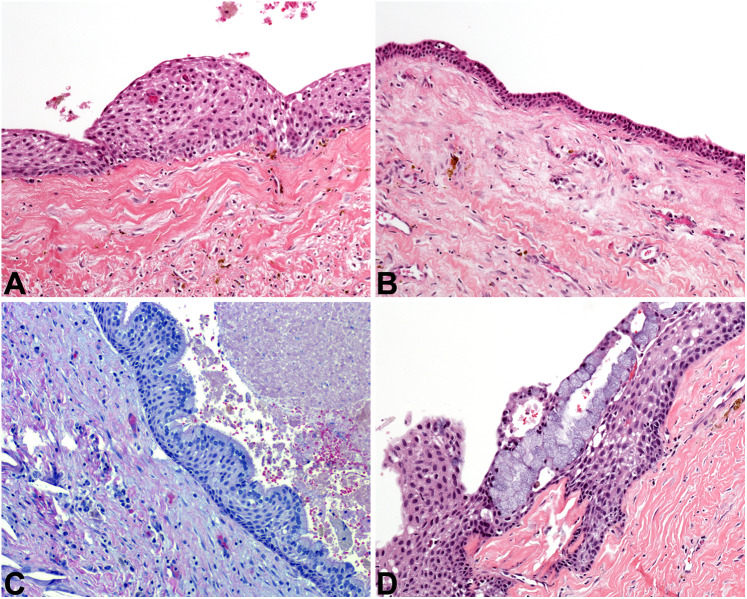
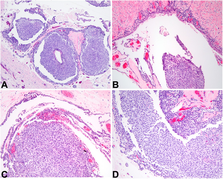
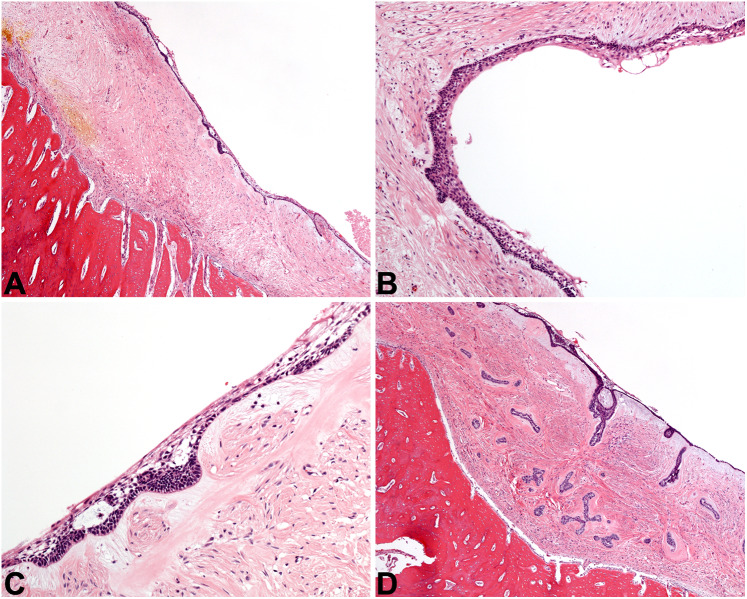
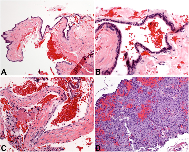
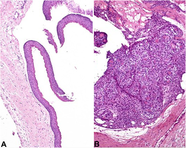
Results: Two clinical cases are presented in parallel and are followed by an illustrated discussion of the ten most likely differential diagnoses that should be considered when confronted with a cystic jaw lesion. Strong emphasis is placed on the histologic differences between these entities, empowering readers to diagnose them with confidence. Perhaps even more importantly, the more common diagnostic pitfalls in gnathic pathology are discussed, recognizing that a definitive diagnosis cannot be rendered in every situation. The histologic diagnoses for the two clinical cases are finally revealed.

Conclusion: Cystic lesions of the maxilla and mandible may be odontogenic or non-odontogenic. The most common cystic lesions are the reactive periapical cyst, and the dentigerous cyst (which is developmental in nature). It is important to note that cystic neoplasms also occur in the jaws, and that the presence of inflammation may obscure the diagnostic histologic features of lesions like odontogenic keratocyst and unicystic ameloblastoma. Ancillary testing is of limited diagnostic value in most scenarios. However, both clinical and radiographic information (such as the location, size, duration, associated symptoms, and morphology of the lesion in its natural habitat) are significantly useful.

Keywords: Cyst; Differential diagnosis; Jaws; Mandible; Maxilla; Odontogenic; Review.


Figures

Fig. 1
Fig. 1
Clinical case descriptions. A Panoramic radiograph of Case 1 shows a radiolucent lesion of the anterior mandible, associated with and displacing two teeth. B At surgery in Case 1, a cystic cavity with focal debris was discovered. C Panoramic radiograph of Case 2 reveals a similar well-defined radiolucent lesion of the anterior mandible, associated with two anterior teeth. D At surgery in Case 2, a solid tumor was “shelled out” of a bony cavity with ease
Fig. 2
Fig. 2
Anatomic locations of gnathic cystic lesions. A A dentigerous relationship is exemplified by the cystic lesion seen here, developing around the crown of an impacted tooth 20 in the left posterior mandible. B This well-defined radiolucent lesion is in a periapical location, directly beneath tooth roots. C At times, multiple cystic lesions may occur, as in this panoramic radiograph of a patient with Gorlin syndrome
Fig. 3
Fig. 3
Dentigerous cyst and periapical cyst. A This DC at low power demonstrates an epithelial lining and a fibrous connective tissue wall (4x). B Odontogenic rests of Malassez may blend with the cystic lining and may proliferate within the cyst wall of DC or PC, and they may be misinterpreted as invasive squamous cell carcinoma. This DC also exhibits mucous metaplasia (10×). C Rushton bodies are curvilinear eosinophilic formations present in the lining of this DC, but these formations are not specific and may also be found in PC (20×). D A PC is depicted, complete with inflammation, histiocytes, and a thinned epithelial lining (20×)
Fig. 4
Fig. 4
Odontogenic keratocyst. A At low power, OKC is overtly cystic, with a lumen filled with pink keratinaceous debris (2x). B Classic OKC lining epithelium is 6–8 cell layers thick, with basal palisading and a corrugated parakeratin surface (10×). C A closer view of a “perfect” OKC, exhibiting the diagnostic features of this cyst (20×). D Classic OKC is demonstrated in the upper left corner of this image, but inflammation completely obscures the diagnostic features of the cystic lining at the right (4x)
Fig. 5
Fig. 5
Calcifying odontogenic cyst. A At low power, a cyst lining epithelium that is proliferative and contains amorphous material is noted (2x). B The lining epithelium of COC looks reminiscent of cystic ameloblastoma in its pattern (4x). C The proliferative epithelium of COC classically contains eosinophilic anucleate formations called ghost cells (10×). D Over time, ghost cells coalesce together and create amorphous amphophilic material that slowly calcifies (10×)
Fig. 6
Fig. 6
Glandular odontogenic cyst. A In the lining of this GOC, there are epithelial thickenings or “spheres” (10×). B GOC classically exhibits cuboidal cells with eosinophilic cytoplasm, and microcysts are also visible to the upper left of the image (10×). C This GOC shows prominent papillary tuft formation, complete with a ciliated luminal surface (10×). D Variability in thickness of the epithelial lining and abundant mucous cells are seen in this GOC example (20×)
Fig. 7
Fig. 7
Adenomatoid odontogenic tumor. A At low power, AOT is an encapsulated cystic tumor filled with whorled epithelial tumor cells (4x). B Without the solid tumor in the lumen, this inflamed AOT lining could be easily mistaken for a reactive or developmental cyst (10×). C AOT is cystic and solid, and the solid component shows evenly spaced and bland epithelial cells arranged in a loosely nested pattern (10×). D Duct-like structures and pseudorosettes are often identified in the solid component of AOT (20×)
Fig. 8
Fig. 8
Unicystic ameloblastoma. A At low power, a relatively nondescript cyst lining is seen adjacent to dense bone (4x). B Upon closer examination, there are subtle palisaded basal cells and a very thin stellate-reticulum-like area, topped at the luminal surface by cells with eosinophilic cytoplasm (10×). C These features are more prominent in this portion of the UA lining, highlighting the necessity to evaluate all of the cystic lining (20×). D Small islands and cords of ameloblastic epithelium are seen in the wall of this cystic ameloblastoma, which for the authors pushes this lesion into the conventional ameloblastoma category (4x)
Fig. 9
Fig. 9
Other cystic lesions. A The nasopalatine duct cyst at low power is lined by epithelium of even thickness (4x). B NPDC is lined by cuboidal cells in a simple arrangement, with an associated fibrous cyst wall (20×). C The simple bone cyst is defined by an empty cavity found at surgery, plus this delicate lace-like osteoid with osteoblastic rimming (20×). D The aneurysmal bone cyst is a solid and cellular proliferation of spindled cells admixed with sinusoidal spaces full of blood, graced with giant cells (10×)
Fig. 10
Fig. 10
Case diagnoses. A Case 1 represents and OKC with classic features (10×). B Case 2 is an AOT, with solid tumor whorls (10×)

References

    1. Jones AV, Craig GT, Franklin CD. Range and demographics of odontogenic cysts diagnosed in a UK population over a 30-year period. J Oral Pathol Med. 2006;35(8):500–507. doi: 10.1111/j.1600-0714.2006.00455.x. - DOI - PubMed
    1. Lin HP, Wang YP, Chen HM, Cheng SJ, Sun A, Chiang CP. A clinicopathological study of 338 dentigerous cysts. J Oral Pathol Med. 2013;42(6):462–467. doi: 10.1111/jop.12042. - DOI - PubMed
    1. Zhang LL, Yang R, Zhang L, Li W, MacDonald-Jankowski D, Poh CF. Dentigerous cyst: a retrospective clinicopathological analysis of 2082 dentigerous cysts in British Columbia, Canada. Int J Oral Maxillofac Surg. 2010;39(9):878–882. doi: 10.1016/j.ijom.2010.04.048. - DOI - PubMed
    1. Takeda Y, Oikawa Y, Furuya I, Satoh M, Yamamoto H. Mucous and ciliated cell metaplasia in epithelial linings of odontogenic inflammatory and developmental cysts. J Oral Sci. 2005;47(2):77–81. doi: 10.2334/josnusd.47.77. - DOI - PubMed
    1. Johnson NR, Gannon OM, Savage NW, Batstone MD. Frequency of odontogenic cysts and tumors: a systematic review. J Investig Clin Dent. 2014;5(1):9–14. doi: 10.1111/jicd.12044. - DOI - PubMed

LinkOut - more resources