

Top 10 Nested Pattern Head and Neck Lesions to Notice
- PMID: 36928740
- PMCID: PMC10063737
- DOI: 10.1007/s12105-023-01534-0
Top 10 Nested Pattern Head and Neck Lesions to Notice
Abstract
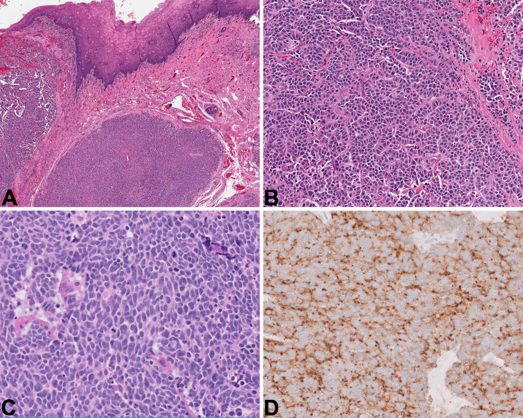
Background: Nested is defined as "cellular clusters arranged in small groupings with intervening vascular or stromal networks, lacking lumens or glandular formation." Using this definition, multiple neoplastic and non-neoplastic lesions of the head and neck come into the differential. We have broadly organized the differential diagnosis of "nested" tumors into entities with neuroendocrine differentiation, squamous differentiation, thyroid follicular cell differentiation, and other lesions.
Methods: Review.
Results: Many different entities have a nested appearance and the morphologic, immunohistochemical, clinical, and radiographic features contribute to the differential diagnosis. The different tumors covered in this review include neuroendocrine neoplasms, paraganglioma, middle ear neuroendocrine tumor (formerly known as middle ear adenoma), medullary thyroid carcinoma, poorly differentiated thyroid carcinoma, olfactory neuroblastoma, ectopic pituitary neuroendocrine tumor, hyalinizing trabecular tumor, solid subtype of papillary thyroid carcinoma, solid cell nests/C-cell hyperplasia, necrotizing sialometaplasia, and meningioma.
Conclusion: In this review, we discuss the morphologic and immunohistochemical features of the covered entities as a guide to differential diagnosis when nested-patterned head and neck lesions are encountered.
Keywords: Head and neck; Nested; Neuroendocrine; Thyroid.
© 2023. The Author(s), under exclusive licence to Springer Science+Business Media, LLC, part of Springer Nature.
Conflict of interest statement
All authors certify that they have no affiliations with or involvement in any organization or entity with any financial interest or non-financial interest in the subject matter or materials discussed in this manuscript.
Figures


References
-
- WHO Classification of Tumours Editorial Board. Endocrine and neuroendocrine tumours. 2022 5th ed. Lyon (France): International Agency for Research on Cancer
-
- WHO Classification of Tumours Editorial Board. Head and neck tumours [Internet; beta version ahead of print]. 2022 5th ed. Lyon (France): International Agency for Research on Cancer
Publication types
MeSH terms
LinkOut - more resources
Full Text Sources
Medical
Molecular Biology Databases
