. 2023 Mar 16;80(4):91.
doi: 10.1007/s00018-023-04752-5.

KIF13B mediates VEGFR2 recycling to modulate vascular permeability

Affiliations

KIF13B mediates VEGFR2 recycling to modulate vascular permeability

Hyun-Dong Cho et al. Cell Mol Life Sci. .

Abstract

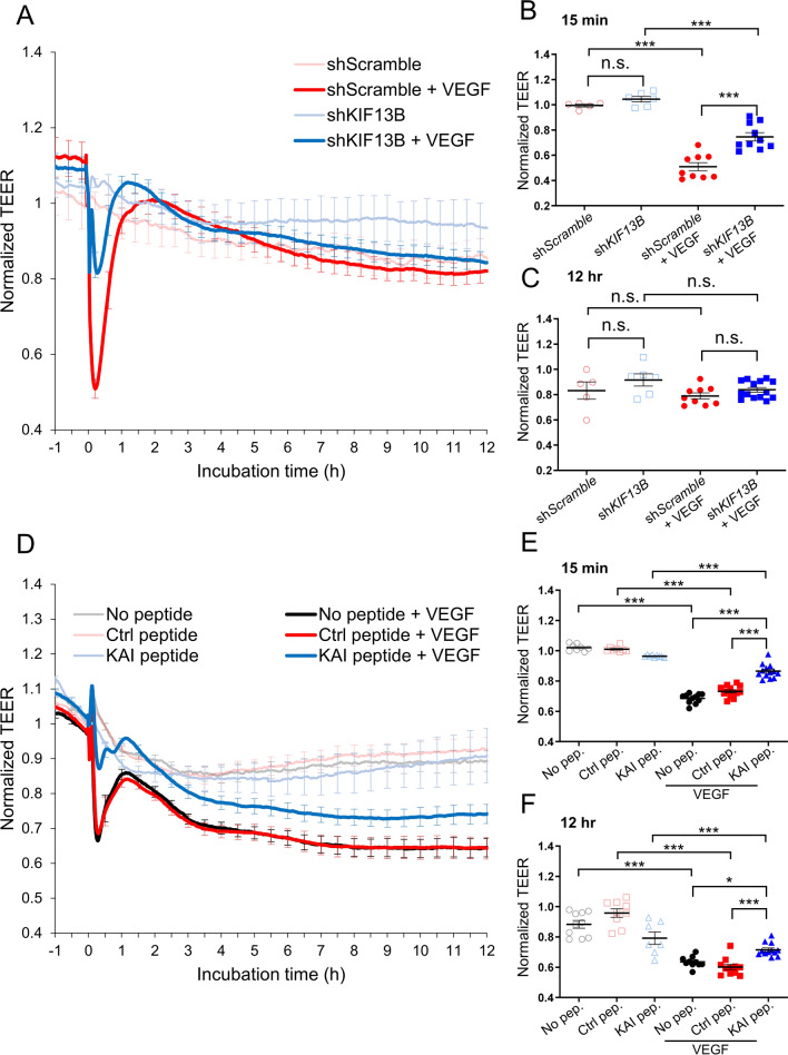
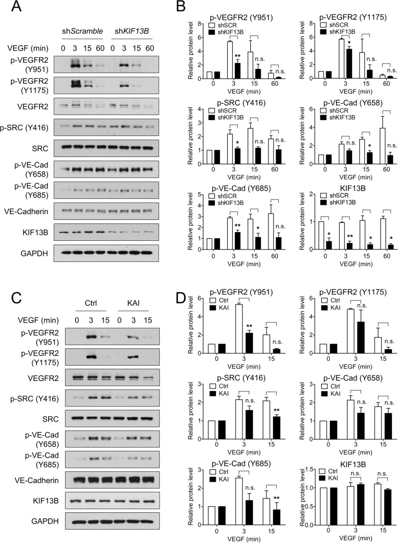
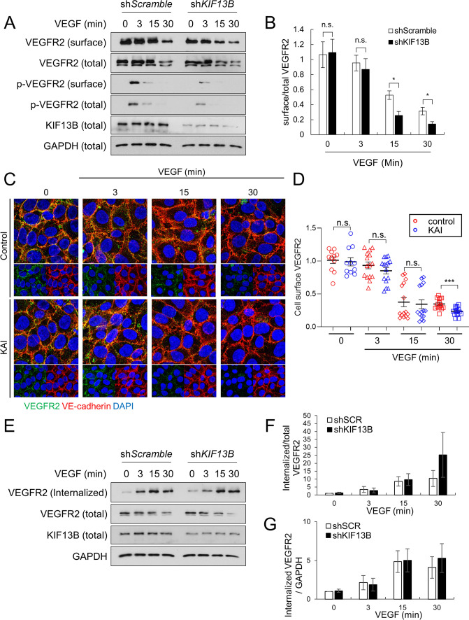
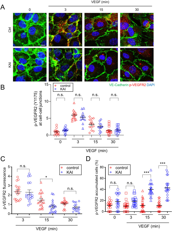
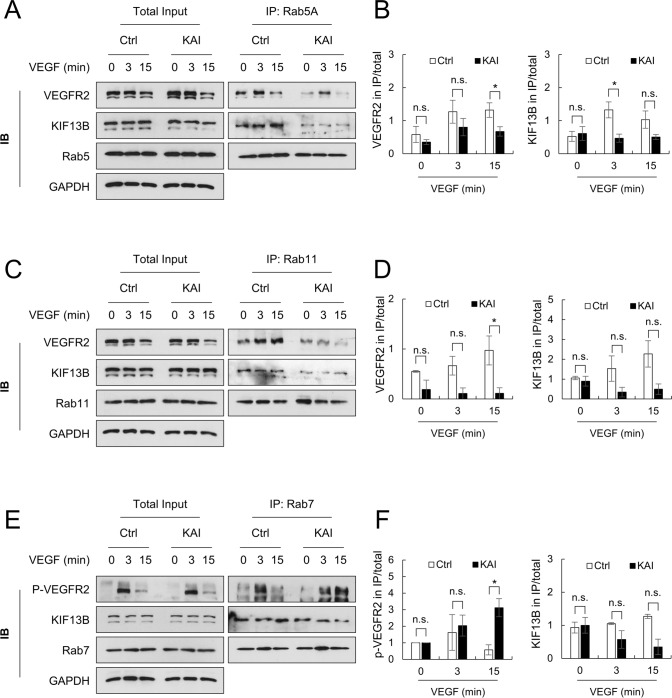
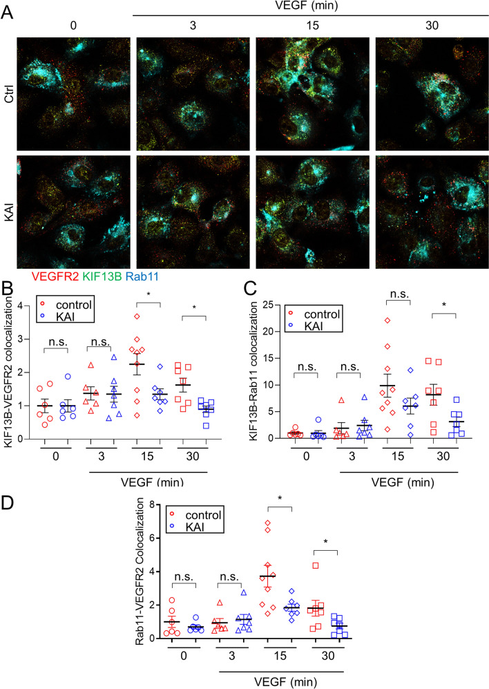
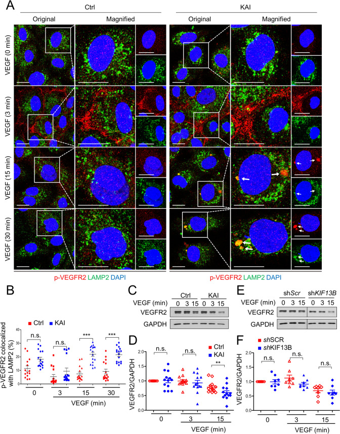
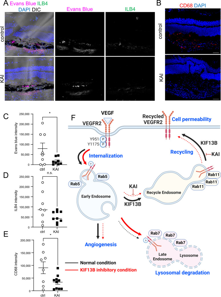
Excessive vascular endothelial growth factor-A (VEGF-A) signaling induces vascular leakage and angiogenesis in diseases. VEGFR2 trafficking to the cell surface, mediated by kinesin-3 family protein KIF13B, is essential to respond to VEGF-A when inducing angiogenesis. However, the precise mechanism of how KIF13B regulates VEGF-induced signaling and its effects on endothelial permeability is largely unknown. Here we show that KIF13B-mediated recycling of internalized VEGFR2 through Rab11-positive recycling vesicle regulates endothelial permeability. Phosphorylated VEGFR2 at the cell-cell junction was internalized and associated with KIF13B in Rab5-positive early endosomes. KIF13B mediated VEGFR2 recycling through Rab11-positive recycling vesicle. Inhibition of the function of KIF13B attenuated phosphorylation of VEGFR2 at Y951, SRC at Y416, and VE-cadherin at Y685, which are necessary for endothelial permeability. Failure of VEGFR2 trafficking to the cell surface induced accumulation and degradation of VEGFR2 in lysosomes. Furthermore, in the animal model of the blinding eye disease wet age-related macular degeneration (AMD), inhibition of KIF13B-mediated VEGFR2 trafficking also mitigated vascular leakage. Thus, the present results identify the fundamental role of VEGFR2 recycling to the cell surface in mediating vascular permeability, which suggests a promising strategy for mitigating vascular leakage associated with inflammatory diseases.

Keywords: Endothelial permeability; Eye diseases; Kinesin; Trafficking; VEGFR2; Vesicle.


Conflict of interest statement

Authors have no conflict of interests that might be perceived to influence the results and/or discussion reported in this article.

Figures

Fig. 1
Fig. 1
KIF13B is essential for VEGF-induced permeability in hREC. A, B, C Transendothelial electrical resistance (TEER) measurement of confluent hRECs transduced with scrambled shRNA (red and pink lines) or shRNA-KIF13B (blue and light blue) expressed as normalized resistance of the TEER basal values. Cells were stimulated with VEGF-A (50 ng/mL) at time zero, and TEER was measured every 30 s over time (4000 Hz of frequency). Normalized resistance changes overtime were shown in graph A. Normalized resistance at 15 min after VEGF-A stimulation and 12 h after VEGF-A stimulation were shown in B, and C as mean ± SE. N = 5, 6, 18, 18, for scrambled shRNA, shRNA-KIF13B, scrambled shRNA + VEGF-A, and shRNA-KIF13B + VEGF-A, respectively. The graphs in B and C show N = 5, 6, 9, 10 and N = 5, 6, 9, 14, respectively, by excluding outliners of mean ± 4 × SE. Note, including outliners and excluding outliners did not affect statistical analyses. One-way ANOVA with post hoc multiple comparisons. ***p < 0.001. DF TEER measurements of hREC treated with KAI (10 µM), an inhibitor for VEGFR2 trafficking, or control peptide (10 µM) followed by VEGF-A (50 ng/mL) stimulation. Changes of normalized resistance over time were shown in graph D. Normalized resistance at 15 min and 12 h after VEGF-A stimulation were shown in E and F as mean ± SE. N = 12, 10, 13, 8, 16, 15, for no peptide, ctrl, KAI, no peptide + VEGF-A, ctrl + VEGF-A, KAI + VEGF-A, respectively. The graphs in E and F show N = 10, 9, 7, 10, 11, 12 and N = 8, 8, 9, 11, 13, 13, respectively, by excluding outliners of mean ± 4 × SE. Note, including outliners and excluding outliners did not affect statistical analyses. One-way ANOVA with post hoc multiple comparisons. *p < 0.05, **p < 0.01
Fig. 2
Fig. 2
KIF13B is required to regulate VEGFR2 signaling pathway. A, B VEGF-induced phosphorylation of signaling molecules in hREC transduced with either scrambled shRNA or shRNA-KIF13B. After stimulation with VEGF-A (50 ng/mL) for indicated time points, cell lysates were analyzed by western blotting for p-VEGFR2 (Y951), p-VEGFR2 (Y1175), VEGFR2, p-SRC (Y416), SRC, p-VE-cadherin (Y658), p-VE-cadherin (Y685), VE-cadherin, KIF13B, and GAPDH (p indicates the phosphorylated form). Representative blots were shown in A. Quantification of blots of phosphorylated proteins expressed relative to total proteins were shown as mean ± SE in graph B. N = 3. One-way ANOVA followed by post hoc multiple comparisons. *p < 0.05, **p < 0.01. No significant difference (n.s.) indicates p > 0.05. C, D VEGF-induced phosphorylation of signaling molecules in hREC treated with either control peptide or KAI (10 µM). After stimulation with VEGF-A (50 ng/mL) for indicated time points, cell lysates were analyzed by western blotting for phosphorylated and total proteins. Representative blots were shown in C. Quantification of phosphorylated proteins relative to total proteins was shown as mean ± SE in graph D. N = 4. One-way ANOVA followed by post hoc multiple comparisons. **p < 0.01. n.s. indicates p > 0.05
Fig. 3
Fig. 3
KIF13B deficiency reduces cell-surface VEGFR2 after VEGF stimulation. A, B Cell surface VEGFR2 levels detected by cell surface biotinylation of hREC transduced with either scrambled shRNA or shRNA-KIF13B, followed by VEGF-A stimulation (50 ng/mL) for indicated time periods. Total lysates (total) and streptavidin pull-down (surface), western blotting for VEGFR, p-VEGFR2 (Y1175) were shown in A. GAPDH was used as a loading control. Quantification of cell surface VEGFR2 was normalized by total VEGFR2, and shown as mean ± SE in graph B. N = 4. One-way ANOVA followed by multiple comparisons. *p < 0.05. n.s. indicates p > 0.05. C, D Immunostaining of cell surface VEGFR2 (green) and VE-cadherin (red) in hREC pretreated with control peptide or KAI peptide followed by VEGF stimulation for indicated times. Goat antibody for extracellular domain of VEGFR2 and mouse antibody for extracellular domain of VE-cadherin were used for staining of cells without detergent-permeabilization. Intensity of VEGFR2 was quantified by Image J and shown as mean ± SE in the graph. N = 11, 11, 15, 15, 15, 15, 15, 15 for control 0 min, KAI 0 min, control 3 min, KAI 3 min, control 15 min, KAI 15 min, control 30 min, KAI 30 min, respectively. N is number of pictures analyzed from 3 independent experiments. Student's t test. *p < 0.05. EG The internalized pool of VEGFR2 after VEGF-A treatment (50 ng/mL) of hREC transduced with either scrambled shRNA or shRNA-KIF13B. Cell surface biotinylation was performed prior to VEGF-A stimulation. At indicated time points, the remaining cell surface biotin was removed, and the internalized pool of VEGFR2 was collected by streptavidin pull-down. Western blotting of the total lysate (total) and streptavidin pull-down (internalized) for VEGFR2 was shown in E. Internalized VEGFR2 was normalized by total VEGFR2 or loading control GAPDH, and shown as mean ± SE in the graph F and G, respectively. N = 3. One-way ANOVA followed by multiple comparisons and Student's t test did not find any statistical significance between shScr and shKIF13B
Fig. 4
Fig. 4
KIF13B regulates endosomal trafficking of phosphorylated VEGFR2. A, B Immunostaining for p-VEGFR2 (Y1175, red) and VE-cadherin (green) in hREC pretreated with either control peptide or KAI (10 µM) and stimulated with VEGF-A (50 ng/mL) for indicated time periods. Scale bars; 10 µm. The intensity of p-VEGFR2 at cell–cell junction (colocalized with VE-cadherin) was quantified by Image J and shown as mean ± SE in graph B. N = 13, 14, 14, 14, 13, 11, 13, 12 for pictures (from 3 independent experiments) in ctrl (0, 3, 15, 30 min), KAI (0, 3, 15, 30 min), respectively. Unpaired t test, n.s. indicates p > 0.05. C The intensity of p-VEGFR2 was quantified by Image J and shown as mean ± SE in graph C. N = 17, 18, 16, 17, 18, 17 (from 3 independent experiments) for pictures in ctrl (3, 15, 30 min), KAI (3, 15, 30 min). One-way ANOVA with post hoc multiple comparisons. *p < 0.05, and n.s. indicates p > 0.05. D The number of cells with p-VEGFR2 accumulation (accumulated particles bigger than 5 µm2) among the total number of the cells was counted and shown as mean ± SE in graph C. N = 17, 17, 20, 18, 17, 18, 20, 19 (from 6 independent experiments) in ctrl (0, 3, 15, 30 min), KAI (0, 3, 15, 30 min), respectively. One-way ANOVA with post hoc multiple comparisons. ***p < 0.001 and n.s. indicates p > 0.05
Fig. 5
Fig. 5
Association of VEGFR2 to Rab family proteins is impaired by KAI, an inhibitor for KIF13B-mediated VEGFR2 trafficking. A, B Association of VEGFR2 and KIF13B with Rab5-positive early endosome in hREC pretreated with either scrambled control peptide or KAI (10 µM). After VEGF-A stimulation (50 ng/mL) for indicated time periods, proteins were co-immunoprecipitated (IP) with Rab5 and analyzed by western blotting. Representative blots were shown in A. Quantification of the proteins co-immunoprecipitated with Rab5 was normalized by proteins in the total lysate and shown as mean ± SE in the graph B. N = 4. Student's t test was performed for statistical analysis in ctrl and KAI groups with the same time point of VEGF stimulation. *p < 0.05. n.s. indicates p > 0.05. C, D Association of VEGFR2 and KIF13B with Rab11-positive recycling vesicles in hREC pretreated with either scrambled control peptide or KAI (10 µM). After VEGF-A stimulation (50 ng/mL) for indicated time points, proteins were co-immunoprecipitated with Rab11 and analyzed by western blotting. Representative blots were shown in C. Quantification of the proteins co-immunoprecipitated with Rab11 was normalized by proteins in the total lysate and shown as mean ± SE in the graph D. N = 4. Student's t test. *p < 0.05. n.s. indicates p > 0.05. E, F Association of phosphorylated VEGFR2 (Y1175), and KIF13B with Rab7-positive late endosome in hREC pretreated with either scrambled control peptide or KAI (10 µM). After VEGF-A stimulation (50 ng/mL) for indicated time points, proteins were co-immunoprecipitated with Rab7, and analyzed by western blotting. Representative blots were shown in E. Quantification of the proteins co-immunoprecipitated with Rab7 was normalized by proteins in the total lysate and shown as mean ± SE in the graph F. N = 4. Student's t test. *p < 0.05. n.s. indicates p > 0.05
Fig. 6
Fig. 6
Inhibition of KIF13B reduced association of VEGFR2 to recycling vesicle. AD Immunostaining for VEGFR2 (red pseudo color, stained with Alexa 647) and KIF13B (green pseudo color, stained with Alexa 594) and fluorescence of mCerulean-Rab11 (cyan) in hREC pretreated with either control peptide or KAI (10 µM), and stimulated with VEGF-A (50 ng/mL) for indicated time periods. The colocalizations of KIF13B and VEGFR2, KIF13B and Rab11, VEGFR2 and Rab11 were quantified by Image J and shown as mean ± SE in graph BD, respectively. N = 6, 6, 9, 7, 6, 7, 7, 7 for pictures (from 3 independent experiments) in ctrl (0, 3, 15, 30 min), KAI (0, 3, 15, 30 min), respectively. Student's t test. *p < 0.05, and n.s. indicates p > 0.05
Fig. 7
Fig. 7
Failure of VEGFR2 trafficking sends p-VEGFR2 to lysosome. A Immunostaining for p-VEGFR2 (Y1175, red) and LAMP2 (green) in hREC pretreated with either control peptide or KAI (10 µM), and stimulated with VEGF-A (50 ng/mL) for indicated time periods. Scale bars; 10 µm. B The colocalization of p-VEGFR2 and LAMP2 was quantified by Image J and shown as mean ± SE in graph B. N = 14, 16, 15, 16, 16, 17, 16 and 16 for pictures in ctrl (0, 3, 15, 30 min), KAI (0, 3, 15, 30 min), respectively, from 3 independent experiments. One-way ANOVA with post hoc multiple comparisons. ***p < 0.001 and n.s. indicates p > 0.05. C, D Degradation of VEGFR2 after VEGF-A stimulation (50 ng/mL) in hREC treated with either scrambled control peptide or KAI peptide (10 µM). Representative blots were shown in C. Quantification of VEGFR2 was normalized by loading control GAPDH, and shown in graph D as mean ± SE. N = 13. T test was performed for statistical analysis in ctrl and KAI groups with the same time point of VEGF stimulation. **p < 0.01. E, F Degradation of VEGFR2 after VEGF-A stimulation (50 ng/mL) in hREC transduced with scrambled shRNA or shRNA-KIF13B. Representative blots were shown in E. Intensity of the bands of VEGFR2 was normalized by loading control GAPDH, and shown in graph F as mean ± SE. N = 8. T test was performed for statistical analysis in scrambled shRNA and shRNA-KIF13B groups with the same time point of VEGF stimulation. N.s. no significant difference
Fig. 8
Fig. 8
Pharmacological inhibition of VEGFR2 trafficking ameliorates vascular leakage in mice. AE Effects of KAI eyedrop on the pathological vascular leakage in mice. After receiving laser burns, mice were treated either control peptide (ctrl) or KAI (5 µg/eye in 5 µl PBS) daily. On day 3, Evans blue was injected i.p., and Evans blue leakage was measured 24 h after injection. Cryosections of the damaged area were also stained with ILB4 (A) and CD68 (B) to visualize neovascularization and macrophage recruitment, respectively. Representative images were shown in A and B. Scale bars; 50 µm. The intensity of the Evans blue extravasation, ILB4, and CD68 was quantified and shown in the graph as mean ± SE in CE. N = 8 and 10 for ctrl and KAI, respectively. F Schematic showing the working model in this study. VEGF induces internalization of phosphorylated VEGFR2 to Rab5-positive early endosomes. Dephosphorylated VEGFR2 is transferred to Rab11-positive recycling vesicle by KIF13B and further trafficked to the cell surface. When KIF13B function is blocked by either knockdown or pharmacological inhibitor KAI, VEGFR2 trafficking is failed, and phosphorylated VEGFR2 is sent from early endosomes to late endosome and lysosome for degradation. Recycling of VEGFR2 is required for VEGF-induced signaling for endothelial permeability. Thus, this pathway can be a therapeutic target for vascular leakage-related diseases such as wet AMD

References

    1. Horiguchi K, Hanada T, Fukui Y, Chishti AH. Transport of PIP3 by GAKIN, a kinesin-3 family protein, regulates neuronal cell polarity. J Cell Biol. 2006;174:425–436. doi: 10.1083/jcb.200604031. - DOI - PMC - PubMed
    1. Claesson-Welsh L, Dejana E, McDonald DM. Permeability of the endothelial barrier: identifying and reconciling controversies. Trends Mol Med. 2020;27:314–331. doi: 10.1016/j.molmed.2020.11.006. - DOI - PMC - PubMed
    1. Duong CN, Vestweber D. Mechanisms ensuring endothelial junction integrity beyond VE-cadherin. Front Physiol. 2020;11:519. doi: 10.3389/fphys.2020.00519. - DOI - PMC - PubMed
    1. Eelen G, Treps L, Li X, Carmeliet P. Basic and therapeutic aspects of angiogenesis updated. Circ Res. 2020;127:310–329. doi: 10.1161/circresaha.120.316851. - DOI - PubMed
    1. Apte RS, Chen DS, Ferrara N. VEGF in signaling and disease: beyond discovery and development. Cell. 2019;176:1248–1264. doi: 10.1016/j.cell.2019.01.021. - DOI - PMC - PubMed

MeSH terms

Substances

LinkOut - more resources