. 2023 Jun 8;141(23):2891-2900.
doi: 10.1182/blood.2022018956.

Efficacy of platelet-inspired hemostatic nanoparticles on bleeding in von Willebrand disease murine models

Affiliations

Efficacy of platelet-inspired hemostatic nanoparticles on bleeding in von Willebrand disease murine models

Stéphanie Roullet et al. Blood. .

Abstract

The lack of innovation in von Willebrand disease (VWD) originates from many factors including the complexity and heterogeneity of the disease but also from a lack of recognition of the impact of the bleeding symptoms experienced by patients with VWD. Recently, a few research initiatives aiming to move past replacement therapies using plasma-derived or recombinant von Willebrand factor (VWF) concentrates have started to emerge. Here, we report an original approach using synthetic platelet (SP) nanoparticles for the treatment of VWD type 2B (VWD-2B) and severe VWD (type 3 VWD). SP are liposomal nanoparticles decorated with peptides enabling them to concomitantly bind to collagen, VWF, and activated platelets. In vitro, using various microfluidic assays, we show the efficacy of SPs to improve thrombus formation in VWF-deficient condition (with human platelets) or using blood from mice with VWD-2B and deficient VWF (VWF-KO, ie, type 3 VWD). In vivo, using a tail-clip assay, SP treatment reduced blood loss by 35% in mice with VWD-2B and 68% in mice with VWF-KO. Additional studies using nanoparticles decorated with various combinations of peptides demonstrated that the collagen-binding peptide, although not sufficient by itself, was crucial for SP efficacy in VWD-2B; whereas all 3 peptides appeared necessary for mice with VWF-KO. Clot imaging by immunofluorescence and scanning electron microscopy revealed that SP treatment of mice with VWF-KO led to a strong clot, similar to those obtained in wild-type mice. Altogether, our results show that SP could represent an attractive therapeutic alternative for VWD, especially considering their long half-life and stability.


Conflict of interest statement

Conflict-of-interest disclosure: A.D., E.G., C.P., and M.B. are employees of Haima Therapeutics LLC. A.S.G. is cofounder and chief scientific adviser for Haima Therapeutics. The remaining authors declare no competing financial interests.

Figures

None
Graphical abstract
Figure 1.
Figure 1.
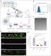
Physical and functional characterization of the SP nanoparticles. (A) Schematic of the manufacturing process for SP nanoparticle with lipids and lipid-peptide conjugates using the thin film rehydration followed by extrusion methodology. (B) Dynamic Light Scattering characterization and (C) Cryo-transmission electron microscopy (Cryo-TEM) imaging of SP nanoparticles indicates a diameter of approximately 150 to 200 nm. (D-E) Representative fluorescence microscopy images and quantitative analysis of imaging data from BioFlux microfluidics studies with platelet suspensions indicate that depletion of platelets from 200 000 per μL (Plt-200K) to 20 000 per μL (Plt-20K) results in drastic reduction of platelet coverage of collagen-coated microfluidic channel surface. Treatment of Plt-20K with CP does not rescue this whereas the treatment of Plt-20K with SP nanoparticles significantly rescues platelet recruitment and coverage on the collagen-coated channel surface (colocalization of Rhodamine B-labeled red fluorescent SP with calcein-stained green fluorescent platelets appear yellow). The particle/platelet ratio was 1000:1. Means ± SD are represented. Statistical analysis was performed using a one-way ANOVA with Tukey's correction. ∗∗P ≤ .01; ∗∗∗P ≤ .001; ∗∗∗∗P ≤ .0001. ns, not significant.
Figure 2.
Figure 2.
In vitro effect of SP nanoparticles added to VWD murine blood in perfusion assays. (A) SPs or CP were added to VWD-2B blood (left panel) or to VWF-KO blood (right panel) at a ratio of 50 particles per platelet. Particle-supplemented blood or unsupplemented blood (untreated) was perfused over AR chips in the T-TAS Plus system. Graphs represent changes in pressure (a measure for thrombus formation) as a function of time. The colored area represents the standard error of the mean for each condition. Statistical analysis was performed using a one-way ANOVA with Tukey's correction. (B-C) SPs or CP were added to VWD-2B murine blood or to VWF-KO murine blood at a ratio of 50 particles per platelet. Particle-supplemented blood or unsupplemented blood (untreated) was perfused over collagen in a parallel flow chamber at 1500 s−1. untreated representative images of thrombus formation are shown in B while percentage platelet coverage, representing adhesion or mean fluorescence intensity as a marker of thrombus size are represented in C. Results obtained with untreated WT murine blood is shown for comparison. Data are presented as mean ± SD, n = 3. Statistical analysis was performed using a one-way ANOVA with Tukey's correction. ∗P ≤ .05; ∗∗P ≤ .01; ∗∗∗∗P ≤ .0001. a.u., arbitrary units; ns, not significant.
Figure 3.
Figure 3.
In vivo effect of SP nanoparticles in VWD-2B and VWF-KO murine models. Mice were injected with CP or SP nanoparticles (2 mg/kg) or not injected. Bleeding was measured for 30 minutes (VWD-2B) (A) or 20 minutes (VWF-KO) (B) after amputation of 3 mm of the tail tip. Blood was collected in warm saline and quantified using a hemoglobin calibration curve. Each dot represents an individual mouse. Data are presented as mean ± SD. Statistical analysis was performed using a one-way ANOVA with Dunnett's correction.∗P ≤ .05; ∗∗∗P ≤ .001; ∗∗∗∗P ≤ .0001. ns, not significant.
Figure 4.
Figure 4.
In vivo effect of various peptide-decorated particles in VWD-2B and VWF-KO murine models. Mice were injected with control particles, SP nanoparticles or particles decorated with various combinations of peptides (2 mg/kg). Bleeding was measured for 30 minutes (VWD-2B) (A) or 20 minutes (VWF-KO) (B) after amputation of 3 mm of the tail tip. Blood was collected in warm saline and quantified using a hemoglobin calibration curve. Each dot represents an individual mouse. Data are presented as mean ± SD. Statistical analysis was performed using a one-way ANOVA with Dunnett's correction. ∗∗P ≤ .01; ∗∗∗P ≤ .001; ∗∗∗∗P ≤ .0001. SP nanoparticles (3 peptides) are represented with the plain gray bar whereas particles decorated with dual peptides appear in diagonally striped bars and single-decorated particles appear in horizontally striped bars.
Figure 5.
Figure 5.
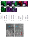
Imaging of the clots obtained after SP treatment of VWF-KO mice. (A) Representative images of the fluorescent staining of clots recovered after amputation of the tail tip of mice with VWF WT and VWF-KO injected with SP (2 mg/kg). Dashes represent the vessel’s edges. Rhodamine-labeled SP are visible in red, fibrin/fibrinogen is visible in magenta and CD41-labeled platelets appear in green. (B) Quantification of the platelets (CD41+) and fibrin area and fluorescence intensity in WT (n = 3) and VWF-KO mice injected with SP (n = 3). Data are presented as mean ± SD. Statistical analysis was performed using a Mann-Whitney test. (C) Scanning electron microscopy of the clot recovered after tail vein transection in mouse with VWF-KO left untreated and treated with SP. In the upper images, black arrows indicate the transection area recognizable by the hair that appear sectioned and the blue arrows point to the clot. In the bottom left image, red arrows show individualized red blood cells. In the bottom right image, blue arrow shows the clot characterized by aggregated red blood cells and red arrows show individualized red blood cells. Two mice per group were imaged and representative images are shown.

References

    1. Denis CV, Susen S, Lenting PJ. von Willebrand disease: what does the future hold? Blood. 2021;137(17):2299–2306. - PubMed
    1. Casari C, Berrou E, Lebret M, et al. von Willebrand factor mutation promotes thrombocytopathy by inhibiting integrin alphaIIbbeta3. J Clin Invest. 2013;123(12):5071–5081. - PMC - PubMed
    1. Federici AB, Mannucci PM, Castaman G, et al. Clinical and molecular predictors of thrombocytopenia and risk of bleeding in patients with von Willebrand disease type 2B: a cohort study of 67 patients. Blood. 2009;113(3):526–534. - PubMed
    1. Kauskot A, Poirault-Chassac S, Adam F, et al. LIM kinase/cofilin dysregulation promotes macrothrombocytopenia in severe von Willebrand disease-type 2B. JCI Insight. 2016;1(16) - PMC - PubMed
    1. Kruse-Jarres R, Johnsen JM. How I treat type 2B von Willebrand disease. Blood. 2018;131(12):1292–1300. - PubMed

Publication types