

Comment
doi: 10.1007/s11739-023-03250-7.
Epub 2023 Mar 16.
Two-years chest-CT follow-up after severe COVID-19 pneumonia
Affiliations
- PMID: 36929349
- PMCID: PMC10018600
- DOI: 10.1007/s11739-023-03250-7
Item in Clipboard
Comment
Two-years chest-CT follow-up after severe COVID-19 pneumonia
Intern Emerg Med.
2023 Jun.
No abstract available
Conflict of interest statement
The authors declare that they have no conflict of interest.
Figures
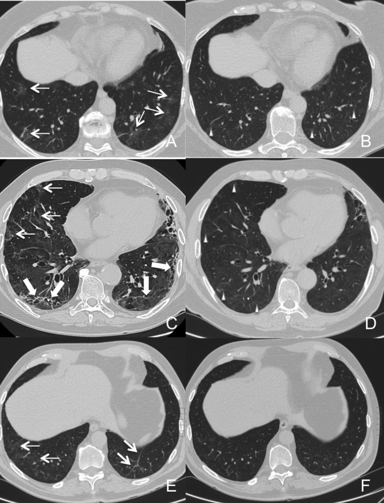
Serial non-contrast axial chest CTs of three studied patients with prior severe COVID-19 pneumonia. Upper line: chest CT of a 61-year-old woman showed ground-glass opacities, reticular alterations and parenchymal bands (Warrick Score: 8; CT score: 5) in both lower lobes at the 6-month CT follow-up (Panel A). At the 2-years follow-up (Panel B) complete resolution of GGO with persistency of reticulations and bands was noticed (Warrick score: 5; CT score: 3). Middle line: panel C shows six-month follow-up chest CT of a 65-year-old woman demonstrating ground-glass opacities, reticular alterations, parenchymal bands and traction bronchiectasis in middle lobe, lingula and in both lower lobes (Warrick score: 20; CT score: 9). At the 2-years follow-up (Panel D) almost complete resolution of traction bronchiectasis, bands and GGO with persistency of reticulations was noted (Warrick score: 13; CT score: 6). Bottom line: panel E shows chest CT of a 57-year-old woman with reticular alterations and parenchymal bands in the lung lower lobes at the 6-month follow-up (Warrick score: 8; CT score: 8). At 2-years follow-up (Panel F) complete resolution of GGO and parenchymal bands with persistency of tiny residual reticulations are observed (Warrick score: 5; CT score: 5).
Comment on
-
Six-month Follow-up Chest CT Findings after Severe COVID-19 Pneumonia.Radiology. 2021 Apr;299(1):E177-E186. doi: 10.1148/radiol.2021203153. Epub 2021 Jan 26. Radiology. 2021. PMID: 33497317 Free PMC article.
References
Publication types
MeSH terms
LinkOut - more resources
Full Text Sources
Medical
