

Noncontact Acoustic Micro-Tapping Optical Coherence Elastography for Quantification of Corneal Anisotropic Elasticity: In Vivo Rabbit Study
- PMID: 36930138
- PMCID: PMC10036949
- DOI: 10.1167/tvst.12.3.15
Noncontact Acoustic Micro-Tapping Optical Coherence Elastography for Quantification of Corneal Anisotropic Elasticity: In Vivo Rabbit Study
Abstract
Purpose: The purpose of this study was to demonstrate accurate measurement of corneal elastic moduli in vivo with noncontact and noninvasive optical coherence elastography.
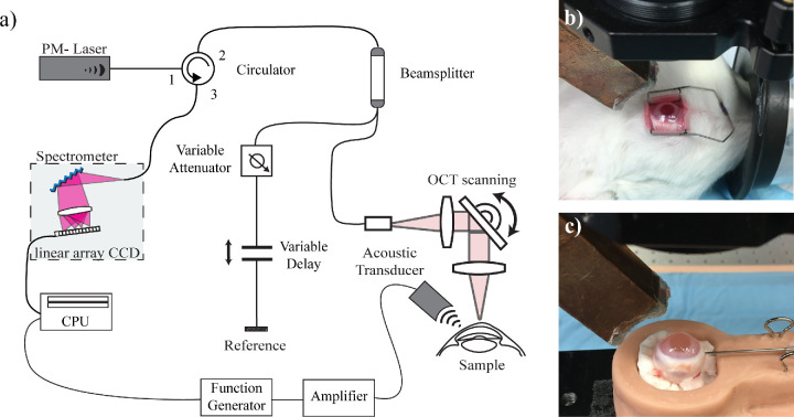
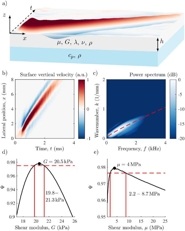
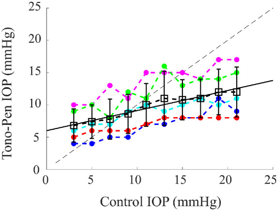
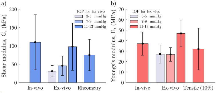
Methods: Elastic properties (in-plane Young's modulus, E, and both in-plane, μ, and out-of-plane, G, shear moduli) of rabbit cornea were quantified in vivo using noncontact dynamic acoustic micro-tapping optical coherence elastography (AµT-OCE). The intraocular pressure (IOP)-dependence of measured mechanical properties was explored in extracted whole globes following in vivo measurement. A nearly incompressible transverse isotropic (NITI) model was used to reconstruct moduli from AµT-OCE data. Independently, cornea elastic moduli were also measured ex vivo with traditional, destructive mechanical tests (tensile extensometry and shear rheometry).
Results: Our study demonstrates strong anisotropy of corneal elasticity in rabbits. The in-plane Young's modulus, computed as E = 3μ, was in the range of 20 MPa to 44 MPa, whereas the out-of-plane shear modulus was in the range of 34 kPa to 261 kPa. Both pressure-dependent ex vivo OCE and destructive mechanical tests performed on the same samples within an hour of euthanasia strongly support the results of AµT-OCE measurements.
Conclusions: Noncontact AµT-OCE can noninvasively quantify cornea anisotropic elastic properties in vivo.
Translational relevance: As optical coherence tomography (OCT) is broadly accepted in ophthalmology, these results suggest the potential for rapid translation of AµT-OCE into clinical practice. In addition, AµT-OCE can likely improve diagnostic criteria of ectatic corneal diseases, leading to early diagnosis, reduced complications, customized surgical treatment, and personalized biomechanical models of the eye.
Conflict of interest statement
Disclosure:
Figures

Update of
-
Non-contact acoustic micro-tapping optical coherence elastography for quantification of corneal anisotropic elasticity: in vivo rabbit study.ArXiv [Preprint]. 2023 Jan 25:arXiv:2301.10652v1. ArXiv. 2023. Update in: Transl Vis Sci Technol. 2023 Mar 1;12(3):15. doi: 10.1167/tvst.12.3.15. PMID: 36748003 Free PMC article. Updated. Preprint.
References
-
- Hatami-Marbini H. Viscoelastic shear properties of the corneal stroma. J Biomech. 2014; 47: 723–728. - PubMed
-
- Sloan SR, Khalifa YM, Buckley MR. The location- and depth-dependent mechanical response of the human cornea under shear loading. Investig Ophthalmol Vis Sci. 2014; 55: 7919–7924. - PubMed
-
- Zeng Y, Yang J, Huang K, Lee Z, Lee X. A comparison of biomechanical properties between human and porcine cornea. J Biomech. 2001; 34: 533–537. - PubMed
-
- Wollensak G, Spoerl E, Seiler T. Stress-strain measurements of human and porcine corneas after riboflavin-ultraviolet-A-induced cross-linking. J Cataract Refract Surg. 2003; 29: 1780–1785. - PubMed
Publication types
MeSH terms
Grants and funding
LinkOut - more resources
Full Text Sources
Research Materials
