

Scleral Proteome in Noninfectious Scleritis Unravels Upregulation of Filaggrin-2 and Signs of Neovascularization
- PMID: 36930145
- PMCID: PMC10036950
- DOI: 10.1167/iovs.64.3.27
Scleral Proteome in Noninfectious Scleritis Unravels Upregulation of Filaggrin-2 and Signs of Neovascularization
Abstract
Purpose: Scleritis is a severe inflammatory ocular disorder with unknown pathogenesis. We investigated healthy sclera as well as sclera affected by noninfectious scleritis for differentially expressed proteins using a mass spectrometry approach.
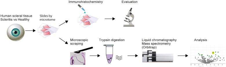
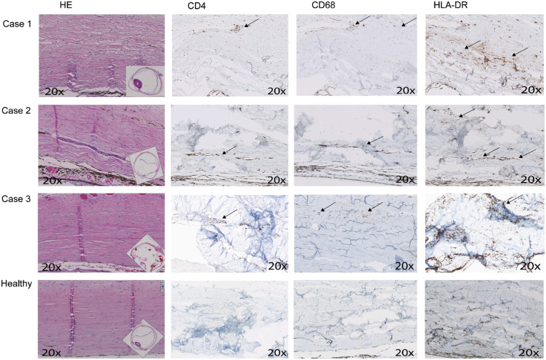
Methods: We collected scleral samples of enucleated eyes due to severe noninfectious scleritis (n = 3), and control scleral tissues (n = 5), all exenterated eyes for eyelid carcinomas (n = 4), or choroidal melanoma (n = 1) without scleral invasion. Samples were prepared for the nano liquid-chromatography mass spectrometer (LC-MS), data were analyzed using proteomics software (Scaffold), and is available via ProteomeXchange (identifier PXD038727). Samples were also stained for immuno-histopathological evaluation.
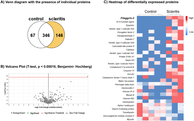
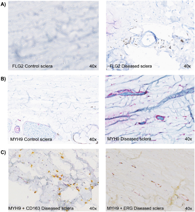
Results: Mass spectrometry identified 629 proteins within the healthy and diseased scleral tissues, whereof collagen type XII, VI, and I were the most abundantly expressed protein. Collagen type II-XII was also present. Filaggrin-2, a protein that plays a crucial role in epidermal barrier function, was found upregulated in all scleritis cases. In addition, other epithelial associated proteins were upregulated (such as keratin 33b, 34, and 85, epiplakin, transglutaminase-3, galectin 7, and caspase-14) in scleritis. Further, upregulated proteins involved in regulation of the cytoskeleton (vinculin and myosin 9), and housekeeping proteins were found (elongation factor-2 and cytoplasmic dynein 1) in our study. Upregulation of filaggrin-2 and myosin-9 was confirmed with immunohistochemistry, the latter protein showing co-localization with the endothelial cell marker ETC-related gene (ERG), indicating neovascularization in scleral tissue affected by scleritis.
Conclusions: We found upregulation of filaggrin-2 and signs of neovascularization in scleral tissue of patients with noninfectious scleritis. Further research, ideally including more scleritis cases, is needed to validate our findings.
Conflict of interest statement
Disclosure:
Figures
Similar articles
-
Alterations in proteome of human sclera associated with primary open-angle glaucoma involve proteins participating in regulation of the extracellular matrix.Mol Vis. 2020 Sep 2;26:623-640. eCollection 2020. Mol Vis. 2020. PMID: 32913388 Free PMC article.
-
Comparative proteome analysis of form-deprivation myopia in sclera with iTRAQ-based quantitative proteomics.Mol Vis. 2021 Sep 1;27:494-505. eCollection 2021. Mol Vis. 2021. PMID: 34526757 Free PMC article.
-
Increased expression of matrix metalloproteinases in vivo in scleritis tissue and in vitro in cultured human scleral fibroblasts.Am J Pathol. 1997 Feb;150(2):653-66. Am J Pathol. 1997. PMID: 9033278 Free PMC article.
-
The spectrum of postoperative scleral necrosis.Surv Ophthalmol. 2013 Nov-Dec;58(6):620-33. doi: 10.1016/j.survophthal.2012.11.002. Epub 2013 Feb 12. Surv Ophthalmol. 2013. PMID: 23410842 Review.
-
Histopathological evaluation of scleritis.J Clin Pathol. 2019 May;72(5):386-390. doi: 10.1136/jclinpath-2018-205360. Epub 2019 Feb 5. J Clin Pathol. 2019. PMID: 30723093 Review.
Cited by
-
Potential Biomarkers for Noninfectious Scleritis Identified by Serum and Tear Fluid Proteomics.Ophthalmol Sci. 2023 Oct 5;4(1):100407. doi: 10.1016/j.xops.2023.100407. eCollection 2024 Jan-Feb. Ophthalmol Sci. 2023. PMID: 38054106 Free PMC article.
References
-
- Wakefield D, Di Girolamo N, Thurau S, Wildner G, McCluskey P.. Scleritis: Immunopathogenesis and molecular basis for therapy. Prog Retinal Eye Res. 2013; 35: 44–62. - PubMed
-
- Hankins M, Margo CE.. Histopathological evaluation of scleritis review. J Clin Pathol. 2019; 72: 386–390. - PubMed
-
- Rao NA, Marak GE, Hidayat AA.. Necrotizing scleritis. A clinico-pathologic study of 41 cases. Ophthalmology. 1985; 92: 1542–1549. - PubMed
Publication types
MeSH terms
Substances
LinkOut - more resources
Full Text Sources
Other Literature Sources
Molecular Biology Databases
