. 2023 Mar 17;80(4):97.
doi: 10.1007/s00018-023-04737-4.

The η-secretase-derived APP fragment ηCTF is localized in Golgi, endosomes and extracellular vesicles and contributes to Aβ production

Affiliations

The η-secretase-derived APP fragment ηCTF is localized in Golgi, endosomes and extracellular vesicles and contributes to Aβ production

Elissa Afram et al. Cell Mol Life Sci. .

Abstract

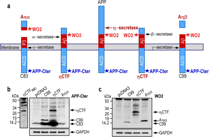
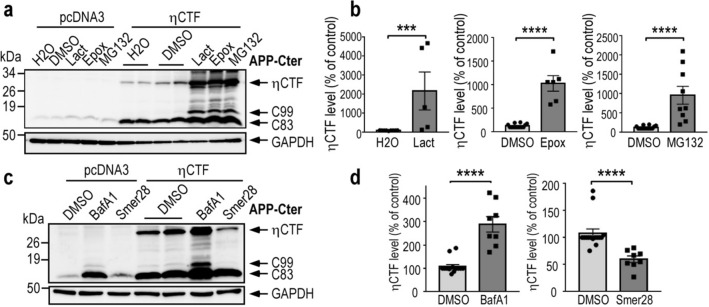
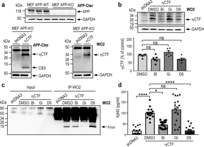
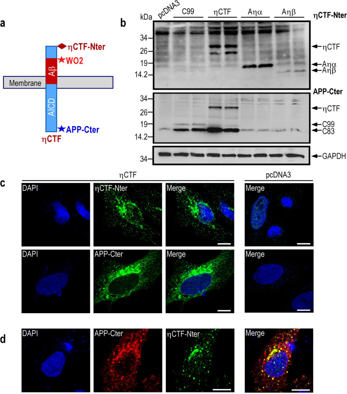
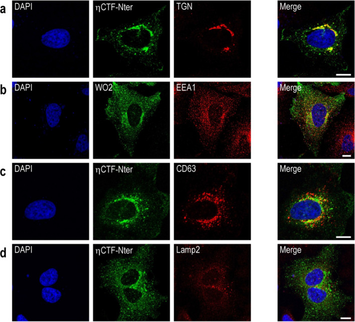
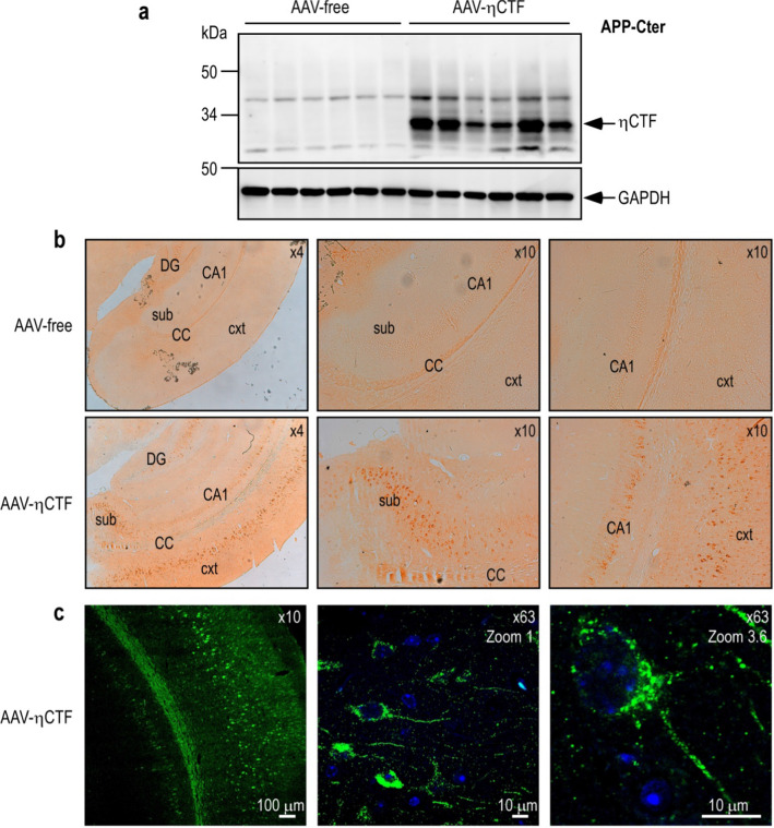
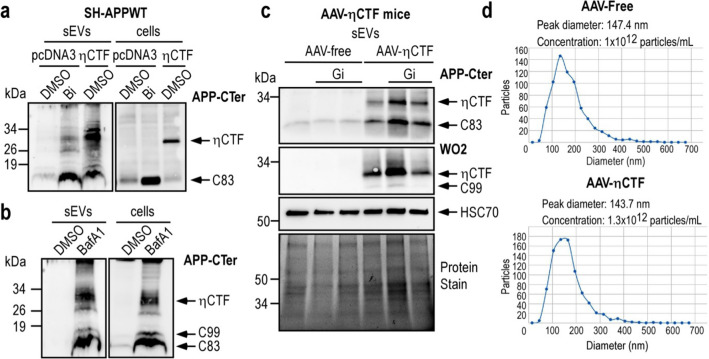
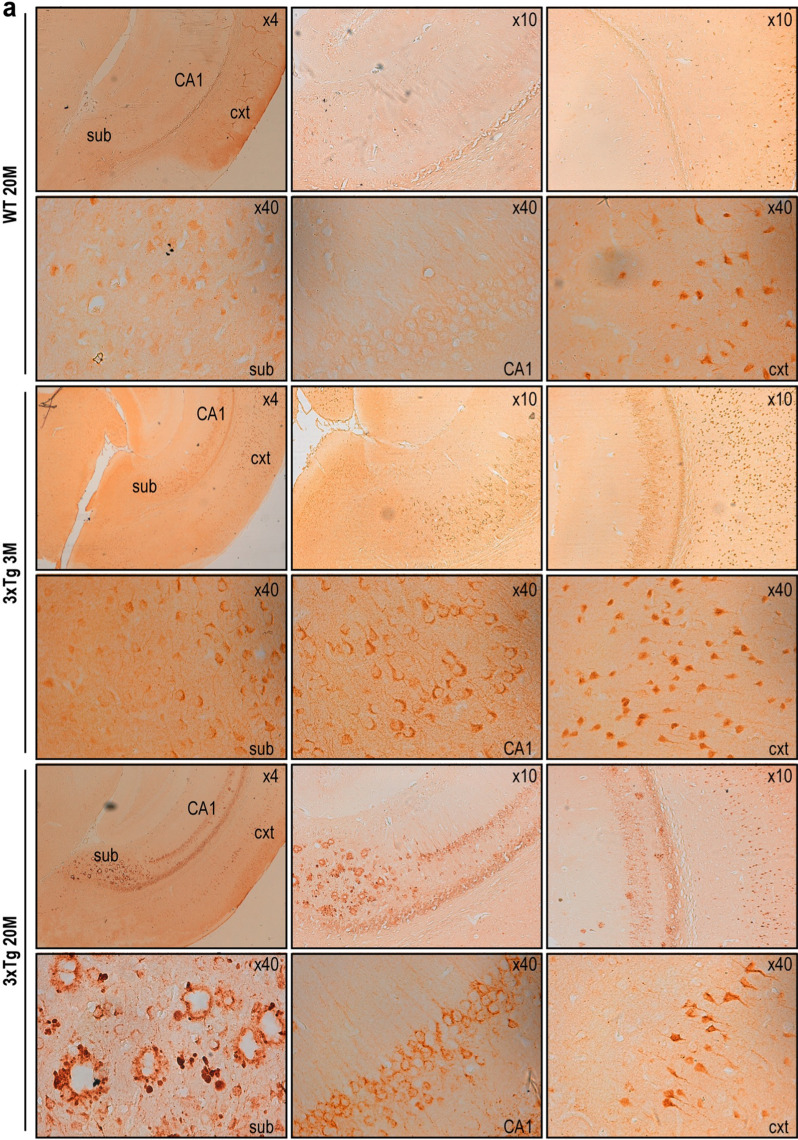
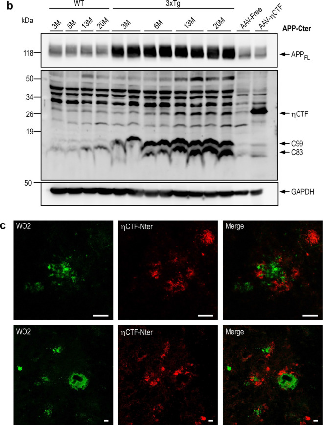
The processing of the amyloid precursor protein (APP) is one of the key events contributing to Alzheimer's disease (AD) etiology. Canonical cleavages by β- and γ-secretases lead to Aβ production which accumulate in amyloid plaques. Recently, the matrix metalloprotease MT5-MMP, referred to as η-secretase, has been identified as a novel APP cleaving enzyme producing a transmembrane fragment, ηCTF that undergoes subsequent cleavages by α- and β-secretases yielding the Aηα and Aηβ peptides, respectively. The functions and contributions of ηCTF and its related fragments to AD pathology are poorly understood. In this study, we designed a novel immunological probe referred to as ηCTF-NTer antibody that specifically interacts with the N-terminal part of ηCTF targeting ηCTF, Aηα, Aηβ but not C99, C83 and Aβ. We examined the fate and localization of ηCTF fragment in various cell models and in mice. We found that overexpressed ηCTF undergoes degradation in the proteasomal and autophagic pathways and accumulates mainly in the Golgi and in endosomes. Moreover, we observed the presence of ηCTF in small extracellular vesicles purified from neuroblastoma cells or from mouse brains expressing ηCTF. Importantly, the expression of ηCTF in fibroblasts devoid on APP leads to Aβ production demonstrating its contribution to the amyloidogenic pathway. Finally, we observed an ηCTF-like immunoreactivity around amyloid plaques and an age-dependent accumulation of ηCTF in the triple-transgenic mouse AD model. Thus, our study suggests that the ηCTF fragment likely contributes to AD pathology by its exosomal spreading and involvement in Aβ production.

Keywords: Alzheimer’s disease; Autophagic degradation; Aβ-production; Endosomes; Extracellular vesicles; ηCTF.


Conflict of interest statement

The authors have nothing to disclose and declare no financial conflict of interest.

Figures

Fig. 1
Fig. 1
Expression and detection of ηCTF in SH-SY5Y cells. a Schematic illustration of APP-derived η-secretase-mediated production of ηCTF and its subsequent cleavages by indicated secretases. Stars indicate the sites of recognition of proteolytic fragments or peptides by WO2 and APP-Cter antibodies. The η-secretase cleaves APP protein to produce a transmembrane fragment called ηCTF that undergoes subsequent cleavages by β and/or α-secretases yielding Aηβ and Aηα respectively. The WO2 antibody targets the amino acid residues 4–10 of Aβ peptide and recognizes full-length APP, ηCTF, C99, Aηα but neither C83 nor Aηβ. The APP-Cter antibody is directed toward the last residues of APP protein and recognizes full-length APP, ηCTF, C99, C83 but neither Aηα nor Aηβ. bc SH-SY5Y cells were transiently transfected with C99-, ηCTF-, Aηα-bearing vectors or empty pcDNA3 vector and analyzed by western blot using APP-Cter (b) or WO2 (c) antibodies. A specific band corresponding to ηCTF is detected around 30 kDa by both APP-Cter and WO2 antibodies. Recombinant ηCTF protein (ηCTFrec) is used as molecular weight control and GAPDH as loading control. All full gels are provided in Sup Fig. 5
Fig. 2
Fig. 2
ηCTF fragment is degraded by both proteasome and autophagic pathways. ad SH-SY5Y cells were transiently transfected with ηCTF or pcDNA3 vectors and treated for 24 h with proteasome inhibitors (a, b, lactacystine (Lact, 5 µM), epoxomicin (Epox, 1 µM), MG132 (5 µM)) or with bafilomycin A1 (BafA1, 100 nM) or Smer28 (50 µM) that blocks or activates autophagy respectively (c, d) then analyzed by western blot using APP-Cter antibody. Histograms in b, d correspond to the quantification of ηCTF immunoreactivity obtained in a, c and are expressed as percent of controls (H2O or DMSO-treated cells) taken as 100. Bars are the means ± SEM of 5–9 independent determinations. ****p < 0.0001 according to Mann–Whitney test. All full gels are provided in Sup Fig. 5
Fig. 3
Fig. 3
ηCTF fragment yields both Aηα and Aβ peptides. a Wild-type (MEF APPwt) and APP/APLPs-deficient mouse embryonic fibroblasts (MEF APPKO) were transiently transfected with ηCTF or pcDNA3 vectors and analyzed by western blot using APP-Cter and WO2 antibodies. GAPDH is used as loading control. bd ηCTF transfected MEF APPKO cells were treated for 24 h with α- β- or γ-secretase inhibitors (Gi:10 µM, Bi:30 µM, D6:1 µM) then analyzed by western blot using WO2 antibody (b). GAPDH is used as loading control. Bars correspond to the quantification of ηCTF immunoreactivity expressed as percent of controls (DMSO-treated cells) taken as 100 and are the means ± SEM of 6 independent determinations. Ns, not statistically significant according to the Tukey one-way ANOVA test (b). Aηα peptides were immunoprecipitated (IP) using WO2 antibody from conditioned medium of MEF APP KO cells expressing or not ηCTF and treated with α- β- or γ-secretase inhibitors. Note that Aηα was not detectable in secretates before immunoprecipitation (Input) (c). Aβ40 levels were measured by ELISA in the conditioned medium of MEF APPKO cells expressing or not ηCTF and treated with α- β- or γ-secretase inhibitors. Bars indicate the concentration of Aβ in pg/ml and are the means ± SEM of 17 independent determinations. ****p < 0.0001, *p < 0.05, ns: not statistically significant according to the Tukey one-way ANOVA test (d). All full gels are provided in Sup Fig. 5
Fig. 4
Fig. 4
Characterization of a new η-CTF-Nter antibody. a Schematic illustration of antibody epitopes on ηCTF fragment. The new ηCTF-Nter antibody is directed towards the free N-terminal epitope of ηCTF. b SH-SY5Y cells were transiently transfected with C99, ηCTF, Aηα, Aηβ or empty pcDNA3 vectors and analyzed by western blot using ηCTF-Nter and APP-Cter antibodies. GAPDH is used as loading control. Note that as expected, ηCTF-Nter antibody recognizes ηCTF, Aηα and Aηβ but neither C99 nor C83 while APP-Cter antibody recognizes ηCTF, C99 and C83 but neither Aηα nor Aηβ. All full gels are provided in Sup Fig. 5. c Hela cells were transiently transfected with ηCTF or empty pcDNA3 vector and analyzed by immunofluorescence using ηCTF-Nter or APP-Cter antibodies as described in Methods. Note that both staining are mostly perinuclear with punctuate intracellular staining. d ηCTF-transfected Hela cells were immunostained with APP-Cter (red) and ηCTF-Nter (green) antibodies. As expected, a part of the APP-Cter staining co-localized with ηCTF-Nter staining (yellow). Nuclei were stained with DAPI. Scale bar is 10 µm. Note that in c, a very faint nuclear label is observed in empty pcDNA3-transfected cells that can be likely accounted for by a very low aspecific ηCTF-Nter background
Fig. 5
Fig. 5
ηCTF fragment is localized in Golgi and endosomes. ad Hela cells were transiently transfected with ηCTF and immunostained with ηCTF-Nter or WO2 (green) antibodies for ηCTF detection and antibodies directed towards TGN-46 (trans-Golgi apparatus, red, a), EEA1 (early endosomes, red, b), CD63 (late endosomes, red, c) or lamp2 (lysosomes, red, d). Note that staining corresponding to ηCTF colocalized mostly with TGN-46 and partially with EEA1 and CD63 antibodies (yellow in merge, ad). Nuclei were stained with DAPI. Scale bar are 10 µm. Note that in b, WO2 was used instead of ηCTF-Nter since ηCTF-Nter and EEA1 are both rabbit antibodies and thus, preventing co-localization study
Fig. 6
Fig. 6
ηCTF expression and in situ localization in AAV-ηCTF mouse brains. ac Wild-type newborn mice were infected with adeno-associated virus expressing ηCTF (AAV-η-CTF) or control empty vector (AAV-free) by intra-cerebro-ventricular (ICV) injection then sacrificed at 3-month-old. Brains were dissected and homogenized for membrane protein purification then analyzed in western blot using APP-Cter antibody. A specific band corresponding to ηCTF is detected around 30 kDa. GAPDH is used as loading control (a). All full gels are provided in Sup Fig. 5. Brain sections were immunostained with ηCTF-Nter antibody and revealed by horseradish peroxidase DAB (b) or by immunofluorescence (c). Brain regions are depicted as cortex (cxt), corpus callosum (CC), subiculum (sub), hippocampal CA1 region (CA1) and dentate gyrus (DG). Specific ηCTF-Nter immunostaining occurs in cortex, subiculum and hippocampus. Confocal images obtained with ηCTF-Nter antibody showed a perinuclear with punctuate intracellular staining (c). Nuclei were stained with DAPI
Fig. 7
Fig. 7
ηCTF fragment is detected in sEVs purified from cells and mouse brains. ab SH-APPWT cells were transiently transfected with ηCTF or empty pcDNA3 vector and treated for 24 h with β-secretase inhibitor (a, Bi, 30 µM), or bafilomycin A1 (b, BafA1, 100 nM). Cell lysates and sEVs were purified from culture media as described in methods and analyzed by western blot using APP-Cter antibody. c sEVs were purified from brain homogenates of 3-month-old AAV-free and AAV-ηCTF mice in the presence or not of the α-secretase inhibitor (Gi:10 µM) and analyzed by western blot using APP-Cter antibody. HSC70 is used as an exosomal marker. Whole loaded proteins were stained by photoactivation using Bio-Rad prestain method (Protein Stain) as loading control. All full gels are provided in Sup Fig. 5. d Concentration and particles size of each brain mouse exosomal purified samples were analyzed in ZetaView instrument (Particle-Metrix) before loading on gels
Fig. 8
Fig. 8
ηCTF fragment accumulates in 3xTgAD brains. ac Brains of wild-type (WT) and triple transgenic (3xTg) females were analyzed at 3-, 6, 13- and 20-month- old by immunohistology (a), western blot (b) or immunofluorescence (c). DAB-immunohistochemical staining is obtained using ηCTF-Nter antibody as described in the Methods. Higher magnification reveals an intracellular labeling clearly observed in cortex, subiculum and hippocampus of 3xTg mice while a weak staining was detected only in the cortex of wild-type mice. In subiculum of 20-month-old 3xTg mice, an extracellular staining is observed around amyloid plaques (a). Brains of wild-type (WT), 3xTg, AAV-free and AAV-ηCTF were homogenized for membrane protein preparation then analyzed in western blot using α-APP-Cter antibody. A specific band corresponding to ηCTF is detected around 30 kDa in wild-type (WT) mice and accumulates in 3xTg mice. GAPDH is used as loading control (b). All full gels are provided in Sup Fig. 5. Confocal images obtained with WO2 (green) and η-CTF-Nter (red) antibodies and merged images from 20-month-old 3xTg hippocampus revealed WO2-positive core plaques surrounded by an ηCTF-Nter-like immunoreactivity (c)
Fig. 8
Fig. 8
ηCTF fragment accumulates in 3xTgAD brains. ac Brains of wild-type (WT) and triple transgenic (3xTg) females were analyzed at 3-, 6, 13- and 20-month- old by immunohistology (a), western blot (b) or immunofluorescence (c). DAB-immunohistochemical staining is obtained using ηCTF-Nter antibody as described in the Methods. Higher magnification reveals an intracellular labeling clearly observed in cortex, subiculum and hippocampus of 3xTg mice while a weak staining was detected only in the cortex of wild-type mice. In subiculum of 20-month-old 3xTg mice, an extracellular staining is observed around amyloid plaques (a). Brains of wild-type (WT), 3xTg, AAV-free and AAV-ηCTF were homogenized for membrane protein preparation then analyzed in western blot using α-APP-Cter antibody. A specific band corresponding to ηCTF is detected around 30 kDa in wild-type (WT) mice and accumulates in 3xTg mice. GAPDH is used as loading control (b). All full gels are provided in Sup Fig. 5. Confocal images obtained with WO2 (green) and η-CTF-Nter (red) antibodies and merged images from 20-month-old 3xTg hippocampus revealed WO2-positive core plaques surrounded by an ηCTF-Nter-like immunoreactivity (c)

References

    1. Hardy J, Allsop D. Amyloid deposition as the central event in the aetiology of Alzheimer’s disease. Trends Pharmacol Sci. 1991;12:383–388. doi: 10.1016/0165-6147(91)90609-V. - DOI - PubMed
    1. Selkoe DJ, Hardy J. The amyloid hypothesis of Alzheimer’s disease at 25 years. EMBO Mol Med. 2016;8:595–608. doi: 10.15252/emmm.201606210. - DOI - PMC - PubMed
    1. Cacace R, Sleegers K, Van Broeckhoven C. Molecular genetics of early-onset Alzheimer’s disease revisited. Alzheimers Dement. 2016;12:733–748. doi: 10.1016/j.jalz.2016.01.012. - DOI - PubMed
    1. Jonsson T, et al. A mutation in APP protects against Alzheimer’s disease and age-related cognitive decline. Nature. 2012;488:96–99. doi: 10.1038/nature11283. - DOI - PubMed
    1. Pardossi-Piquard R, Checler F. The physiology of the beta-amyloid precursor protein intracellular domain AICD. J Neurochem. 2012;120(Suppl 1):109–124. doi: 10.1111/j.1471-4159.2011.07475.x. - DOI - PubMed

MeSH terms

Substances