. 2023 Apr;601(7):1225-1246.
doi: 10.1113/JP283701. Epub 2023 Mar 17.

An evolutionarily conserved pacemaker role for HCN ion channels in smooth muscle

Affiliations

An evolutionarily conserved pacemaker role for HCN ion channels in smooth muscle

Lei Yang et al. J Physiol. 2023 Apr.

Abstract

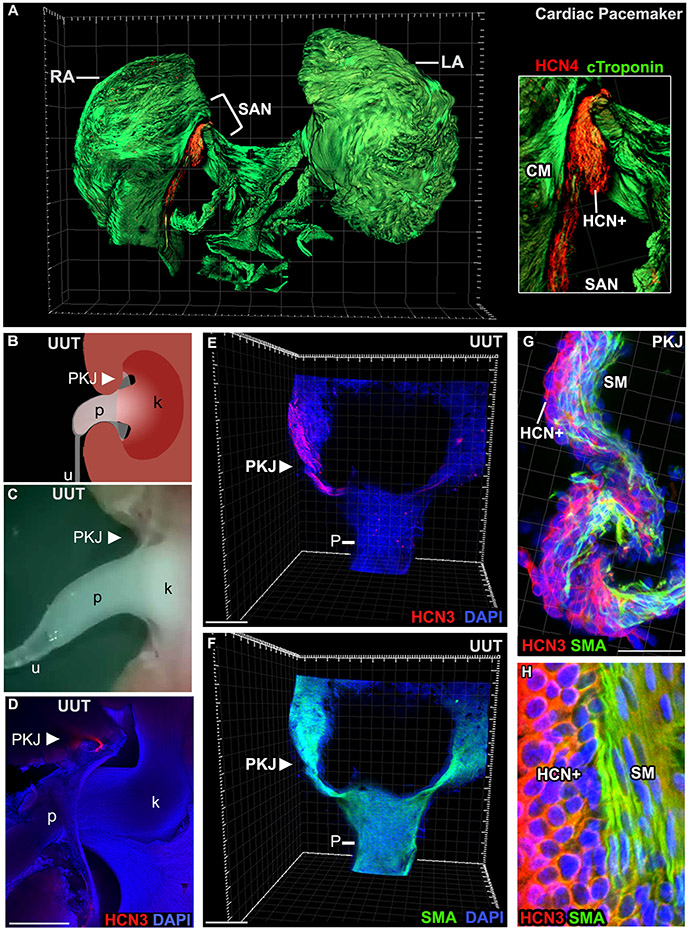
Although hyperpolarization-activated cation (HCN) ion channels are well established to underlie cardiac pacemaker activity, their role in smooth muscle organs remains controversial. HCN-expressing cells are localized to renal pelvic smooth muscle (RPSM) pacemaker tissues of the murine upper urinary tract and HCN channel conductance is required for peristalsis. To date, however, the Ih pacemaker current conducted by HCN channels has never been detected in these cells, raising questions on the identity of RPSM pacemakers. Indeed, the RPSM pacemaker mechanisms of the unique multicalyceal upper urinary tract exhibited by humans remains unknown. Here, we developed immunopanning purification protocols and demonstrate that 96% of isolated HCN+ cells exhibit Ih . Single-molecule STORM to whole-tissue imaging showed HCN+ cells express single HCN channels on their plasma membrane and integrate into the muscular syncytium. By contrast, PDGFR-α+ cells exhibiting the morphology of ICC gut pacemakers were shown to be vascular mural cells. Translational studies in the homologous human and porcine multicalyceal upper urinary tracts showed that contractions and pacemaker depolarizations originate in proximal calyceal RPSM. Critically, HCN+ cells were shown to integrate into calyceal RPSM pacemaker tissues, and HCN channel block abolished electrical pacemaker activity and peristalsis of the multicalyceal upper urinary tract. Cumulatively, these studies demonstrate that HCN ion channels play a broad, evolutionarily conserved pacemaker role in both cardiac and smooth muscle organs and have implications for channelopathies as putative aetiologies of smooth muscle disorders. KEY POINTS: Pacemakers trigger contractions of involuntary muscles. Hyperpolarization-activated cation (HCN) ion channels underpin cardiac pacemaker activity, but their role in smooth muscle organs remains controversial. Renal pelvic smooth muscle (RPSM) pacemakers trigger contractions that propel waste away from the kidney. HCN+ cells localize to murine RPSM pacemaker tissue and HCN channel conductance is required for peristalsis. The HCN (Ih ) current has never been detected in RPSM cells, raising doubt whether HCN+ cells are bona fide pacemakers. Moreover, the pacemaker mechanisms of the unique multicalyceal RPSM of higher order mammals remains unknown. In total, 97% of purified HCN+ RPSM cells exhibit Ih . HCN+ cells integrate into the RPSM musculature, and pacemaker tissue peristalsis is dependent on HCN channels. Translational studies in human and swine demonstrate HCN channels are conserved in the multicalyceal RPSM and that HCN channels underlie pacemaker activity that drives peristalsis. These studies provide insight into putative channelopathies that can underlie smooth muscle dysfunction.

Keywords: HCN channels; Ih funny current; PDGFR-α; pacemakers; peristalsis; renal pelvis; smooth muscle.


Figures

Figure 1.
Figure 1.. Cardiac and RPSM pacemaker tissues contain an analogous tissue architecture consisting of an HCN+ cell layer that makes up part of the musculature.
A) Confocal imaging of whole adult murine atria immunolabeled for c-Troponin (green) to mark the myocardium and HCN4 to mark the SAN pacemaker (Inset, high magnification of the SAN). See supplemental movie 1. B) Schematic of the murine UUT, which is a continuous muscular organ with a flared proximal end termed the renal pelvis (p) and a more narrowed distal end, termed the ureter (u). RPSM pacemaker tissue localizes to the site where the pelvis joins the kidney (k), termed the pelvis kidney junction (PKJ). C) Representative still image of supplemental movie 2 showing live peristaltic contractions of the murine UUT. D) Cryosection of murine UUT immunostained for HCN3 (red) and DAPI. E-F) Imaging of whole proximal UUTs immunolabeled for HCN3 (E, red) and SMA (F, green). Low magnification analyses (E, F) revealed HCN+ cell tissue layer localized to the PKJ pacemaker. High magnification analyses showed that the HCN+ tissue layer was integrated into UUT musculature (G; see supplementary movie 3), with HCN+ cells and smooth muscle cells forming adjacent tissue layers (H).
Figure 2.
Figure 2.. PDGFR-α cells of the kidney are vascular associated mural cells.
The cell identity of PDGFR-α+ cells in the kidney were ascertained. Tissues were immunostained for the vasculature (vascular endothelium, endomucin, green) and PDGFR-α (red). PDGFR-α cells were established to be vascular associate mural cells that overlay the vasculature in of the kidney. Brackets in top panels mark regions assayed at higher magnification in bottom panels. rt, renal tubules.
Figure 3.
Figure 3.. PDGFR-α+ cells of the UUT are vascular associated mural cells.
A-N) The cell identity of PDGFR-α+ cells was assayed in the intact UUT. Three independent representative samples are shown (N=1, A-D; N=2, E-H; N=3, I-N). UUTs were immunolabeled for the vasculature (vascular endothelium, endomucin, green) and PDGFR-α (red). PDGFR-α+ cells proved to be vascular associated mural cells, overlaying the vascular tree of the UUT. Arrows point to small caliber vessels lacking PDGFR-α+ mural cell coverage (F-H, and L-N). Brackets in A, E, and K mark PKJ tissue that was assayed at higher magnification (B-D; F-H; and L-N). pelvis-kidney junction, PKJ; ureter pelvis junction, UPJ.
Figure 4.
Figure 4.. PDGFR-α cells of the distal UUT are vascular associated mural cells.
The cell identity of PDGFR-α+ cells in the distal UUT were assayed. Tissues were immunostained for the vasculature (vascular endothelium, endomucin, green) and PDGFR-α (red). PDGFR-α cells were established to be vascular associate mural cells that overlay the vasculature of the distal UUT. Brackets in top panels mark regions assayed at higher magnification in bottom panels. upj, ureter-pelvis junction
Figure 5.
Figure 5.. Super resolution imaging reveals HCN ion channels on the plasma membrane of PKJ cells with a compact, round morphology.
A-D) Super resolution imaging of PKJ tissues immunolabeled for HCN3. (A) TIRF images of PKJ tissue immunolabeled for HCN3 (inset, DAPI). (B-D) STORM analyses of TIRF images (HCN3, red; blue, DAPI). E, F) Low (E) and high (F) magnification phase contrast images of primary cells freshly dissociated from the PKJ. G) IF of primary cells isolated from the PKJ (HCN3, red; DAPI, blue). HCN3+ cells exhibited a morphology consistent with small, compact round cell types observed by phase microscopy. H-J) Super resolution imaging of compact, rounded primary cells freshly isolated from the PKJ and immunolabeled for HCN3. (H) TIRF images of primary cells immunolabeled for HCN3. (I and J) STORM analyses of TIRF images (HCN3, red).
Figure 6.
Figure 6.. PKJ cells exhibit Ih current.
A-D) Freshly isolated primary cells of the PKJ with the compact rounded morphology of HCN3+ cells were used for whole-cell voltage-clamp recording. (A) Typical current traces in response to hyperpolarization of the membrane potential from −50 mV. (B) voltage-dependence of Ih, measured as the difference between the current at the end of the voltage step and at the beginning (after decay of the capacitance transient), normalized to the value at −130 mV. Data represent mean ± SD for 8 cells. (C) Voltage-dependent activation of Ih in PKJ cells. The voltage was clamped to values between −30 and −150 mV for 5 or 10 sec, and then held at −130 mV for 500 msec. (D) Data are plotted according to eq. 1 and normalized to values at −130 mV, V1/2 = −100 mV and k = 22. Data represent mean ± SD (E) Percentage of cells with measurable Ih in unselected primary cell populations (left diagram) and in primary cells purified by immunopanning with an anti-HCN3 antibody (right diagram).
Figure 7.
Figure 7.. Ih current exhibited by PKJ cells is inhibited by HCN channel blockers.
A-C) Ih expressed by cells of the PKJ is inhibited by the HCN channel antagonist ZD7288. (A) Representative trace of Ih before and 13 min after application of 100μM ZD7288. (B) Mean fractional change in Ih at V = −150 mV after inhibition with ZD7288 (mean ± SD for n = 3 cells, *p= 0.03). Currents were measured 3 times under basal condition to establish a baseline. Ih values for the last measurement before and 10-15 min after ZD7288 addition were normalized to the baseline average. (C) Mean values of Ih in immunopanned cells pre-incubated with or without 100μM ZD7288. (mean ± SD n = 10 cells, *p= 0.04). (D) Block of Ih by 3 mM Cs+, which was partially reversible upon washout.
Figure 8.
Figure 8.. Calyces serve as the origin of peristalsis, elicit pacemaker depolarizations, and exhibit a conserved tissue architecture consisting of an HCN+ tissue layer.
A-D) Representative still frames of live imaging of human multicalyceal UUT peristalsis via real-time fluoroscopic recordings. See supplemental movie 9 of live recording. Arrow head indicates origin of contraction at the calyx (A); arrows indicate point of contraction over time (B and C; D, isochronal map of contraction over time, from red initiation site to yellow). E-I) Representative still frames of live imaging of porcine UUT peristalsis. See supplemental movie 10 of live recording. Brackets indicate point of contraction over time (F-H; I, isochronal map of contraction over time, from red initiation site to yellow). J) Fluorimetric IF staining of 1mm thick porcine UUT sections labeled for HCN3 (K, red). K-M) Standard IF of calyx cryosections labeled for HCN3 (red, L and N), SMA (green, M and N) and uroplakin (blue, L-N). N) Confocal imaging of calyx tissue immunolabeled for HCN3 (red), SMA (green), and DAPI (blue). O) Detection of spontaneous pacemaker membrane depolarizations elicited at the calyx (top trace) via dual emission ratiometric optical mapping (bottom shows adjacent regions lacked pacemaker depolarizations). k, kidney; c, calyx; p, pelvis; u, ureter; pa, papilla; up, uroplakin; sma, smooth muscle actin.
Figure 9.
Figure 9.. HCN channel conductance underlies pacemaker activity that triggers multicalyceal UUT peristalsis.
A-G) Direct visualization of electrical and contractile excitation in control porcine UUTs. (A) Pacemaker depolarizations detected at the calyx of control porcine UUTs by dual emission ratiometric optical mapping. (B-G) Live imaging of coordinated, proximal to distal peristaltic contractions of control porcine UUTs (representative still images of supplemental movie 11, brackets mark site of contraction; G isochronal map of contraction over time, red initiation to yellow). H-N) Direct visualization of electrical and contractile excitation in porcine UUTs upon HCN channel block. (H) Pacemaker depolarizations were not detected in porcine UUTs inhibited with 100 μM ZD7288. (I-N) Live imaging of twitch like contractile activity in porcine UUTs inhibited with 100 μM ZD7288 (Supplemental movie 12, before inhibition; Supplemental movie 13, after inhibition; brackets mark the site of twitching; N, isochronal map of contraction over time, red initiation site to yellow). pa, papillae; p, renal pelvis.
Figure 10.
Figure 10.. Validation of a robust HCN+ cell population in pacemaker tissues of the unicalyceal and multicalyceal UUT.
A-C) Whole tissue imaging of UUT tissue by IF staining for an intracellular epitope of HCN3 (A, HCN3, red; DAPI) and SMA (B, SMA, green; DAPI). C) High magnification analyses of PKJ shown in A and B. D) Fluoriometric imaging of HCN3 in the multicalyceal UUT. E, F) IF of calyx tissues for HCN3 and smooth muscle tissue layers (E), as well as single cell populations (F).

References

    1. Arena S, Iacona R, Impellizzeri P, Russo T, Marseglia L, Gitto E & Romeo C. (2016). Physiopathology of vesico-ureteral reflux. Ital J Pediatr 42, 103. - PMC - PubMed
    1. Barres BA. (2014). Designing and troubleshooting immunopanning protocols for purifying neural cells. Cold Spring Harbor protocols 2014, 1342–1347. - PubMed
    1. Bassols A, Costa C, Eckersall PD, Osada J, Sabria J & Tibau J. (2014). The pig as an animal model for human pathologies: A proteomics perspective. Proteomics Clinical applications 8, 715–731. - PubMed
    1. Benzoni P, Bertoli G, Giannetti F, Piantoni C, Milanesi R, Pecchiari M, Barbuti A, Baruscotti M & Bucchi A. (2021). The funny current: Even funnier than 40 years ago. Uncanonical expression and roles of HCN/f channels all over the body. Prog Biophys Mol Biol. - PubMed
    1. Biel M, Wahl-Schott C, Michalakis S & Zong X. (2009). Hyperpolarization-activated cation channels: from genes to function. Physiological reviews 89, 847–885. - PubMed

Publication types

Substances