. 2023 Jun 1;72(6):781-794.
doi: 10.2337/db22-0619.

Transcriptomic Profiling Reveals Chemokine CXCL1 as a Mediator for Neutrophil Recruitment Associated With Blood-Retinal Barrier Alteration in Diabetic Retinopathy

Affiliations

Transcriptomic Profiling Reveals Chemokine CXCL1 as a Mediator for Neutrophil Recruitment Associated With Blood-Retinal Barrier Alteration in Diabetic Retinopathy

Finny Monickaraj et al. Diabetes. .

Abstract

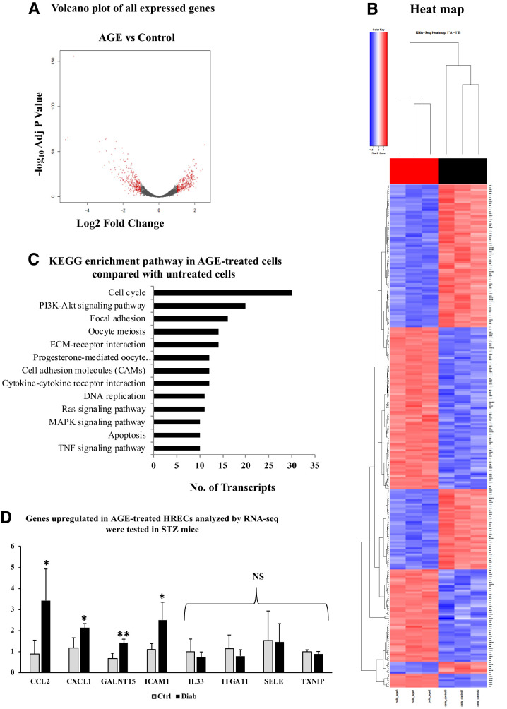
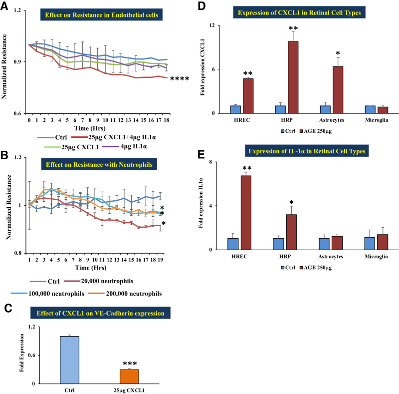
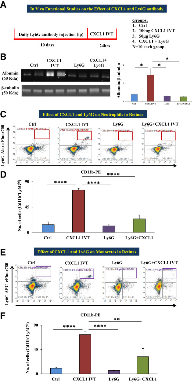
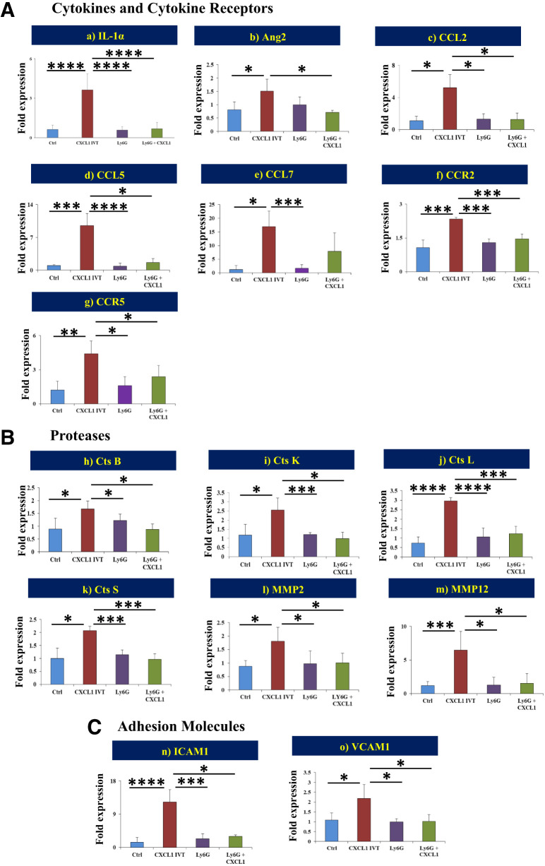
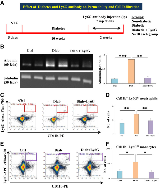
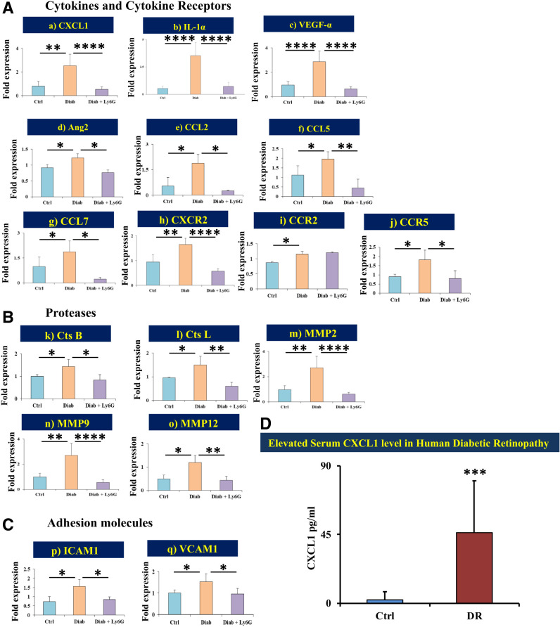
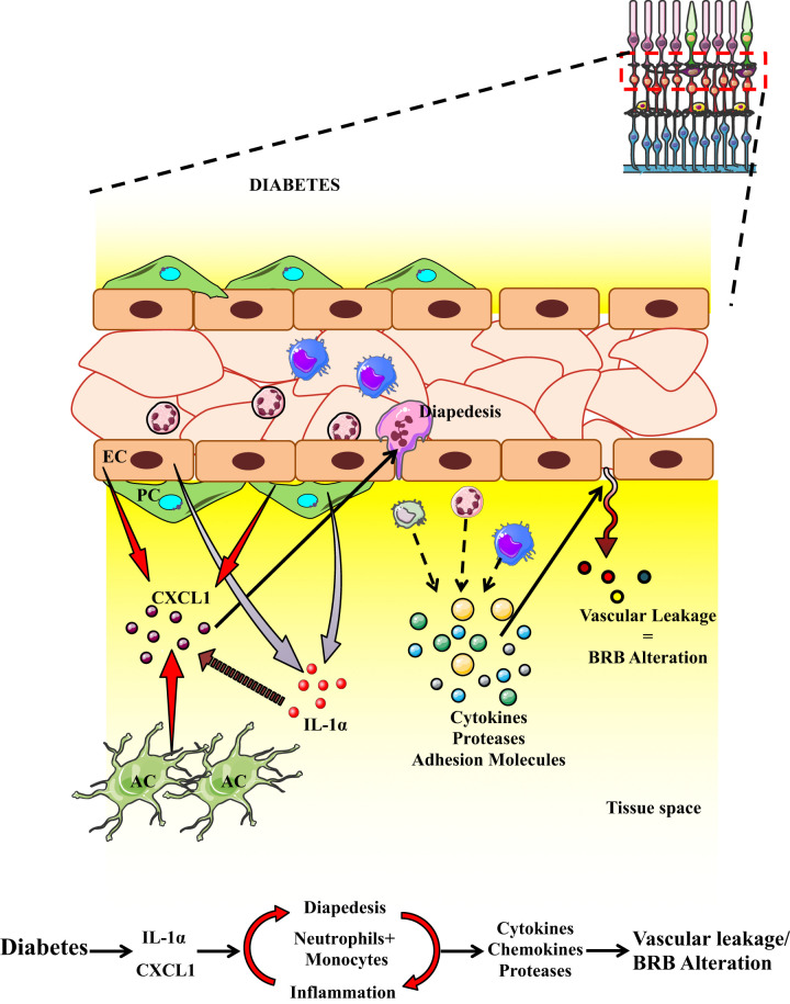
Inflammation plays an important role in the pathogenesis of diabetic retinopathy (DR). To precisely define the inflammatory mediators, we examined the transcriptomic profile of human retinal endothelial cells exposed to advanced glycation end products, which revealed the neutrophil chemoattractant chemokine CXCL1 as one of the top genes upregulated. The effect of neutrophils in the alteration of the blood-retinal barrier (BRB) was further assessed in wild-type C57BL/6J mice intravitreally injected with recombinant CXCL1 as well as in streptozotocin-induced diabetic mice. Both intravitreally CXCL1-injected and diabetic animals showed significantly increased retinal vascular permeability, with significant increase in infiltration of neutrophils and monocytes in retinas and increased expression of chemokines and their receptors, proteases, and adhesion molecules. Treatment with Ly6G antibody for neutrophil depletion in both diabetic mice as well as CXCL1-injected animals showed significantly decreased retinal vascular permeability accompanied by decreased infiltration of neutrophils and monocytes and decreased expression of cytokines and proteases. CXCL1 level was significantly increased in the serum samples of patients with DR compared with samples of those without diabetes. These data reveal a novel mechanism by which the chemokine CXCL1, through neutrophil recruitment, alters the BRB in DR and, thus, serves as a potential novel therapeutic target.

Article highlights: Intravitreal CXCL1 injection and diabetes result in increased retinal vascular permeability with neutrophil and monocyte recruitment. Ly6G antibody treatment for neutrophil depletion in both animal models showed decreased retinal permeability and decreased cytokine expression. CXCL1 is produced by retinal endothelial cells, pericytes, and astrocytes. CXCL1 level is significantly increased in serum samples of patients with diabetic retinopathy. CXCL1, through neutrophil recruitment, alters the blood-retinal barrier in diabetic retinopathy and, thus, may be used as a therapeutic target.


Conflict of interest statement

Duality of Interest. No potential conflicts of interest relevant to this article were reported.

Figures

Figure 1
Figure 1
A: Volcano plot of fold change of transcripts derived using edgeR. AGE-treated HRECs compared with control. Red circles at the left side of the plot indicate downregulated genes, and those at the right side indicate upregulated genes. B: Hierarchical cluster heat map of DEGs in AGE-treated and control (untreated) cells. C: KEGG signaling enrichment pathways with genes of fold change of 2 and P value of ≤0.05 in AGE-treated retinal endothelial cells compared with control. D: RT-PCR validation of top 10 upregulated genes from the RNA sequencing (RNA-seq) data on STZ-induced diabetic (2 months of diabetes) and nondiabetic mouse retinas (n = 5 each). Relative gene expression levels of CCL2, CXCL1, GALNT15, and ICAM1 were significantly increased in the retinas of diabetic mice in comparison with those of nondiabetic mice. mRNA levels of IL33, ITGA11, SELE, and TXNIP did not show any significant change in the diabetic retinas compared with nondiabetic retinas. All genes were normalized to 18S. Data are presented as mean ± SD. Diab, positive for diabetes; ECM, extracellular matrix; MAPK, mitogen-activated protein kinase; NS, nonsignificant; TNF, tumor necrosis factor. *P < 0.05, **P < 0.01.
Figure 2
Figure 2
In vitro functional studies of the effect of CXCL1 and neutrophils. A: HRECs treated with a combination of 25 μg recombinant CXCL1 plus 4 μg IL-1α showed significantly decreased resistance compared with cells grown on normal growth medium; cells treated with 4 μg IL-1α alone did not show any significant change in resistance in comparison with untreated cells (control). B: Coculture of HRECs with 250 μg AGE–pretreated human neutrophils of different numbers: 20,000, 100,000, and 200,000 cells showed a significant decrease in the monolayer resistance in comparison with untreated cells (control). Decreased resistance implies increased permeability. C: mRNA expression of adherent junction molecule VE-cadherin was significantly decreased with 25 μg recombinant CXCL1 treatment compared with untreated cells (control). D: mRNA expression of CXCL1 in different retinal cells in treatment with 250 μg AGE–treated HRECs, human retinal pericytes (HRPs), and human retinal astrocytes (HRAs) were significantly increased in comparison with cells grown on respective normal growth medium (control), except microglia. E: mRNA expression of IL-1α in different retinal cells in treatment with 250 μg AGE. HRECs and HRPs were significantly increased in comparison with cells grown on respective normal growth medium (control), except HRAs and microglia. Data are presented as mean ± SD. *P < 0.05, **P < 0.01, ***P < 0.001, ****P < 0.0001.
Figure 3
Figure 3
In vivo functional studies of the effect of CXCL1 and Ly6G antibody. A: A schematic representation of the experimental design and groups are depicted. B: Representative Western blot images of albumin and β-tubulin; the right-hand side histogram shows band intensity of albumin, a marker to determine vascular permeability, which showed significantly increased level in the retinas of the animals that received an intravitreal injection of 5 μL of 100 ng recombinant CXCL1 (CXCL1 IVT) compared with animals that received vehicle injection (distilled water); the albumin level was significantly decreased in the retinas of animals treated with 50 μg Ly6G or Ly6G + CXCL1 IVT in comparison with CXCL1 IVT animals. C: Representative dot plots of flow cytometric analysis for neutrophils (gated for Cd11b+/Ly6Ghi in red box at the top right quadrant) of differently treated animal groups: control (vehicle), CXCL1 IVT, Ly6G, and Ly6G + CXCL1 IVT. D: Total number of neutrophils was significantly increased in CXCL1 IVT animal retinas compared with vehicle-injected animal retinas; the neutrophil count was significantly decreased in Ly6G or Ly6G + CXCL1 IVT in comparison with CXCL1 IVT animals. E: Flow cytometric dot plots depict the gating of monocytes (Cd11b+Ly6Chi in purple box at the top right quadrant) in the four different animal groups as mentioned above. F: Monocyte count was significantly increased in the retinas of the CXCL1 IVT group compared with control retinas. Retinas of Ly6G- or Ly6G + CXCL1 IVT–treated groups had a significantly lower monocyte count in comparison with CXCL1 IVT animals. Data are presented as mean ± SD. *P = 0.05, **P < 0.01, ****P < 0.0001.
Figure 4
Figure 4
Gene expression levels of proinflammatory cytokines, receptors, proteases, and adhesion molecules. mRNA expression levels of multiple genes from cytokines and cytokine receptors (A) [IL-1α (a), Ang2 (b), CCL2 (c), CCL5 (d), CCL7 (e), CCR2 (f), and CCR5 (g)], proteases (B) [cathepsin (Cts) B (h), Cts K (i), Cts L (j), Cts S (k), MMP2 (l), and MMP12 (m)], and adhesion molecules (C) [ICAM1 (n) and VCAM1 (o)] were statistically compared between the different treatment groups: 1) control versus CXCL1 IVT, 2) CXCL1 IVT versus Ly6G, and 3) CXCL1 IVT versus Ly6G + CXCL1 IVT. Data are presented as mean ± SD. *P < 0.05, ***P < 0.001, ****P < 0.0001.
Figure 5
Figure 5
A: Schematic representation of the experimental design and groups are depicted. B: Representative Western blot images of albumin and β-tubulin; the histogram on the right-hand side shows significantly increased albumin level in the retinas of diabetic animals compared with that in nondiabetic animals. Meanwhile, the albumin level was significantly decreased in diabetic + Ly6G-treated animal retinas compared with diabetic animal retinas. C: Representative dot plots of flow cytometric analysis for neutrophils (gated for Cd11b+/Ly6Ghi in red box at the top right quadrant) of different experimental groups: control (vehicle), diabetic, and diabetic + Ly6G. D: Total number of neutrophils was significantly increased in the retinas of diabetic animals compared with nondiabetic animal retinas; the neutrophil count was significantly decreased in Ly6G antibody–treated diabetic animal retinas compared with retinas of diabetic animals that did not receive this treatment. E: Representative dot plots of flow cytometric analysis for monocytes (gated for Cd11b+/Ly6Chi in purple box at the top right quadrant) of different experimental groups. F: Total number of monocytes was significantly increased in the retinas of diabetic animals compared with nondiabetic (control) animal retinas. Ly6G antibody treatment in diabetic animals significantly reduced the influx of both monocytes compared with in diabetic animals that did not receive this treatment. Data are presented as mean ± SD. *P < 0.05, **P < 0.01, ***P < 0.001. Diab, positive for diabetes.
Figure 6
Figure 6
Gene expression levels of proinflammatory cytokines, receptors, proteases, and adhesion molecules. mRNA expression levels of multiple genes from cytokines and cytokine receptors (A) [CXCL1 (a), IL-1α (b), VEGF-α (c), Ang2 (d), CCL2 (e), CCL5 (f), CCL7 (g), CXCR2 (h), CCR2 (i), and CCR5 (j)], proteases (B) [cathepsin (Cts) B (k), Cts L (l), MMP2 (m), MMP9 (n), and MMP12 (o)], and adhesion molecules (C) [ICAM1 (p) and VCAM1 (q)] were statistically compared between the different treatment groups: 1) control versus diabetes and 2) diabetic versus diabetic + Ly6G. D: Human serum CXCL1. Age-adjusted mean values of CXCL1 level measured by ELISA from human serum samples showed a significantly elevated level in patients with DR compared with participants without diabetes. Data are presented as mean ± SD. *P < 0.05, **P < 0.01, ***P < 0.001, ****P < 0.0001. Diab, positive for diabetes.
Figure 7
Figure 7
Schematic diagram highlighting the effect of CXCL1 and neutrophils. Increased neutrophil and monocyte presence leads to expression of chemokines, cytokines, and proteases, which leads to increased endothelial permeability and alteration of the BRB.

References

    1. Tang J, Kern TS. Inflammation in diabetic retinopathy. Prog Retin Eye Res 2011;30:343–358 - PMC - PubMed
    1. Das A, McGuire PG, Rangasamy S. Diabetic macular edema: pathophysiology and novel therapeutic targets. Ophthalmology 2015;122:1375–1394 - PubMed
    1. Rangasamy S, McGuire PG, Franco Nitta C, Monickaraj F, Oruganti SR, Das A. Chemokine mediated monocyte trafficking into the retina: role of inflammation in alteration of the blood-retinal barrier in diabetic retinopathy. PLoS One 2014;9:e108508. - PMC - PubMed
    1. Navaratna D, McGuire PG, Menicucci G, Das A. Proteolytic degradation of VE-cadherin alters the blood-retinal barrier in diabetes. Diabetes 2007;56:2380–2387 - PubMed
    1. Rangasamy S, Srinivasan R, Maestas J, McGuire PG, Das A. A potential role for angiopoietin 2 in the regulation of the blood-retinal barrier in diabetic retinopathy. Invest Ophthalmol Vis Sci 2011;52:3784–3791 - PMC - PubMed

Publication types