. 2023 Aug 22;7(16):4233-4246.
doi: 10.1182/bloodadvances.2022008735.

Individual phosphatidylinositol transfer proteins have distinct functions that do not involve lipid transfer activity

Affiliations

Individual phosphatidylinositol transfer proteins have distinct functions that do not involve lipid transfer activity

Liang Zhao et al. Blood Adv. .

Abstract

Platelets use signal transduction pathways facilitated by class I phosphatidylinositol transfer proteins (PITPs). The 2 mammalian class I PITPs, PITPα and PITPβ, are single PITP domain soluble proteins that are encoded by different genes and share 77% sequence identity, although their individual roles in mammalian biology remain uncharacterized. These proteins are believed to shuttle phosphatidylinositol and phosphatidylcholine between separate intracellular membrane compartments, thereby regulating phosphoinositide synthesis and second messenger formation. Previously, we observed that platelet-specific deletion of PITPα, the predominantly expressed murine PITP isoform, had no effect on hemostasis but impaired tumor metastasis formation and disrupted phosphoinositide signaling. Here, we found that mice lacking the less expressed PITPβ in their platelets exhibited a similar phenotype. However, in contrast to PITPα-null platelet lysates, which have impaired lipid transfer activity, PITPβ-null platelet lysates have essentially normal lipid transfer activity, although both isoforms contribute to phosphoinositide synthesis in vitro. Moreover, we found that platelet-specific deletion of both PITPs led to ex vivo platelet aggregation/secretion and spreading defects, impaired tail bleeding, and profound tumor dissemination. Our study also demonstrated that PITP isoforms are required to maintain endogenous phosphoinositide PtdInsP2 levels and agonist-stimulated second messenger formation. The data shown here demonstrate that the 2 isoforms are functionally overlapping and that a single isoform is able to maintain the homeostasis of platelets. However, both class I PITP isoforms contribute to phosphoinositide signaling in platelets through distinct biochemical mechanisms or different subcellular domains.


Conflict of interest statement

Conflict-of-interest disclosure: The authors declare no competing financial interests.

The current affiliation of S.H.M is Department of Internal Medicine, University of Michigan, Ann Arbor, Michigan.

Figures

None
Graphical abstract
Figure 1.
Figure 1.
Platelet-specific loss of both PITPs impairs ex vivo platelet aggregation/secretion and spreading. (A) Western blot analysis of PITP expression in human platelets compared with their mouse counterparts. (B) Western blot–based densitometry quantification of individual PITP isoforms in human platelets. (C) Schematic representation of the conditional targeting strategy for Pitpβ. A 1.89 kb genomic DNA of PITPβ (which includes exons 4-6) was targeted by the insertion of loxP recombination sites. The Neo cassette was removed by crossing with the FRT mice before further crossing with PF4-Cre transgenic mice. (D) Western blot of platelet lysates demonstrating the specific deletion of the PITPβ isoform in Pitpβfl/flPf4-Cre+ mice. (E) Complete blood count analyses show mild thrombocytopenia in Pitpβfl/flPf4-Cre+ mice and more severe thrombocytopenia in Pitpαfl/flfl/flPf4-Cre+ mice compared with their respective littermate controls (n = 6 for Pitpβfl/flPf4-Cre- mice; n = 8 for Pitpβfl/flPf4-Cre+ mice; n = 13 for Pitpαfl/flfl/flPf4-Cre- mice; and n = 9 for Pitpαfl/flfl/flPf4-Cre+ mice; error bars are standard deviation(s.d.); P values are shown obtained from unpaired t test). (F,G) Ex vivo analysis of platelet aggregation and dense granule secretion. PITPβ-null platelets aggregate normally, but dense granule secretion was impaired in response to low-dose thrombin (0.05 U/mL) and collagen (10 μg/mL), as measured by adenosine triphosphate release (F). Deleting both PITP isoforms increased the severity of aggregation and secretion defects (G). Adenosine triphosphate secretion traces start at 100% and trend downward, and aggregation traces start at 0% and trend upward. Traces are representative of 5 separate experiments per condition. (H) Spreading of PITPβ-null and PITPα/β-null platelets on fibrinogen after stimulation with thrombin (0.025 U/mL) revealed that PITPα/β-null platelets had a spreading defect, whereas PITPβ-null platelets spread normally. (I) PITPα/β-null platelet spreading was quantified as the total cumulative area of platelets per field (n = 3 per group). (J) The number of adherent PITPα/β-null platelets was quantified as the average number per field under a 100× microscope. (n = 5 per group; error bars represent s.d.; P values are shown, unpaired t test).
Figure 2.
Figure 2.
Mice lacking platelet PITPs have prolonged tail bleeding time, but no defects in laser-induced in vivo thrombosis. The laser-induced injury model demonstrates normal in vivo thrombosis and platelet secretion in the Pitpαfl/flfl/flPf4-Cre+ mice. (A) Representative images show platelet accumulation (CD41, red) and P-selectin exposure (green [overlay of red/green is yellow]) 3 minutes after laser-induced injury to the cremaster arterioles. Images are binary representations of 2D confocal fluorescence images overlaid on the bright-field. White arrows indicate the direction of flow; scale bar, 10 μm. (B) Graphs of the CD41+ area over time (left, mean ± standard error of the mean), median CD41+ area over time (middle), and CD41 peak area (right, lines represent median ± interquartile range). (C) Graphs of the P-selectin–positive area over time (left, mean ± standard error of the mean), the median P-selectin–positive area over time (middle), and P-selectin peak area (right, lines are median ± interquartile range). n = 20 thrombi in 4 wild-type mice and n = 29 thrombi in 4 Pitpαfl/flfl/flPf4-Cre+ mice. Statistical analysis were performed using a two-tailed Mann-Whitney test. (D,E) Tail bleeding times in mice lacking PITPβ (D) or both PITP isoforms compared with their littermate controls (E). Tail bleeding was normal in mice with platelets lacking PITPβ (NS, Mann-Whitney test; n = 52 for Pitpβfl/flPf4-Cre- mice and n = 58 for Pitpβfl/flPf4-Cre+ mice). When both PITP isoforms were deleted in platelets, there was a mild increase in bleeding time (P = .0013 using two-tailed Mann-Whitney test; n = 57 for Pitpαfl/flfl/flPf4-Cre- mice and n = 54 for Pitpαfl/flfl/flPf4-Cre+ mice).
Figure 3.
Figure 3.
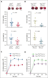
Platelets lacking PITPβ or both PITP isoforms are less susceptible to tumor metastasis. (A,B) Lungs harvested from Pitpβfl/flPf4-Cre- and Pitpβfl/flPf4-Cre+ mice (A) or Pitpαfl/flfl/flPf4-Cre- and Pitpαfl/flfl/flPf4-Cre+ mice (B) 2 weeks after tail vein injection with B16F10 melanoma cells demonstrated that loss of PITP impairs metastasis. Representative lungs are shown at 2 weeks after tumor cell injection (top); the number of tumor nodules on the lung surface 2 weeks after tumor cell injection (middle); and wet lung weights 3 weeks after tumor injection (bottom). For tumor nodule counting, n = 21 lungs for Pitpβfl/flPf4-Cre- mice, n = 19 for Pitpβfl/flPf4-Cre+ mice, and n = 13 for both Pitpαfl/flfl/flPf4-Cre- mice and Pitpαfl/flfl/flPf4-Cre+ mice. For lung weight, n = 21 lungs for Pitpβfl/flPf4-Cre- mice, n = 20 for Pitpβfl/flPf4-Cre+ mice, n = 17 for Pitpαfl/flfl/flPf4-Cre- mice, and n = 18 for Pitpαfl/flfl/flPf4-Cre+ mice. Statistical analysis was performed using an unpaired t test. Black scale bars represent 10 mm. (C,D) Ex vivo adhesion of PITPβ-null (C) or PITPα/β-null (D) platelets to a tissue-cultured tumor cell monolayer was impaired compared with wild-type controls. Error bars represent s.d.; n = 3 for each genotype.
Figure 4.
Figure 4.
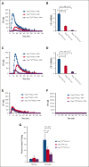
Loss of PITP in platelets impairs thrombin generation and Annexin V binding. (A-D) Representative kinetics of thrombin generation induced by BF610 tumor cells (A,B) or TF (C,D) in platelet-rich plasma (PRP) from Pitpβfl/flPf4-Cre- mice (wild-type control, navy trace), Pitpβfl/flPf4-Cre+ mice (PITPβ-null, red trace), and Pitpαfl/flfl/flPf4-Cre+ mice (PITPα/β-null, purple trace). Endogenous thrombin potential is shown as the mean value of total thrombin induced by B16F10 tumor cells (B) or by TF (D) over 90 minutes of reaction time in PRP containing PITPβ-null platelets (red bars), PITPα/β-null platelets (purple bars), and their wild-type controls (navy bars). (E,F) Platelet-poor plasma (PPP) was used as a control to demonstrate the platelet-intrinsic nature of thrombin generation upon stimulation with B16F10 tumor cells (E) or TF (F). n = 3 mice per group. Statistical analysis was performed using an unpaired t test. Error bars are s.d. (G) Platelets lacking PITPβ and both PITP isoforms have an impaired ability to bind Annexin V after activation by the combination of 5 μg/mL collagen and 0.05 U/mL thrombin. The mean values are averaged from 4 independent experiments. Data were analyzed using unpaired t test. Error bars are s.d.
Figure 5.
Figure 5.
Mass spectrometry analysis of endogenous phosphoinositide and IP3 production in murine platelets. (A) Endogenous levels of PtdInsP(PIP) in platelets lacking a single isoform of PITPα, PITPβ, or both. The total PIP response include PtdIns(3)P, PtdIns(4)P, and PtdIns(5)P in all fractions, including C38:4, C38:3, and C36:2. (B) PtdInsP2 production in platelets with the deletion of either a single isoform or both PITPs. The total PtdInsP2 response include PtdIns(3,4)P2, PtdIns(3,5)P2, and PtdIns(4,5)P2 in all the fractions. The assay was repeated 3 times for each group. The data represent endogenous phosphoinositide levels in 5 × 106 cells and normalized by adding known amounts of phosphoinositide as an internal control (mean ± s.d.). IP3 production was impaired in thrombin-stimulated (1 U/mL for 1 minute) PITPβ-null (C, red trace) platelets and PITPα/β-null (D, teal trace) platelets compared with wild-type littermate controls.
Figure 6.
Figure 6.
PITPβ does not have transfer activity but has cofactor activity. (A) In vitro [3H]-labeled PtdIns transfer activity from microsomes (permeabilized HL60 cells) to liposomes (PC:PI :: 98:2) is mediated by platelet PITPα, but not PITPβ. (B.C) Lipid kinase assays were performed to determine the effects of platelet PITPα (left) and PITPβ (right) on PtdInsP synthesis in vitro, before and after thrombin stimulation (3 minutes: time of thrombin stimulation [1 U/mL]). This assay, which does not require transfer activity, demonstrated that both PITP isoforms are required for phospholipid kinases to generate phosphoinositides. Phosphoinositide production in PITP-null platelets was restored by the addition of rPITPα and rPITPβ. ∗∗P < .01.
Figure 7.
Figure 7.
Fractioned distribution of PITPα and PITPβ within the platelets. (A) Representative immunoblot and (B) densitometry quantification of wild-type platelets indicates that PITPα is mostly distributed in the cytosol, whereas PITPβ has a disproportionate amount of total protein in the membrane and cytoskeleton (n = 3 separate experiments) in both resting and thrombin-activated platelets (1 U/mL for 1 minute). (C) Representative immunoblot of the fractioned distribution of PITPα in platelets lacking PITPβ. (D) Densitometry quantification of PITPα fractioned distribution in platelets lacking PITPβ. (E) Distribution of PITPβ in platelets lacking PITPα (bottom). (F) Fractioned distribution of PITPα and PITPβ in resting (left) and thrombin-activated (right) human platelets. (G) Densitometry quantification of PITPα and PITPβ in different cellular fractions of resting human platelets. All densitometry data from 3 separate experiments were summed and the percentage relative to the total single PITP isoform was plotted for each fraction. (H) In this model, PITPα serves the traditional role of transferring PtdIns from 1 subcellular compartment to another, such as the plasma membrane, and PITPβ, in turn, serves as a cofactor for PI kinase–mediated PIP synthesis.

References

    1. Cockcroft S. The diverse functions of phosphatidylinositol transfer proteins. Curr Top Microbiol Immunol. 2012;362:185–208. - PubMed
    1. Yoder MD, Thomas LM, Tremblay JM, Oliver RL, Yarbrough LR, Helmkamp GM., Jr. Structure of a multifunctional protein. Mammalian phosphatidylinositol transfer protein complexed with phosphatidylcholine. J Biol Chem. 2001;276(12):9246–9252. - PubMed
    1. Cockcroft S, Carvou N. Biochemical and biological functions of class I phosphatidylinositol transfer proteins. Biochim Biophys Acta. 2007;1771(6):677–691. - PubMed
    1. Ile KE, Kassen S, Cao C, et al. Zebrafish class 1 phosphatidylinositol transfer proteins: PITPbeta and double cone cell outer segment integrity in retina. Traffic. 2010;11(9):1151–1167. - PMC - PubMed
    1. Morgan CP, Allen-Baume V, Radulovic M, Li M, Skippen A, Cockcroft S. Differential expression of a C-terminal splice variant of phosphatidylinositol transfer protein beta lacking the constitutive-phosphorylated Ser262 that localizes to the Golgi compartment. Biochem J. 2006;398(3):411–421. - PMC - PubMed

Publication types