. 2023:6:0056.
doi: 10.34133/research.0056. Epub 2023 Mar 9.

Restoration of Motor Function through Delayed Intraspinal Delivery of Human IL-10-Encoding Nucleoside-Modified mRNA after Spinal Cord Injury

Affiliations

Restoration of Motor Function through Delayed Intraspinal Delivery of Human IL-10-Encoding Nucleoside-Modified mRNA after Spinal Cord Injury

László Gál et al. Research (Wash D C). 2023.

Abstract

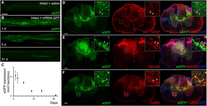
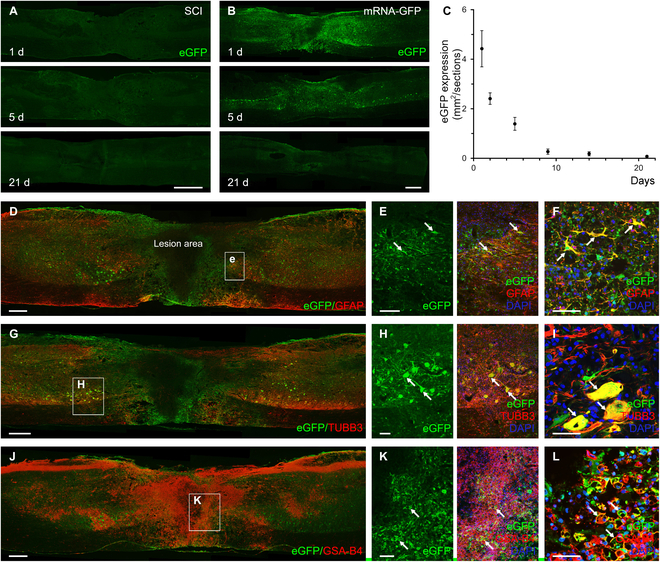
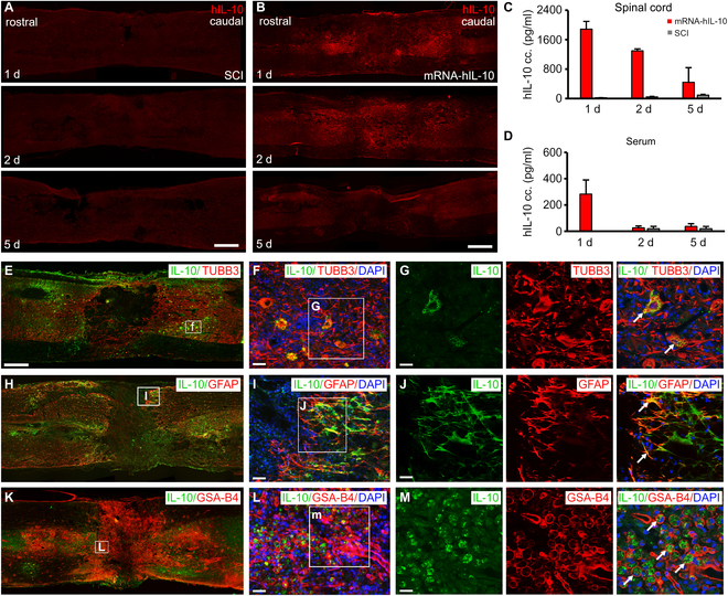
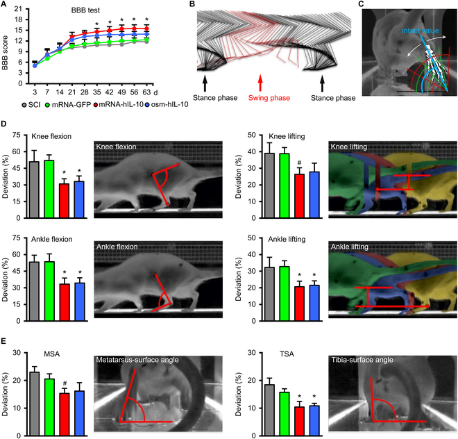
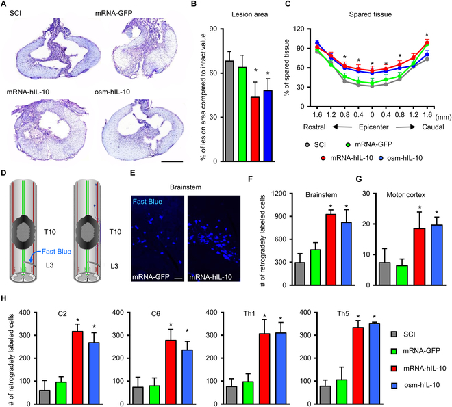
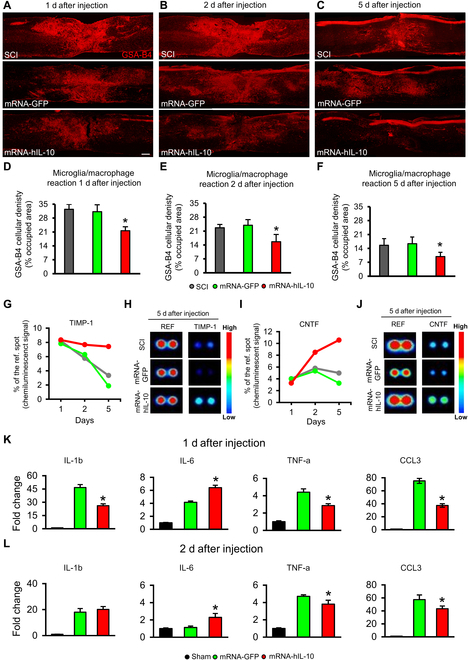
Efficient in vivo delivery of anti-inflammatory proteins to modulate the microenvironment of an injured spinal cord and promote neuroprotection and functional recovery is a great challenge. Nucleoside-modified messenger RNA (mRNA) has become a promising new modality that can be utilized for the safe and efficient delivery of therapeutic proteins. Here, we used lipid nanoparticle (LNP)-encapsulated human interleukin-10 (hIL-10)-encoding nucleoside-modified mRNA to induce neuroprotection and functional recovery following rat spinal cord contusion injury. Intralesional administration of hIL-10 mRNA-LNP to rats led to a remarkable reduction of the microglia/macrophage reaction in the injured spinal segment and induced significant functional recovery compared to controls. Furthermore, hIL-10 mRNA treatment induced increased expression in tissue inhibitor of matrix metalloproteinase 1 and ciliary neurotrophic factor levels in the affected spinal segment indicating a time-delayed secondary effect of IL-10 5 d after injection. Our results suggest that treatment with nucleoside-modified mRNAs encoding neuroprotective factors is an effective strategy for spinal cord injury repair.


Figures

Fig. 1.
Fig. 1.
eGFP expression in intact rat spinal cords following intraspinal delivery of mRNA-LNP encoding eGFP. (A) No eGFP expression can be seen in the intact rat spinal cord without intraspinal administration of mRNA-LNP encoding eGFP. (B) eGFP expression in parasagittal sections of rat spinal cord 1, 5, and 21 d after the intraspinal injection of mRNA-LNP. The extent of eGFP-positive area is very limited 21 d after mRNA-LNP administration. (C) Quantification of the fluorescent eGFP signal after intraspinal injection of 3.0-μg mRNA-LNP. Note the marked drop in the size of eGFP+ area after 5 d. (D and E) Cross-sections of spinal cord show immunhistochemically detected eGFP in astrocytes (GFAP+) (D), in neurons (TUBB3+) (E), and in microglia/macrophages (GSA-B4+) (F) 1 d after the intraspinal mRNA-LNP administration. Data represent the mean ± SEM in (C) (n = 4; biologically independent experiments). Arrowheads show the colocalized cells. Scale bars in (A) = 1 mm and in (D) to (F) = 200 μm. DAPI, 4′,6-diamidino-2-phenylindole.
Fig. 2.
Fig. 2.
Protein production from eGFP mRNA-LNP in injured rat spinal cord after intralesional delivery. (A) No eGFP expression is seen in the untreated SCI group. SCI animals received spinal cord contusion injury and 3-μl saline 7 d after injury. (B) eGFP expression in injured spinal cords followed up to 21 d after intraspinal mRNA-LNP encoding eGFP administration. (C) Quantification of eGFP expression in parasagittal sections of spinal cords at various time points in the mRNA-GFP animals. The eGFP expression drops dramatically after 5 d. (D to L) Astrocytes (GFAP+) (D to F), neurons (TUBB3+) (G to I), and microglia/macrophages (GSA-B4+) (J to L) expressed eGFP 1 d after intraspinal delivery of mRNA-LNP encoding eGFP. (F, I, and L) Higher magnification clearly shows the presence of eGFP in the cytoplasm of astrocytes, neurons, and microglia/macrophages. Arrows show GFAP-, TUBB3-, and GSA-B4-positive cells colocalizing eGFP. Data represent the mean ± SEM in (C) (n = 4; biologically independent experiments). Scale bars in (A) and (B) = 1 mm, in (D), (G), and (J) = 500 μm, in (E), (H), and (K) = 100 μm, and in (F), (I), and (L) = 50 μm.
Fig. 3.
Fig. 3.
hIL-10 expression in injured spinal cords following intralesional delivery of mRNA-LNP encoding hIL-10. (A) No hIL-10 expression was detected in the injured rat spinal cord (SCI group). SCI animals received 3 μl of saline intralesionally 7 d after the spinal cord contusion injury. (B) hIL-10 expression detected in paramedian sagittal sections of injured rat spinal cords 1, 2, and 5 d after intraspinal delivery of mRNA-LNP encoding hIL-10. (C) and (D) Production of hIL-10 in injured spinal cord and serum after hIL-10 mRNA-LNP delivery determined by ELISA. (E to G) Rat neurons express hIL-10 1 d after intralesional mRNA-LNP administration. (H to J) Rat astrocytes produced hIL-10 in the close vicinity of the lesion 1 d after mRNA-LNP delivery. (K to M) GSA-B4-positive cells colocalized with hIL-10 in the lesion area 1 d after mRNA-LNP delivery. Data represent the mean ± SEM; (C and D) n = 4, biologically independent experiments. Arrows show the colocalized cells. Scale bar in (A) and (B) = 800 μm, in (E) = 750 μm, in (F) and (L) = 30 μm, in (I) 25 = μm, and in (G), (J), and (M) = 20 μm.
Fig. 4.
Fig. 4.
Delayed intraspinal administration of mRNA-LNP encoding hIL-10 improves locomotor function. (A) Open-field locomotor test (BBB) shows significant improvement of hIL-10-treated animals (mRNA-hIL-10 and osm-hIL-10 group) compared with their controls. Asterisks indicate significant difference between the hIL-10-treated animals (mRNA-hIL-10 and osm-hIL-10 group) and SCI and mRNA-GFP groups at various time points. (B) The image shows every position of the measured bones during 1 intact step cycle from the lateral aspect. The step cycle can be divided into stance phase (black) and swing phase (red). (C) The measurement of the rear-view parameters is based on the angle enclosed by a selected bone and the floor plate. The intact value is displayed in blue, while green and red angles represent the deviations followed by contusion injury, respectively. White arrows show the deviations in both directions. (D and E) Kinematic analysis of the animals in the various groups 9 weeks after injury. Note the significantly improved parameters of the hIL-10-treated animals (mRNA-hIL-10 and osm-hIL-10 group) compared with SCI and mRNA-GFP groups. Data represent the mean ± SEM. (A, D, and E) n = 8, biologically independent experiments. *P < 0.05 and indicates significant difference among SCI, mRNA-GFP vs. mRNA-hIL-10 and osm-hIL-10 groups. #P < 0.05 and shows significant difference between SCI, mRNA-GFP vs. mRNA-IL10 group. Data were analyzed using the 2-way ANOVA (A) or 1-way ANOVA with LSD multiple comparisons tests (D and E).
Fig. 5.
Fig. 5.
Delayed intraspinal administration of mRNA-LNP encoding hIL-10 induces tissue sparing. (A) Representative images of cresyl-violet-stained sections taken at 100 μm rostrally from the lesion epicenter. (B) Quantification of lesion area shows that hIL-10 treatment resulted in significantly reduced size of injury following SCI. (C) Improved tissue sparing can be seen rostral and caudal to lesion epicenter in hIL-10-treated groups (mRNA-hIL-10 and osm-hIL-10) compared with SCI and mRNA-GFP control animals. (D) Schematic image shows the retrograde labeling procedure. FB crystals were placed into the right hemisection gap of the L3 spinal segment. (E) Retrogradely labeled neurons are shown in the brainstem of mRNA-GFP and mRNA-IL10 animals. (F to H) Quantification of retrogradely labeled neurons in the brainstem (F), in the motor cortex (G) and in various spinal segments rostrally from the contusion injury (H). Data represent the mean ± SEM. (A, B, C, F, G, and H) n = 4, biologically independent experiments. *P < 0.05 and indicates significant difference between SCI, mRNA-GFP vs. mRNA-hIL-10, and osm-hIL-10 groups. Data were analyzed using 1-way ANOVA with LSD multiple comparisons test (A, B, C, F, G, and H). Scale bar in (A) = 500 μm and in (E) = 100 μm.
Fig. 6.
Fig. 6.
Microglia/macrophage and cytokine changes after mRNA LNP encoding hIL-10 treatment in the injured spinal cord. (A to C) Representative images of paramedian sagittal spinal cord sections show the GSA-B4 reactivity 1, 2, and 5 d after intralesional delivery of saline (A), mRNA-LNP encoding eGFP (B), and mRNA-LNP encoding hIL-10 (C) in the lesion area. (D to F) Quantification of microglia/macrophage (GSA-B4) density in the sagittal sections of the spinal cord revealed significantly decreased level of GSA-B4 at all examined time points in the hIL-10 mRNA-treated group (mRNA-hIL-10) compared with the SCI and mRNA-GFP groups. (G to J) Rodent cytokine level changes were assessed by using the Proteome Profiler array (ARY008) in the spinal cords of SCI, mRNA-GFP, and mRNA-hIL-10 groups, comparing the relative levels of 29 rat cytokines. The chemiluminescence signal of spots were compared with reference spots and expressed as % of those. Higher chemiluminescence signals of tissue inhibitor of matrix metalloproteinase 1 (TIMP-1) (G) and ciliary neurotrophic factor (CNTF) (I) were detected in the mRNA-hIL-10 group compared with the SCI and mRNA-GFP groups 2 and 5 d after the mRNA-LNP treatment. (H) and (J) show representative images of reference control, TIMP-1, and CNTF spots 5 d after the saline or mRNA-LNP injection. (K to L) Rat cytokine changes 1 and 2 d after mRNA-LNP administration detected by PCR analysis. Quantification of IL-6 mRNA in the spinal cord showed significantly increased levels, while that of TNF-a and CCL3 mRNAs revealed significantly decreased levels at both examined time points in the hIL-10 mRNA-treated group (mRNA-hIL-10) compared with the mRNA-GFP group. IL1-b mRNA levels were significantly decreased on day 1 but showed nonsignificant changes on day 2 after treatment in the hIL-10 mRNA-treated animals. Data were analyzed by using 1-way ANOVA with LSD multiple comparisons test. Data represent the mean ± SEM. (D to F) n = 4; (K and L) n = 3 each group, biologically independent experiments. (D to F) *P < 0.05 indicates significant difference between SCI and mRNA-GFP vs. mRNA-hIL-10. (K and L) * indicates statistically significant difference (P < 0.05) between mRNA-GFP vs. mRNA-hIL-10 groups. Scale bar in (A) = 200 μm.

Similar articles

Cited by

References

    1. Ahuja CS, Fehlings M. Concise review: Bridging the gap: Novel Neuroregenerative and neuroprotective strategies in spinal cord injury. Stem Cells Transl Med. 2016;5:914–924. - PMC - PubMed
    1. Ulndreaj A, Badner A, Fehlings MG. Promising neuroprotective strategies for traumatic spinal cord injury with a focus on the differential effects among anatomical levels of injury. F1000Res. 2017;6:Article 1907. - PMC - PubMed
    1. Alizadeh A, Dyck SM, Karimi-Abdolrezaee S. Traumatic spinal cord injury: An overview of pathophysiology, models and acute injury mechanisms. Front Neurol. 2019;10:Article 282. - PMC - PubMed
    1. Li X, Li M, Tian L, Chen J, Liu R, Ning B. Reactive astrogliosis: Implications in spinal cord injury progression and therapy. Oxidative Med Cell Longev. 2020;2020:Article 9494352. - PMC - PubMed
    1. Smith DR, Dumont CM, Park J, Ciciriello AJ, Guo A, Tatineni R, Cummings BJ, Anderson AJ, Shea LD. Polycistronic delivery of IL-10 and NT-3 promotes oligodendrocyte myelination and functional recovery in a mouse spinal cord injury model. Tissue Eng Part A. 2020;26(11–12):672–682. - PMC - PubMed

LinkOut - more resources