. 2023 Apr;299(4):104616.
doi: 10.1016/j.jbc.2023.104616. Epub 2023 Mar 16.

Molecular basis of Q-length selectivity for the MW1 antibody-huntingtin interaction

Affiliations

Molecular basis of Q-length selectivity for the MW1 antibody-huntingtin interaction

Jose M Bravo-Arredondo et al. J Biol Chem. 2023 Apr.

Abstract

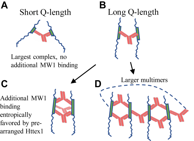
Huntington's disease is caused by a polyglutamine (polyQ) expansion in the huntingtin protein. Huntingtin exon 1 (Httex1), as well as other naturally occurring N-terminal huntingtin fragments with expanded polyQ are prone to aggregation, forming potentially cytotoxic oligomers and fibrils. Antibodies and other N-terminal huntingtin binders are widely explored as biomarkers and possible aggregation-inhibiting therapeutics. A monoclonal antibody, MW1, is known to preferentially bind to huntingtin fragments with expanded polyQ lengths, but the molecular basis of the polyQ length specificity remains poorly understood. Using solution NMR, electron paramagnetic resonance, and other biophysical methods, we investigated the structural features of the Httex1-MW1 interaction. Rather than recognizing residual α-helical structure, which is promoted by expanded Q-lengths, MW1 caused the formation of a new, non-native, conformation in which the entire polyQ is largely extended. This non-native polyQ structure allowed the formation of large mixed Httex1-MW1 multimers (600-2900 kD), when Httex1 with pathogenic Q-length (Q46) was used. We propose that these multivalent, entropically favored interactions, are available only to proteins with longer Q-lengths and represent a major factor governing the Q-length preference of MW1. The present study reveals that it is possible to target proteins with longer Q-lengths without having to stabilize a natively favored conformation. Such mechanisms could be exploited in the design of other Q-length specific binders.

Keywords: Huntington’s disease; MW1; antibody; biomarkers; electron paramagnetic resonance; polyglutamine; protein aggregation; protein conformation; protein-aggregation inhibitors.


Conflict of interest statement

Conflict of interest The authors declare no conflict of interest with the contents of this article.

Figures

Figure 1
Figure 1
MW1 preferentially binds to monomeric Httex1.A, HEK293T cells were transfected with Httex1Q72RFP and stained with the MW1 antibody. Twenty-four hours after transfection, bright and round Q72RFP (red) aggregates were seen in some cells (asterisks, middle panel). These aggregates showed very low reactivity with MW1 (green, see left panel). Other cells with lower Q72RFP expression showed a diffuse pattern within the cytoplasm. These cells showed more overlap with MW1, as can be seen in the orange signal in the right panel. B, the conformational specificity of MW1 binding was tested using dot blotting. Monomeric Httex1(46Q) or different fibril types of Httex1(Q46) as indicated in the figure were spotted on nitrocellulose membrane (5 ng each). Fluorescently labeled antibody (IRDye-800 anti-mouse IgG) was used as secondary antibody to detect MW1. The top row shows binding in the presence of both the primary and the secondary antibody while the bottom row shows the control in the presence of the secondary antibody alone. No binding was observed in the presence of the secondary antibody alone. Httex1, Huntingtin exon 1.
Figure 2
Figure 2
Mapping of the MW1 binding sites in Httex1(Q46 and Q25) using EPR.A, representative EPR spectra for sites in the N17, polyQ, and PRD of Httex1(Q46), and Httex1(Q25) in the absence or presence of varying amounts of MW1 antibody. 10 μM of Httex1 were used with in the presence of 0 (gray), 10 (blue), 20 (green), and 30 (red) μM MW1. Arrows indicate the immobile components that appear in the spectra of the 35R1 derivatives in the presence of MW1. B and C, the changes in the EPR central line amplitude are expressed by the ratio of the amplitude of the spin labeled Httex1Q46 (B) or Httex1Q25 (C) in the presence or absence of MW1. The x-axis indicates the specific labeling position. Spectra are provided in Figures 1A and S1. EPR, electron paramagnetic resonance; Httex1, Huntingtin exon 1; polyQ, polyglutamine; PRD, proline-rich domain.
Figure 3
Figure 3
Effect of MW1 binding on the solution NMR spectrum of the Httex1(Q46) polyQ region.A, two-dimensional 15N-1HN correlation spectra of 14 μM 15N-labeled Trx-Httex1(Q46) were recorded in the absence (top panel) or presence (bottom panel) of 28 μM MW1 at 10 °C and 700 MHz. Spectral regions centering on the HN backbone and H2N side-chain signals of polyQ are shown. B, the intensity ratios of the depicted signals in the presence or absence of MW1, termed I/I0, were quantified for molar Httex1(Q46):MW1 ratios of 1:0 and 1:2. Control refers to a signal from the linker connecting Trx and Httex1(Q46). Httex1, Huntingtin exon 1; polyQ, polyglutamine.
Figure 4
Figure 4
Spin-label intramolecular distances from four-pulse DEER experiments.A, C, and E, represent dipolar evolution data for Httex1(Q46) 25R1-35R1, 35R1-45R1, and 40R1-50R1 monomers. B, D, and F, represent the respective data for Httex1(Q46) 25R1-35R1, 35R1-45R1, and 40R1-50R1 monomers in the presence of the antibody MW1. The black traces in the left panels show the experimental data, while the red curves are from fits obtained based on Gaussian distributions. The gray traces in the panels on the left, A, C, and E, represent raw data without background correction. No background correction was required for the data obtained in the presence of MW1, indicating that the spin labeled sites are kept far apart from each other (on the EPR scale) by the antibodies. DEER, pulsed EPR; EPR, electron paramagnetic resonance; Httex1, Huntingtin exon 1.
Figure 5
Figure 5
Gel filtration chromatography for mixtures of MW1 and Httex1 with different Q-lengths. The complexes were incubated at 1:1 for Httex1Q16, 0.5:1 and 1:1 for both constructs Httex1Q25 and Httex1Q46. The concentration for Httex1 was 10 μM for all experiments. The vertical lines indicate the approximate molecular weights estimated based on molecular weight standards. Httex1, Huntingtin exon 1.
Figure 6
Figure 6
Isothermal titration calorimetry of Trx-Httex1 with MW1.A and B, Trx-Httex1(Q46) (A) and Trx-Httex1(Q25) (B) at concentrations of 0.4 μM were titrated with MW1 at 25 °C in 20 mM NaH2PO4/Na2HPO4 pH 7.4, 150 mM NaCl solution. Httex1, Huntingtin exon 1.
Figure 7
Figure 7
Schematic Model illustrating different modes of binding and binding stoichiometries for Httex1 of different Q-lengths in the presence of MW1.A, in case of short Q-length proteins, only one MW1 protein (red) can bind to the polyQ region (green). Due to the bidentate nature of the MW1 antibody, one antibody can bind up to two polyQ regions. BD, longer Q-lengths can accommodate additional MW1 antibodies, causing the formation of larger complexes that are favored entropically. Httex1, Huntingtin exon 1; polyQ, polyglutamine.

References

    1. Arrasate M., Finkbeiner S. Protein aggregates in Huntington's disease. Exp. Neurol. 2012;238:1–11. - PMC - PubMed
    1. Walker F.O. Huntington's disease. Lancet. 2007;369:218–228. - PubMed
    1. Hoffner G., Djian P. Polyglutamine aggregation in huntington disease: does structure determine toxicity? Mol. Neurobiol. 2015;52:1297–1314. - PubMed
    1. Mangiarini L., Sathasivam K., Seller M., Cozens B., Harper A., Hetherington C., et al. Exon 1 of the HD gene with an expanded CAG repeat is sufficient to cause a progressive neurological phenotype in transgenic mice. Cell. 1996;87:493–506. - PubMed
    1. Miller J., Arrasate M., Brooks E., Libeu C.P., Legleiter J., Hatters D., et al. Identifying polyglutamine protein species in situ that best predict neurodegeneration. Nat. Chem. Biol. 2011;7:925–934. - PMC - PubMed

Publication types

MeSH terms

LinkOut - more resources