. 2023 May:71:101706.
doi: 10.1016/j.molmet.2023.101706. Epub 2023 Mar 15.

Inceptor correlates with markers of prostate cancer progression and modulates insulin/IGF1 signaling and cancer cell migration

Affiliations

Inceptor correlates with markers of prostate cancer progression and modulates insulin/IGF1 signaling and cancer cell migration

Katharina Wissmiller et al. Mol Metab. 2023 May.

Abstract

Objective: The insulin/insulin-like growth factor 1 (IGF1) pathway is emerging as a crucial component of prostate cancer progression. Therefore, we investigated the role of the novel insulin/IGF1 signaling modulator inceptor in prostate cancer.

Methods: We analyzed the expression of inceptor in human samples of benign prostate epithelium and prostate cancer. Further, we performed signaling and functional assays using prostate cancer cell lines.

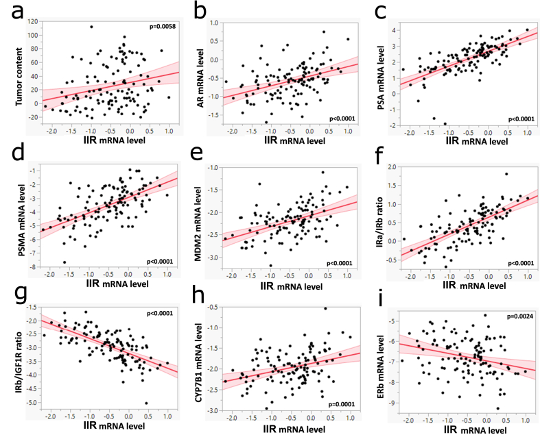
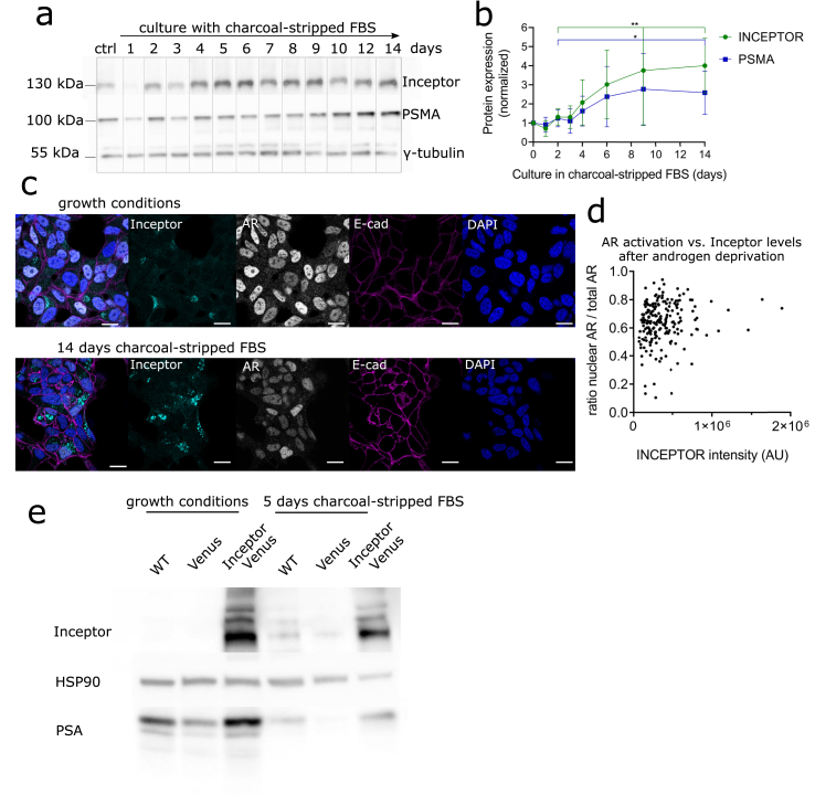
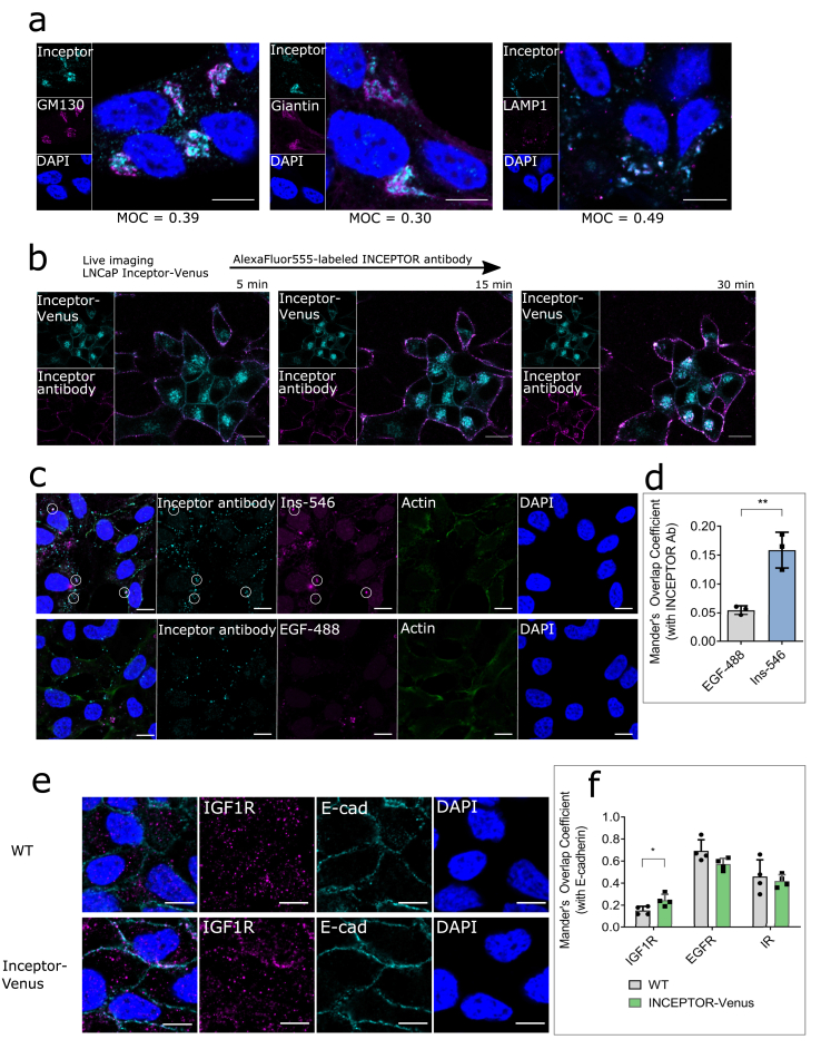
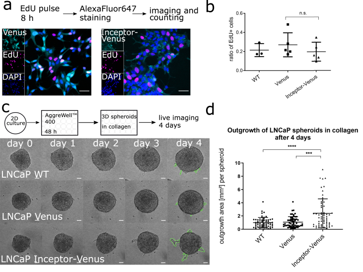
Results: We found that inceptor was expressed in human benign and malignant prostate tissue and its expression positively correlated with various genes of interest, including genes involved in androgen signaling. In vitro, total levels of inceptor were increased upon androgen deprivation and correlated with high levels of androgen receptor in the nucleus. Inceptor overexpression was associated with increased cell migration, altered IGF1R trafficking and higher IGF1R activation.

Conclusions: Our in vitro results showed that inceptor expression was associated with androgen status, increased migration, and IGF1R signaling. In human samples, inceptor expression was significantly correlated with markers of prostate cancer progression. Taken together, these data provide a basis for investigation of inceptor in the context of prostate cancer.

Keywords: Androgen; IGF1R; Insulin; Signaling; Trafficking.


Figures

Figure 1
Figure 1
Inceptor is expressed in healthy murine prostate epithelium and human prostate cancer cell lines. a. Immunohistochemical staining of WT mouse prostate with antibodies against inceptor, Cytokeratin 8/18 marking luminal cells and Cytokeratin 5 marking basal cells. Image shows a section of dorsal prostate, representative of 14 slices from two mice, taken from the dorsal, ventral or anterior prostate. Scale bar: 100 μm (upper row), 20 μm (lower row, zoomed in). b. Immunohistochemical staining of human prostate with antibodies against inceptor (cyan), Cytokeratin 8/18 (magenta), and Cytokeratin 5 (red). Scale bar: 50 μm. c. WT mouse prostate stained for inceptor, Cytokeratin 8/18 and Serotonin (5-HT) expressed in neuroendocrine cells. Image shows a section of dorsal prostate, representative of 8 slices from teo mice, taken from the dorsal, ventral or anterior prostate. Scale bar: 100 μm (upper row), 20 μm (lower row, zoomed in). d,e. Expression levels of inceptor in different human prostate cancer cell lines, determined by qPCR and Western blot. Densitometric quantification of the Western blot is shown on the right, values are normalized on γ-tubulin as loading control (n = 3).
Figure 2
Figure 2
Inceptor expression correlates with carcinogenic markers in human prostate samples. a. The mRNA expression of IIR in human benign prostate tissues (n = 71) and prostate cancer samples (n = 50) were determined with real-time PCR and normalized to ubiquitin c. Multivariate linear regression models were adjusted to age and BMI. Log transformed mRNA level of inceptor was correlated with tumor content (in %). b,c,d,e,h,i. Correlation of log transformed mRNA levels of inceptor with the indicated genes. f,g. Ratios of log transformed mRNA levels of key players of the Ins/IGF1 system, plotted against inceptor expression.
Figure 3
Figure 3
Inceptor expression is dependent on the hormone status of prostate cancer cells. a. Expression of inceptor, PSMA and AR after up to 14 days of culture in RPMI 1640 + 10% charcoal-stripped FBS (androgen deprivation conditions). The control was cultured in the same medium for 14 days, with the addition of 10 nM dihydrotestosterone. b. Densitometric quantification of a, normalized to γ-tubulin as loading control (selected time points from n = 5 independent experiments). Values show fold change compared to control. P = 0.0049 for inceptor and p = 0.0441 for PSMA, determined by unpaired student's t test. c. LNCaP cells in normal growth medium (RPMI 1640 + 10% FBS) or RPMI 1640 + 10% charcoal-stripped FBS were stained for inceptor, AR and E-cadherin. Laser and detector settings were kept constant between both images (scale bar: 20 μm). d. Quantification of fluorescence intensity of nuclear AR vs. total AR, shown in dependence of inceptor intensity (Figure 3 c, left). Single cells from two independent experiments were analyzed, after 14 days in charcoal-stripped FBS (>100 cells/n), p = 0.038 (linear regression). e. LNCaP WT, LNCaP venus or LNCaP inceptor-venus cells were cultured in normal growth medium (RPMI 1640 + 10% FBS) or RPMI 1640 + 10% charcoal-stripped FBS for 5 days.
Figure 4
Figure 4
Inceptor is involved in the trafficking of IR/IGF1R. a. Staining of LNCaP cells for inceptor and GM130 (cis-Golgi), Giantin (medial Golgi) or LAMP1 (Lysosome) to determine the subcellular localization of inceptor. The Mander's overlap coefficient was calculated from >80 cells/n (n = 3). Scale bar: 10 μm. b. LNCaP cells expressing inceptor-venus were pulse-labeled with an AlexaFluor555-conjugated inceptor antibody. Internalization of the antibody was tracked in live imaging. Images are representative for two independent experiments (>60 cells/n). Scale bar: 10 μm. c. Endocytosis assay in LNCaP C4-2 cells incubated with 10 μg/ml inceptor antibody (rat) and 100 nM AlexaFluor546-labeled insulin or 1 μg/ml AlexaFluor488-labeled EGF for 60 min, before fixation and staining with an AlexaFluor488 or 555-labeled secondary anti-rat antibody. Scale bar: 10 μm. d. Quantification of c. The Mander's Overlap Coefficient was calculated from >50 cells/n, n = 3. P = 0.0049 (unpaired student's t test). e. Staining of WT and inceptor overexpressing LNCaP cells using an IGF1R or EGFR antibody, along with an E-cadherin antibody. Scale bar: 10 μm. f. Quantification of e (including IR and EGFR, not shown in d). The Mander's Overlap Coefficient was calculated from >40 cells/n, n = 4 (p = 0.0403, unpaired student's t test).
Figure 5
Figure 5
Inceptor modulates Insulin/IGF1 signaling in prostate cancer cells. a. Co-IP using an IGF1R antibody or IR antibody or beads only (control) in LNCaP cells overexpressing inceptor-venus. To show total levels of each protein, 20 μg lysate were loaded (input), while for each IP 400 μg lysate were used. Image is representative for 4 indepenent experiments. b. Signaling assay in LNCaP cells overexpressing inceptor-venus, compared to venus only and WT cells. The cells were growth factor starved in serum-free RPMI 1640 for 4 h, before induction with 10 nM insulin, 10 nM IGF1 or 10 nM EGF for 30 min, and subsequent lysis for Western blot. c. Densitometric quantification of b (n = 4). Values were normalized to tubulin as loading control, the graph shows fold change compared to WT LNCaP. pIR/pIGF1R (+IGF1): p = 0.0017; IGF1R levels: p = 0.0636; EGFR levels: p = 0.0491 (unpaired student's t test).
Figure 6
Figure 6
Inceptor overexpression induces migration in prostate cancer cells. a. Proliferation assay of stable LNCaP cell lines overexpressing inceptor-venus or Venus only. b. Quantification of a, by image segmentation of EdU vs. DAPI. >5000 cells/n were counted; n = 5 for Venus and inceptor-venus overexpressing cells, n = 3 for WT cells. P = 0.3381, determined by unpaired student's t test. c. Migration of LNCaP spheroids in a collagen matrix (experimental scheme shown above). Scale bar: 50 μm. d. Quantification of c by measuring the outgrowth area (as indicated in green color in c) of 60 spheroids for each cell line from three independent experiments. P < 0.0001 as determined by unpaired student's t test; two outliers were removed, based on Grubb's outlier test.

References

    1. Belfiore A., Malaguarnera R. Insulin receptor and cancer. Endocr Relat Cancer. 2011;18(4):R125–R147. - PubMed
    1. Crawley D., Chamberlain F., Garmo H., Rudman S., Zethelius B., Holmberg L., et al. A systematic review of the literature exploring the interplay between prostate cancer and type two diabetes mellitus. Ecancermedicalscience. 2018;12:802. - PMC - PubMed
    1. Bensimon L., Yin H., Suissa S., Pollak M.N., Azoulay L. Type 2 diabetes and the risk of mortality among patients with prostate cancer. Cancer Causes Control. 2014;25(3):329–338. - PubMed
    1. Cai H., Xu Z., Xu T., Yu B., Zou Q. Diabetes mellitus is associated with elevated risk of mortality amongst patients with prostate cancer: a meta-analysis of 11 cohort studies. Diabetes Metabol Res Rev. 2015;31(4):336–343. - PubMed
    1. Currie C.J., Poole C.D., Jenkins-Jones S., Gale E.A.M., Johnson J.A., Morgan C.L. Mortality after incident cancer in people with and without type 2 diabetes: impact of metformin on survival. Diabetes Care. 2012;35(2):299–304. - PMC - PubMed