. 2023 Mar;11(3):e006364.
doi: 10.1136/jitc-2022-006364.

Serpin B9 controls tumor cell killing by CAR T cells

Affiliations

Serpin B9 controls tumor cell killing by CAR T cells

Thomas Kimman et al. J Immunother Cancer. 2023 Mar.

Abstract

Background: Initial clinical responses with gene engineered chimeric antigen receptor (CAR) T cells in cancer patients are highly encouraging; however, primary resistance and also relapse may prevent durable remission in a substantial part of the patients. One of the underlying causes is the resistance mechanisms in cancer cells that limit effective killing by CAR T cells. CAR T cells exert their cytotoxic function through secretion of granzymes and perforin. Inhibition of granzyme B (GrB) can underlie resistance to T cell-mediated killing, and it has been shown that serine proteinase inhibitor serpin B9 can effectively inhibit GrB. We aimed to determine whether expression of serpin B9 by cancer cells can lead to resistance toward CAR T cells.

Methods: Serpin B9 gene and protein expression were examined by R2 or DepMap database mining and by western blot or flow cytometric analysis, respectively. Coculture killing experiments were performed with melanoma cell line MeWo, diffuse large B cell lymphoma (DLBCL) cell line OCI-Ly7 or primary chronic lymphocytic leukemia (CLL) cells as target cells and natural killer cell line YT-Indy, CD20 CAR T cells or CD19 CAR T cells as effector cells and analyzed by flow cytometry.

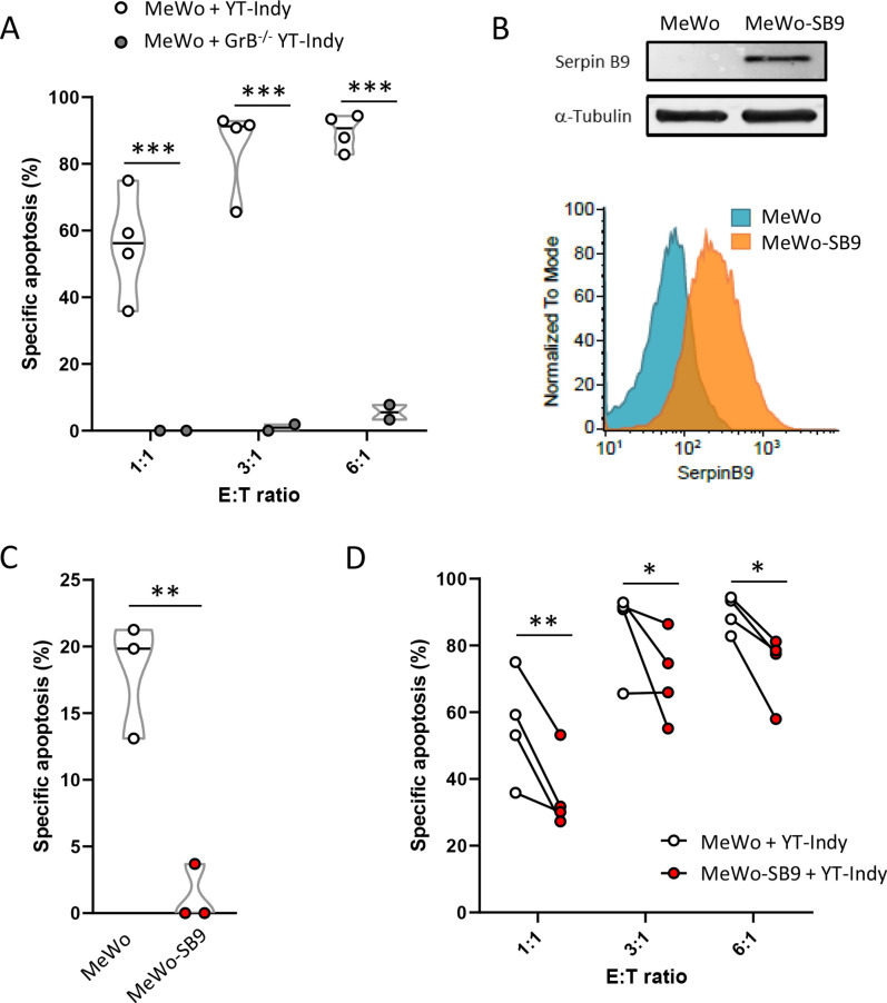
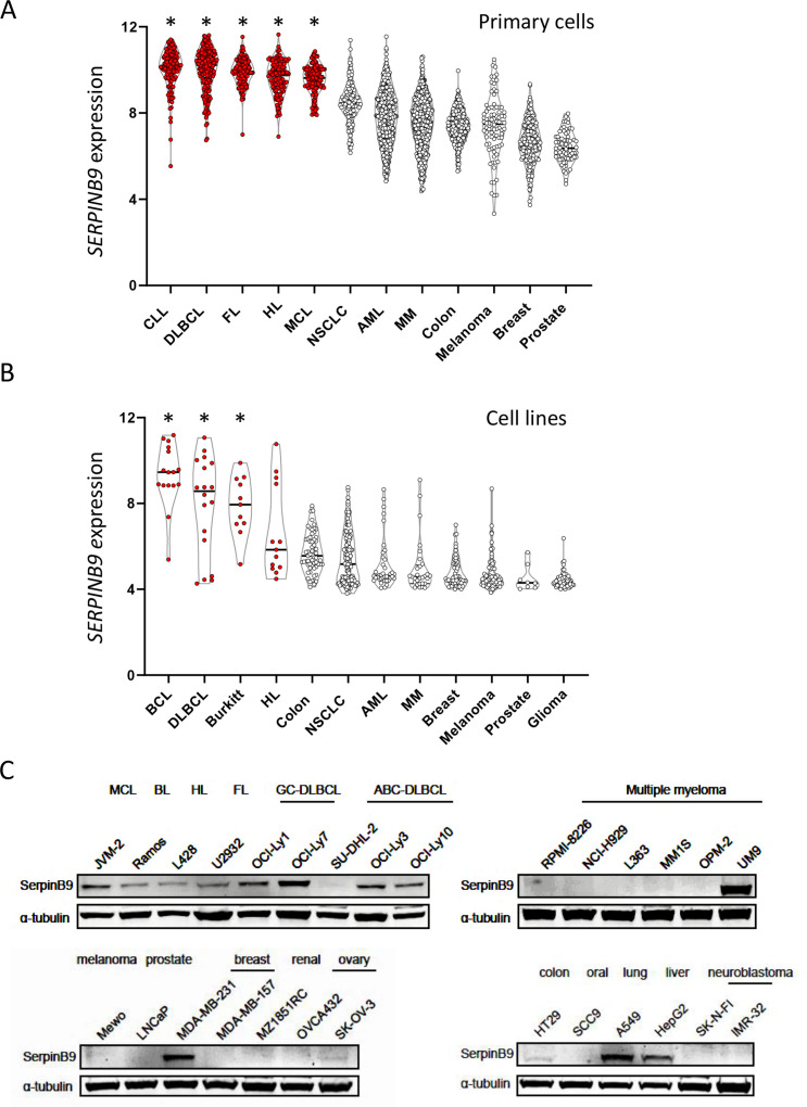
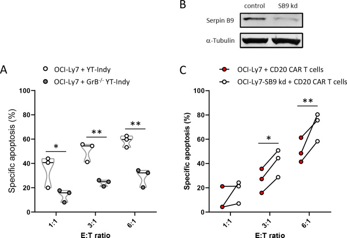
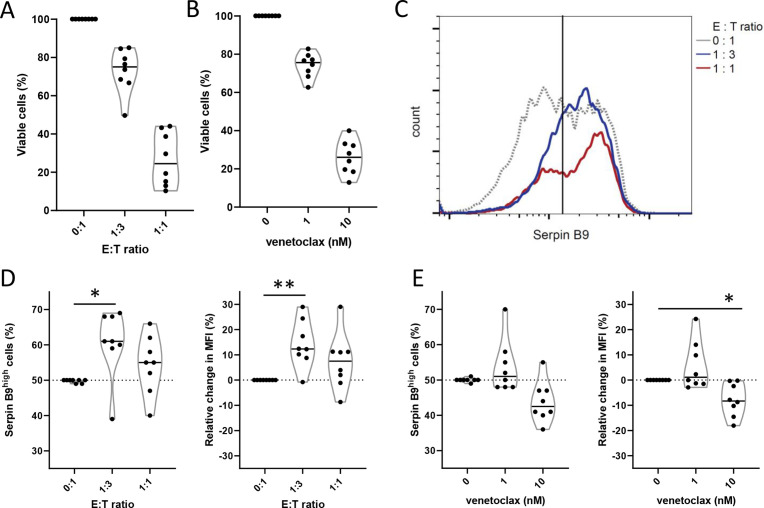
Results: Serpin B9 protein expression was previously shown to be associated with clinical outcome in melanoma patients and in line with these observations we demonstrate that enforced serpin B9 expression in melanoma cells reduces sensitivity to GrB-mediated killing. Next, we examined serpin B9 expression in a wide array of primary tumor tissues and human cell lines to find that serpin B9 is uniformly expressed in B-cell lymphomas and most prominently in DLBCL and CLL. Subsequently, using small interfering RNA, we silenced serpin B9 expression in DLBCL cells, which increased their sensitivity to CD20 CAR T cell-mediated killing. In addition, we showed that co-ulture of primary CLL cells with CD20 CAR T cells results in selection of serpin B9-high CLL cells, suggesting these cells resist CAR T-cell killing.

Conclusions: Overall, the data indicate that serpin B9 is a resistance mediator for CAR T cell-mediated tumor cell killing that should be inhibited or bypassed to improve CAR T-cell responses.

Keywords: Cytotoxicity, Immunologic; Hematologic Neoplasms; Immunotherapy, Adoptive.


Conflict of interest statement

Competing interests: VP received royalty payments related to venetoclax. VP, ZS, and TK are inventors on a patent for improving cytotoxicity of gene engineered T and NK cells. MCM received honoraria from Medscape, Jansen Cilag, and BMS. ZS and JK are inventors on different patents for γδ T -cell receptor sequences, recognition mechanisms, and isolation strategies. JK is scientific cofounder and shareholder of Gadeta. The remaining authors declare no competing interests.

Figures

Figure 1
Figure 1
Serpin B9 expression constrains sensitivity to GrB-induced cell death in human melanoma cells. (A) Melanoma MeWo cells were cocultured with wild-type NK YT-Indy cells or GrB−/− YT-Indy cells at indicated E:T cell ratios. Specific apoptosis of target cells was measured after 24 hours of culture. Data represent two to four independent experiments. (B) Serpin B9 protein expression in mock-transduced (MeWo) and serpin B9-overexpressing (MeWo-SB9) cells, as determined by immunoblotting (top) and flow cytometry (bottom). (C) Specific apoptosis of MeWo or MeWo-SB9 cells after 24 hours of treatment with 200 nM recombinant GrB through SLO-mediated pores in the cell membrane. Data represent three independent experiments. (D) Specific apoptosis of MeWo or MeWo-SB9 cells after 24 hours of coculture with YT-Indy cells at the indicated E:T ratios. Data represent four independent experiments every circle (A, C), and connecting line (D) represents one individual experiment. Two-way analysis of variance with Sidak multiple testing correction (A) and (D) and unpaired Student’s t-test (D) were performed to test statistical significance. *P<0.05, **P<0.01, ***P<0.001. E:T, effector-to-target; GrB, granzyme B; GrB−/−, granzyme B-deficient; NK, natural killer; SLO, streptolysin O.
Figure 2
Figure 2
Serpin B9 is uniformly expressed in malignant B cells and in a subset of solid cancers. (A, B) Violin plots showing SERPINB9 mRNA transcript expression in malignant cells. Samples are represented by individual data points, where red dots indicate B-cell malignancies. Data are retrieved from R2 databases (https://hgserver1.amc.nl/cgi-bin/r2/main.cgi). (A) Expression in datasets representing primary cells from CLL (n=130), DLBCL (n=223), FL (n=149), classical HL (n=130), MCL (n=122), NSCLC (n=120), AML (n=460), MM (n=542), colon cancer (n=315), melanoma (n=87), breast cancer (n=204) and prostate cancer (n=72) patients. (B) expression in human cell lines representing mixed mature B cell lymphomas (Bcl, n=16), DLBCL (n=18), Burkitt’s lymphoma (Burkitt, n=11), HL (n=13), colon cancer (n=61), NSCLC (n=134), AML (n=38), MM (n=29), breast cancer (n=59), melanoma (n=62), prostate cancer (n=8), and glioma (n=65). Data are retrieved from https://depmap.org/portal/. (C) Serpin B9 protein expression as measured by western blot in indicated human cancer cell lines and relative to expression of α-tubulin as control. One-way analysis of variance with Tukey’s multiple testing correction was performed in (A, B) to test statistical significance, where *p<0.001. Indicated statistical differences are in comparison with each individual non-malignant B-cell group (white dots). AML, acute myeloid leukemia; CLL, chronic lymphocytic leukemia; DLBCL, diffuse large B-cell lymphoma; FL, follicular lymphoma; HL, Hodgkin’s lymphoma; MCL, mantle cell lymphoma; MM, multiple myeloma; NSCLC, non-small-cell lung carcinoma.
Figure 3
Figure 3
Serpin B9 controls CAR T-cell killing of OCI-Ly7 DLBCL cells. (A) Specific apoptosis of OCI-Ly7 cells after 24 hours of coculture with WT NK YT-Indy cells or GrB−/− YT-Indy cells at indicated effector (E) to target (T) cell ratios. data represent three independent experiments. (B) representative western blot staining of serpin B9 protein expression in OCI-Ly7 cells after siRNA-mediated SB9 kd or cells treated with non-targeting siRNA (control). (C) Specific apoptosis of WT or SB9 kd OCI-Ly7 cells, 4 hours after coculture with anti-CD20 CAR expressing primary T cells. Connected data points show three independent experiments with primary T cells isolated from different healthy donors per experiment at indicated E:T cell ratios. Two-way analysis of variance with Sidak post hoc test was performed to test statistical significance. *P<0.05, **P<0.01. CAR, chimeric antigen receptor; DLBCL, diffuse large B-cell lymphoma; E:T, effector-to-target; GrB−/−, granzyme B-deficient; NK, natural killer; SB9 kd, knockdown of serpin B9; WT, wild type.
Figure 4
Figure 4
Serpin B9-high primary CLL cells are selected on coculture with CD20 CAR T cells. (A, B) Viable primary CLL cells were measured after 48 hours of coculture with CD20 CAR T cells at indicated E:T cell ratios (A) or after treatment with indicated concentration of BCL-2-inhibitor venetoclax (B). (C) Serpin B9 protein expression was measured by intracellular flow cytometry in CLL cells after 48 hours of coculture (E:T of 1:3 for the blue line and 1:1 for the red line). The black line demarcates the median in serpin B9 expression of control CLL cells (dotted line). (D) Percentage of primary CLL cells above the median of control cells (C) (left panel) or the change in MFI (right panel), after 48 hours of coculture with CD20 CAR T cells. (E) Percentage of primary CLL cells above the median of control cells (C) (left panel) or the change in MFI (right panel), after 48 hours of treatment with venetoclax. Every dot represents one patient sample. Two-way analysis of variance with Sidak post hoc tests were performed to test statistical significance. *P<0.05, **P<0.01. CAR, chimeric antigen receptor; CLL, chronic lymphocytic leukemia; E:T, effector-to-target; MFI, mean fluorescence intensity.

Similar articles

Cited by

References

    1. Rosenberg SA, Restifo NP. Adoptive cell transfer as personalized immunotherapy for human cancer. Science 2015;348:62–8. 10.1126/science.aaa4967 - DOI - PMC - PubMed
    1. June CH, O’Connor RS, Kawalekar OU, et al. . Car T cell immunotherapy for human cancer. Science 2018;359:1361–5. 10.1126/science.aar6711 - DOI - PubMed
    1. Lesch S, Benmebarek M-R, Cadilha BL, et al. . Determinants of response and resistance to CAR T cell therapy. Semin Cancer Biol 2020;65:80–90. 10.1016/j.semcancer.2019.11.004 - DOI - PubMed
    1. Shadman M, Gopal AK, Smith SD, et al. . CD20 targeted CAR-T for high-risk B-cell non-hodgkin lymphomas. Blood 2019;134(Supplement_1):3235. 10.1182/blood-2019-125102 - DOI
    1. Martínez-Lostao L, Anel A, Pardo J. How do cytotoxic lymphocytes kill cancer cells? Clin Cancer Res 2015;21:5047–56. 10.1158/1078-0432.CCR-15-0685 - DOI - PubMed

Publication types

LinkOut - more resources