

Synthesis and Biological Activities of 11- and 12-Substituted Benzophenanthridinone Derivatives as DNA Topoisomerase IB and Tyrosyl-DNA Phosphodiesterase 1 Inhibitors
- PMID: 36932053
- PMCID: PMC10233710
- DOI: 10.1002/cmdc.202200593
Synthesis and Biological Activities of 11- and 12-Substituted Benzophenanthridinone Derivatives as DNA Topoisomerase IB and Tyrosyl-DNA Phosphodiesterase 1 Inhibitors
Abstract
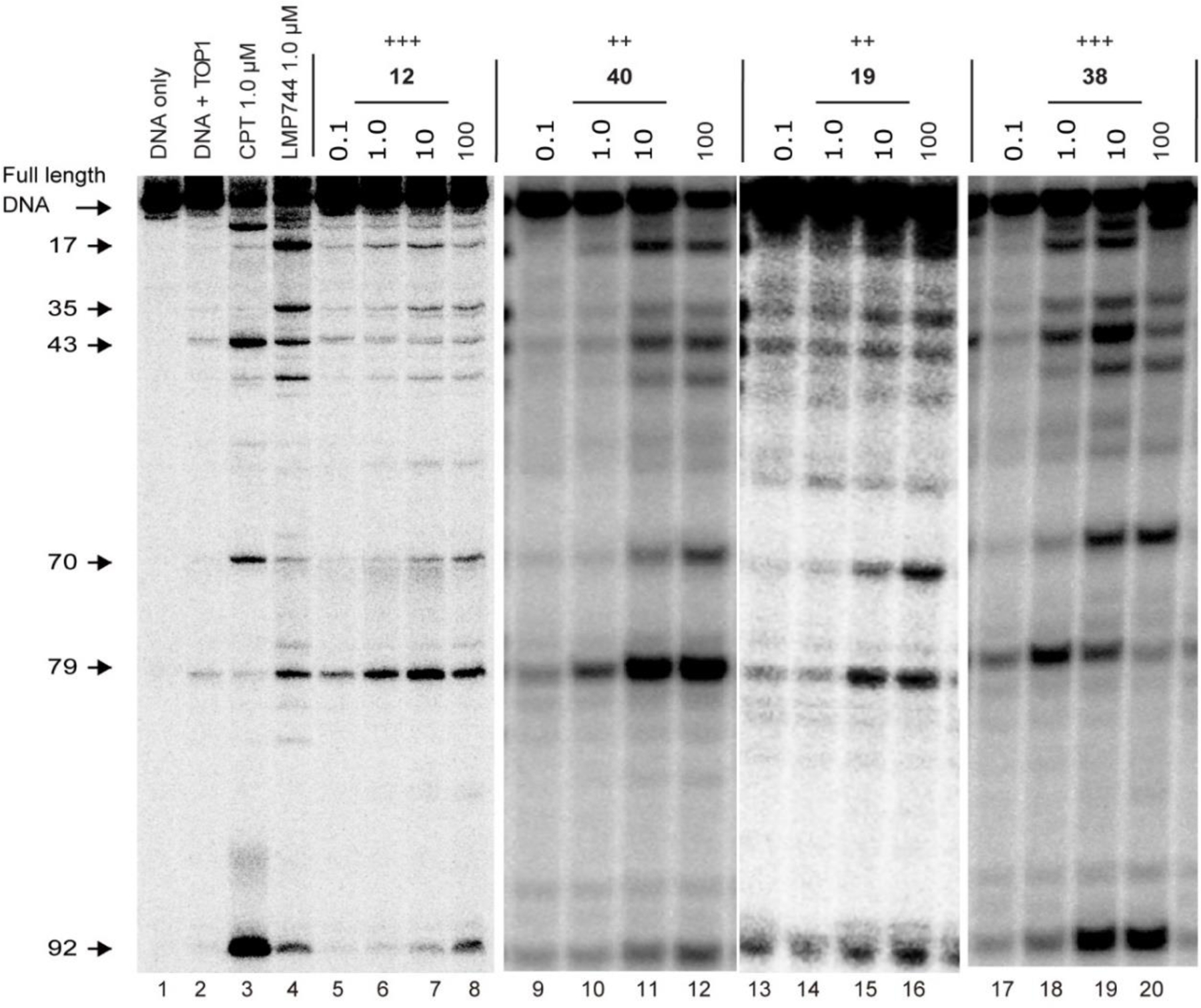
Herein, a series of 11- or 12-substituted benzophenanthridinone derivatives was designed and synthesized for the discovery of dual topoisomerase IB (TOP1) and tyrosyl-DNA phosphodiesterase 1 (TDP1) inhibitors. Enzyme-based assays indicated that two compounds 12 and 38 showed high TOP1 inhibitory potency (+++), and four compounds 35, 37, 39 and 43 showed good TDP1 inhibition with IC50 values ranging from 10 to 18 μM. 38 could induce cellular TOP1cc formation, resulting in the highest cytotoxicity against HCT-116 cells (0.25 μM). The most potent TDP1 inhibitor 43 (10 μM) could induce cellular TDP1cc formation and enhance topotecan-induced DNA damage and showed strong synergistic cytotoxicity with topotecan in both MCF-7 and MCF-7/TDP1 cells.
Keywords: DNA repair; anticancer agents; inhibitors; topoisomerase; tyrosyl-DNA phosphodiesterase.
© 2023 Wiley-VCH GmbH.
Figures


Similar articles
-
Synthesis of Methoxy-, Methylenedioxy-, Hydroxy-, and Halo-Substituted Benzophenanthridinone Derivatives as DNA Topoisomerase IB (TOP1) and Tyrosyl-DNA Phosphodiesterase 1 (TDP1) Inhibitors and Their Biological Activity for Drug-Resistant Cancer.J Med Chem. 2021 Jun 10;64(11):7617-7629. doi: 10.1021/acs.jmedchem.1c00318. Epub 2021 May 19. J Med Chem. 2021. PMID: 34008967 Free PMC article.
-
Synthesis of 11-aminoalkoxy substituted benzophenanthridine derivatives as tyrosyl-DNA phosphodiesterase 1 inhibitors and their anticancer activity.Bioorg Chem. 2022 Jun;123:105789. doi: 10.1016/j.bioorg.2022.105789. Epub 2022 Apr 4. Bioorg Chem. 2022. PMID: 35429714 Free PMC article.
-
Discovery, Synthesis, and Evaluation of Oxynitidine Derivatives as Dual Inhibitors of DNA Topoisomerase IB (TOP1) and Tyrosyl-DNA Phosphodiesterase 1 (TDP1), and Potential Antitumor Agents.J Med Chem. 2018 Nov 21;61(22):9908-9930. doi: 10.1021/acs.jmedchem.8b00639. Epub 2018 Oct 31. J Med Chem. 2018. PMID: 30336023 Free PMC article.
-
Tyrosyl-DNA Phosphodiesterase 1 (Tdp1) inhibitors.Expert Opin Ther Pat. 2011 Sep;21(9):1285-92. doi: 10.1517/13543776.2011.604314. Expert Opin Ther Pat. 2011. PMID: 21843105 Free PMC article. Review.
-
Natural Products and Their Derivatives as Inhibitors of the DNA Repair Enzyme Tyrosyl-DNA Phosphodiesterase 1.Int J Mol Sci. 2023 Mar 17;24(6):5781. doi: 10.3390/ijms24065781. Int J Mol Sci. 2023. PMID: 36982848 Free PMC article. Review.
Cited by
-
Advancing Topoisomerase Research Using DNA Nanotechnology.Small Methods. 2025 Jun;9(6):e2401113. doi: 10.1002/smtd.202401113. Epub 2024 Nov 11. Small Methods. 2025. PMID: 39526512 Free PMC article. Review.
-
New 5-Hydroxycoumarin-Based Tyrosyl-DNA Phosphodiesterase I Inhibitors Sensitize Tumor Cell Line to Topotecan.Int J Mol Sci. 2023 May 23;24(11):9155. doi: 10.3390/ijms24119155. Int J Mol Sci. 2023. PMID: 37298106 Free PMC article.
References
Publication types
MeSH terms
Substances
Grants and funding
LinkOut - more resources
Full Text Sources
Research Materials
