. 2023 Mar 17;14(1):1430.
doi: 10.1038/s41467-023-37148-x.

A mycobacterial effector promotes ferroptosis-dependent pathogenicity and dissemination

Affiliations

A mycobacterial effector promotes ferroptosis-dependent pathogenicity and dissemination

Lihua Qiang et al. Nat Commun. .

Abstract

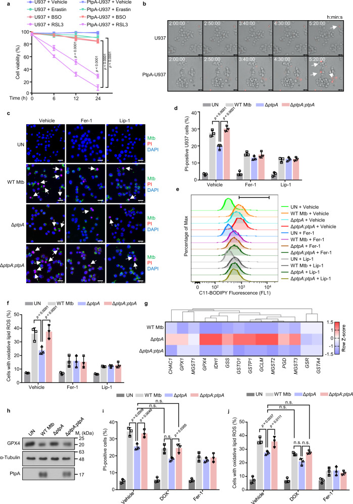
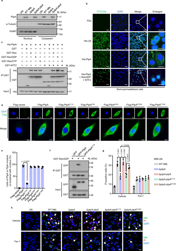
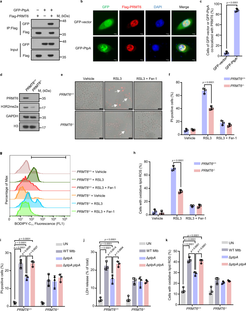
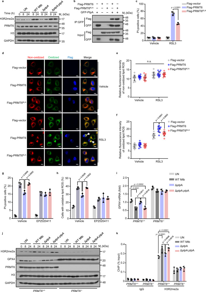
Ferroptosis is a lipid peroxidation-driven and iron-dependent programmed cell death involved in multiple physical processes and various diseases. Emerging evidence suggests that several pathogens manipulate ferroptosis for their pathogenicity and dissemination, but the underlying molecular mechanisms remain elusive. Here, we identify that protein tyrosine phosphatase A (PtpA), an effector secreted by tuberculosis (TB)-causing pathogen Mycobacterium tuberculosis (Mtb), triggers ferroptosis to promote Mtb pathogenicity and dissemination. Mechanistically, PtpA, through its Cys11 site, interacts with host RanGDP to enter host cell nucleus. Then, the nuclear PtpA enhances asymmetric dimethylation of histone H3 arginine 2 (H3R2me2a) via targeting protein arginine methyltransferase 6 (PRMT6), thus inhibiting glutathione peroxidase 4 (GPX4) expression, eventually inducing ferroptosis to promote Mtb pathogenicity and dissemination. Taken together, our findings provide insights into molecular mechanisms of pathogen-induced ferroptosis, indicating a potential TB treatment via blocking Mtb PtpA-host PRMT6 interface to target GPX4-dependent ferroptosis.


Conflict of interest statement

The authors declare no competing interests.

Figures

Fig. 1
Fig. 1. Mtb PtpA induces GPX4-dependent ferroptosis.
a Cell viability of U937 cells by Cell Counting Kit-8 (CCK-8) assay. WT U937 cells and U937 cells stably expressing PtpA (PtpA-U937) were treated with vehicle, 10 μM Erastin, 50 μM BSO, or 2 μM RSL3 for the indicated time periods. b Frames of Supplementary Movies 1 and 2 showing propidium iodide (PI)-positive WT U937 and PtpA-U937 cells treated with 2 μM RSL3. Dead cells (red) were stained with PI. Scale bars, 25 μm. c Confocal microscopic analysis for cell death of U937 cells. Cells were uninfected (UN) or infected with WT Mtb, Mtb ΔptpA, or Mtb ΔptpA:ptpA strain at an MOI of 10 for 24 h with the treatment of vehicle, Fer-1, or Lip-1. Mtb strains (green) were stained with Alexa FluorTM 488 succinimidyl ester, dead cells (red) were stained with PI, and nuclei (blue) were stained with DAPI. Scale bars, 25 μm. d Quantification of PI-positive infected U937 cells treated as in (c). e Flow cytometry for measurement of lipid peroxides with 1.5 μM BODIPY C11 lipid probe in U937 cells treated as in (c). f Quantification of cells with oxidative lipid ROS treated as in (c). g Heat map of genes related to GSH metabolism in U937 cells infected with WT Mtb, Mtb ΔptpA, or Mtb ΔptpA:ptpA strain at an MOI of 10 for 24 h. The genes indicated in red and blue represent upregulated and down-regulated genes, respectively. h Immunoblot analysis of GPX4, α-Tubulin, and PtpA in U937 cells treated as in (g). i Quantification of PI-positive infected U937 cells. Cells were uninfected or infected with WT Mtb, Mtb ΔptpA, or Mtb ΔptpA:ptpA strain at an MOI of 10 for 24 h with the treatment of 1 μM DOX or 10 μM Fer-1. j Quantification of cells with oxidative lipid ROS treated as in (i). Error bars are means ± SD of three groups. Statistical significance was determined using two-way ANOVA (Tukey’s multiple comparisons test). n.s., not significant. Source data are provided as a Source Data file.
Fig. 2
Fig. 2. Mtb PtpA, through a non-canonical RanGDP-binding site, enters host cell nucleus to induce ferroptosis.
a Immunoblot analysis of the cytosolic fraction and the pellets containing the nuclei in U937 cells. Cells were uninfected or infected with WT Mtb, Mtb ΔptpA, or Mtb ΔptpA:ptpA strain at an MOI of 20 for 8 h. b Confocal microscopic analysis for protein nuclear localization in digitonin-treated semipermeable U937 cells. Cells were incubated with FITC-labeled dextrans (FDs, a negative control), His-Ub, or His-PtpA with or without GST-RanGDP and GST-NTF2 in the presence of 10 mg/mL U937 cytosol and an energy-regenerating mixture. His-tagged proteins (green) were stained with anti-His antibody, and nuclei (blue) were stained with DAPI. Scale bars, 25 μm. c Pull-down assay of purified His-PtpA by GST, GST-Ran, GST-RanGDP, GST-RanGTP with or without GST-NTF2. d Confocal microscopic analysis for nuclear localization of PtpA or PtpA mutants. A549 Cells were transfected with empty vector, or vectors encoding Flag-PtpA, Flag-PtpAC11A, Flag-PtpAI15A, Flag-PtpAL62A, Flag-PtpAD126A, or Flag-PtpAF135A. Flag-tagged proteins (green) were stained with anti-Flag antibody, and nuclei (blue) were stained with DAPI. Scale bars, 7.5 μm. e Quantification of cells of PtpA or PtpA mutants co-localized with host cell nuclei treated as in (d). f Pull-down assay of GFP, GFP-PtpA, or GFP-PtpAC11A by GST-RanGDP. g Quantification of PI-positive infected U937 cells. Cells were incubated with 5 μg/mL PI for 20 min after being infected with WT Mtb, Mtb ΔptpA, Mtb ΔptpA:ptpA, Mtb ΔptpA:ptpAC11A, or Mtb ΔptpA:ptpAD126A strain at an MOI of 10 for 24 h with the treatment of vehicle or Fer-1. Uninfected cells were used as the control. h Confocal microscopic analysis for cell death of U937 cells treated as in (g). Mtb strains (green) were stained with Alexa FluorTM 488 succinimidyl ester, dead cells (red) were stained with PI, and nuclei (blue) were stained with DAPI. Scale bars, 25 μm. Error bars are means ± SD of three groups. Statistical significance was determined using one-way ANOVA (Dunnett’s multiple comparisons test) and two-way ANOVA (Tukey’s multiple comparisons test). Source data are provided as a Source Data file.
Fig. 3
Fig. 3. Nuclear Mtb PtpA promotes ferroptosis via targeting PRMT6.
a Co-immunoprecipitation of GFP-PtpA from the lysates of HEK293T cells co-transfected with Flag-PRMT6. b Confocal microscopic analysis for the subcellular localization of GFP or GFP-PtpA (green) with Flag-PRMT6 in A549 cells. Flag-tagged proteins (red) were stained with anti-Flag antibody, and nuclei (blue) were stained with DAPI. Scale bars, 8 μm. c Percentage of cells of GFP or GFP-PtpA co-localized with PRMT6 treated as in (b). d Immunoblot analysis of PRMT6, H3R2me2a, GAPDH, and histone H3 in PRMT6+/+ and PRMT6−/− U937 cells. e Representative phase-contrast images of PI-positive PRMT6+/+ or PRMT6−/− U937 cells treated with vehicle, 2 μM RSL3, or 10 μM Fer-1 for 8 h. Dead cells (red) were stained with PI. Scale bars, 25 μm. f Quantification of PI-positive PRMT6+/+ or PRMT6−/− U937 cells treated as in (e). g Flow cytometry analysis for lipid peroxides using 1.5 μM BODIPY C11 lipid probe in U937 cells. Cells were treated with vehicle, 2 μM RSL3, or 10 μM Fer-1 for 8 h. h The percentage of cells with oxidative lipid ROS treated as in (g). i Quantification of PI-positive PRMT6+/+ or PRMT6−/− U937 cells uninfected or infected with WT Mtb, Mtb ΔptpA, or Mtb ΔptpA:ptpA strain at an MOI of 10 for 24 h. j LDH release of PRMT6+/+ or PRMT6-/- U937 cells treated as in (i). k The percentage of PRMT6+/+ or PRMT6−/− U937 cells with oxidative lipid ROS. Cells were uninfected or infected with WT Mtb, Mtb ΔptpA, or Mtb ΔptpA:ptpA strain at an MOI of 10 for 24 h, and incubated with 1.5 μM BODIPY C11 lipid probe for 20 min. Error bars are means ± SD of three groups. Statistical significance was determined using unpaired two-sided Student’s t test, one-way ANOVA (Dunnett’s multiple comparisons test) and two-way ANOVA (Tukey’s multiple comparisons test). Source data are provided as a Source Data file.
Fig. 4
Fig. 4. Mtb PtpA inhibits GPX4 expression by promoting PRMT6-mediated H3R2me2a.
a Immunoblot analysis of H3R2me2a, PRMT6, H3, and GAPDH in U937 cells. Cells were uninfected or infected with WT Mtb, Mtb ΔptpA, or Mtb ΔptpA:ptpA strain at an MOI of 10 for 24 h. b Co-immunoprecipitation of Flag-PRMT6 or Flag-PRMT6KLA from the lysates of HEK293T cells co-transfected with GFP-PtpA. c Quantification of PI-positive A549 cells. Cells transfected with empty vector, or vectors encoding GFP-PRMT6 or GFP-PRMT6KLA were treated with vehicle or 4 μM RSL3 for 8 h. d Confocal microscopic analysis for lipid peroxides using BODIPY C11 lipid probe in A549 cells. Cells transfected with empty vector, or vectors encoding Flag-PRMT6 or Flag-PRMT6KLA were treated with vehicle or 4 μM RSL3 for 8 h, and were then treated with 1.5 μM BODIPY C11 lipid probe for additional 20 min. Flag-tagged proteins (blue) were stained with anti-Flag antibodies. Red and green fluorescence represent the non-oxidized and oxidized BODIPY C11, respectively. Scale bars, 10 μm. e, f Quantification of the fluorescence intensity of non-oxidized (e) and oxidized BODIPY C11 (f) in cells treated as in (d). g Quantification of PI-positive infected U937 cells. Cells were uninfected or infected with WT Mtb, Mtb ΔptpA, or Mtb ΔptpA:ptpA strain as in a after being treated with vehicle or 20 μM EPZ020411. h Quantification of cells with oxidative lipid ROS treated as in (g). i RT-qPCR analysis for the mRNA of GPX4 in PRMT6+/+ or PRMT6−/− U937 cells uninfected or infected with WT Mtb, Mtb ΔptpA, or Mtb ΔptpA:ptpA strain as in (a). j Immunoblot analysis of H3R2me2a, GPX4, PRMT6, H3, and GAPDH in PRMT6+/+ or PRMT6−/− U937 cells treated as in (i). k ChIP-qPCR analysis of the GPX4 promoter in PRMT6+/+ or PRMT6−/− U937 cells using the H3R2me2a antibody. Error bars are means ± SD of three groups. Statistical significance was determined using two-way ANOVA (Tukey’s multiple comparisons test). n.s., not significant. Source data are provided as a Source Data file.
Fig. 5
Fig. 5. Mtb PtpA enhances methyltransferase activity of PRMT6.
a Immunoblot analysis of H3R2me2a, H3, PRMT6, and GST-PtpA in HEK293T cells. Cell lysates were incubated with 0.1, 0.2, 0.4, or 0.8 μM GST-PtpA for 2 h. b Schematic representation of PtpA truncated mutants including PtpA, PtpAΔ1–50, PtpAΔ51–70, PtpAΔ70–122, and PtpAΔ122–163. c Co-immunoprecipitation of Flag-PRMT6 and H3 from the lysates of HEK293T cells co-transfected with GFP-PtpA, GFP-PtpAΔ1–50, GFP-PtpAΔ51–70, GFP-PtpAΔ70–122, or GFP-PtpAΔ122–163. d Immunoblot analysis of H3R2me2a, PRMT6, H3, and GAPDH in PRMT6+/+ or PRMT6−/− U937 cells. Cells were uninfected or infected with WT Mtb, Mtb ΔptpA, Mtb ΔptpA:ptpA, Mtb ΔptpA:ptpAΔ1–50, or Mtb ΔptpA:ptpAΔ122–163 strain at an MOI of 10 for 24 h. e Quantification of PI-positive cells. A549 cells were transfected with GFP, GFP-PtpA, GFP-PtpAΔ1–50, or GFP-PtpAΔ122–163 for 24 h, and were then treated with vehicle or 2 μM RSL3 for 8 h. f Confocal microscopic analysis for cell death in A549 cells treated as in (e). g RT-qPCR analysis of GPX4 mRNA in PRMT6+/+ or PRMT6−/− U937 cells. Cells were uninfected or infected with WT Mtb, Mtb ΔptpA, Mtb ΔptpA:ptpA, or Mtb ΔptpA:ptpAΔ1–50 strain at an MOI of 10 for 24 h. h Quantification of PI-positive PRMT6+/+ or PRMT6−/− U937 cells treated as in (g). i LDH release of PRMT6+/+ or PRMT6−/− U937 cells treated as in (g). j Quantification of PRMT6+/+ or PRMT6−/− U937 cells with oxidative lipid ROS treated as in (g). Error bars are means ± SD of three groups. Statistical significance was determined using two-way ANOVA (Tukey’s multiple comparisons test). Source data are provided as a Source Data file.
Fig. 6
Fig. 6. Mtb PtpA induces ferroptosis to promote Mtb pathogenicity and dissemination in vivo.
a Hematoxylin and eosin (H&E) of lung sections from mice uninfected or infected with WT Mtb, Mtb ΔptpA, Mtb ΔptpA:ptpA, or Mtb ΔptpA:ptpAΔ1–50 strain for 5 weeks. Scale bars, 200 μm. Daily intraperitoneal injections of vehicle or Lip-1 (3 mg/kg) were administered to mice from day 15 after Mtb infection (n = 4). b Quantification of inflammatory areas in lungs from mice treated as in (a). c Mycobacterial survival in the lungs from mice treated as in (a). d Mycobacterial survival in the spleen from mice as in (a). e Immunofluorescence of H3R2me2a in lung sections from mice treated as in (a). Mtb strains (red) were stained with anti-Mtb antibody, H3R2me2a (Green) were stained with anti-H3R2me2a antibody, and nuclei (blue) were stained with DAPI. Scale bars, 200 μm. f Quantification of the mean intensity of H3R2me2a in lung sections from mice treated as in (e). g Immunohistochemical staining of 4-HNE in lung sections from mice treated as in (a). Scale bars, 20 μm. h Quantification of 4-HNE expression in lung sections from mice treated as in (g). i Proposed model depicting Mtb PtpA-induced ferroptosis as well as pathogen pathogenicity and dissemination. Error bars are means ± SD of four mice per group and each represents data from two separate experiments. Statistical significance was determined using two-way ANOVA (Tukey’s multiple comparisons test). Source data are provided as a Source Data file.

References

    1. Jorgensen I, Rayamajhi M, Miao EA. Programmed cell death as a defence against infection. Nat. Rev. Immunol. 2017;17:151–164. doi: 10.1038/nri.2016.147. - DOI - PMC - PubMed
    1. Stephenson HN, Herzig A, Zychlinsky A. Beyond the grave: when is cell death critical for immunity to infection? Curr. Opin. Immunol. 2016;38:59–66. doi: 10.1016/j.coi.2015.11.004. - DOI - PubMed
    1. Stockwell BR, et al. Ferroptosis: a regulated cell death nexus linking metabolism, redox biology, and disease. Cell. 2017;171:273–285. doi: 10.1016/j.cell.2017.09.021. - DOI - PMC - PubMed
    1. Xie Y, et al. Ferroptosis: process and function. Cell Death Differ. 2016;23:369–379. doi: 10.1038/cdd.2015.158. - DOI - PMC - PubMed
    1. Dixon SJ, et al. Ferroptosis: an iron-dependent form of nonapoptotic cell death. Cell. 2012;149:1060–1072. doi: 10.1016/j.cell.2012.03.042. - DOI - PMC - PubMed

Publication types

MeSH terms