Review
. 2023 Mar 17;8(1):129.
doi: 10.1038/s41392-023-01379-7.

Kidney fibrosis: from mechanisms to therapeutic medicines

Affiliations
Review

Kidney fibrosis: from mechanisms to therapeutic medicines

Rongshuang Huang et al. Signal Transduct Target Ther. .

Abstract

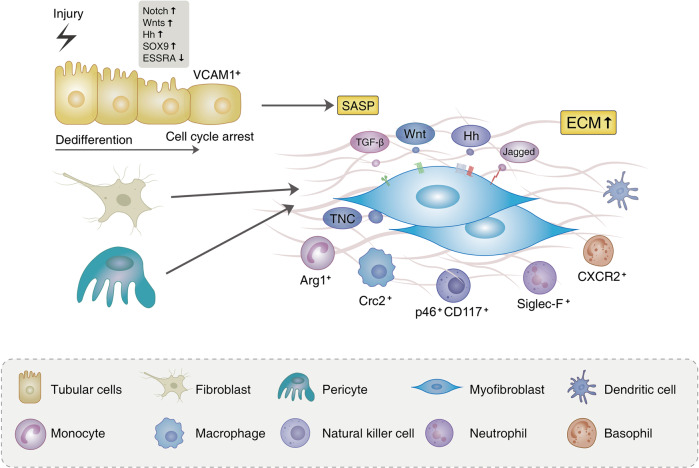
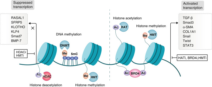
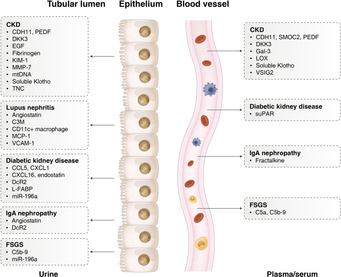
Chronic kidney disease (CKD) is estimated to affect 10-14% of global population. Kidney fibrosis, characterized by excessive extracellular matrix deposition leading to scarring, is a hallmark manifestation in different progressive CKD; However, at present no antifibrotic therapies against CKD exist. Kidney fibrosis is identified by tubule atrophy, interstitial chronic inflammation and fibrogenesis, glomerulosclerosis, and vascular rarefaction. Fibrotic niche, where organ fibrosis initiates, is a complex interplay between injured parenchyma (like tubular cells) and multiple non-parenchymal cell lineages (immune and mesenchymal cells) located spatially within scarring areas. Although the mechanisms of kidney fibrosis are complicated due to the kinds of cells involved, with the help of single-cell technology, many key questions have been explored, such as what kind of renal tubules are profibrotic, where myofibroblasts originate, which immune cells are involved, and how cells communicate with each other. In addition, genetics and epigenetics are deeper mechanisms that regulate kidney fibrosis. And the reversible nature of epigenetic changes including DNA methylation, RNA interference, and chromatin remodeling, gives an opportunity to stop or reverse kidney fibrosis by therapeutic strategies. More marketed (e.g., RAS blockage, SGLT2 inhibitors) have been developed to delay CKD progression in recent years. Furthermore, a better understanding of renal fibrosis is also favored to discover biomarkers of fibrotic injury. In the review, we update recent advances in the mechanism of renal fibrosis and summarize novel biomarkers and antifibrotic treatment for CKD.


Conflict of interest statement

The authors declare no competing interests.

Figures

Fig. 1
Fig. 1
Origination and activation of myofibroblasts. In tubulointerstitium, injury results in epithelial dedifferentiation, which is characterized by the upregulation of Notch, Wnt, Hedgehog (Hh), and SOX9 pathways. Persistent damage leads to cycle arrest and senescence of tubular epithelial cells, accompanying the secretion of profibrotic factors and senescence-associated secretory phenotype (SASP). Injured VCAM-1+ tubules secrete paracrine mediators such as TGF-β, Hh, and Wnt ligands, which impact interstitial pericytes and fibroblasts to activate myofibroblast differentiation, proliferation, and ECM accumulation. Of note, the different population of immune cell including macrophage, lymphocyte, neutrophil, and basophil also have been found in the interstitial space. And these cells expressing specific markers play an important part in kidney fibrosis
Fig. 2
Fig. 2
Histone modification and DNA methylation in kidney fibrosis. Suppression of antifibrotic genes (e.g., RASAL1, KLOTHO, KLF4, Smad7) could be accomplished by DNA methyltransferase (DNMT)-governed DNA methylation, HDACs-induced deacetylation, and histone methyltransferase (HMT)-driven methylation (e.g., H3K9me, H3K27me). On the contrary, HATs-mediated histone acetylation, HMT-ruled H3K4me, and BET proteins are involved in profibrotic gene transcriptional activation (TGF-β, Smad3, α-SMA, Snail, Twist, STAT3, et al.) α-SMA alpha-smooth-muscle actin, BMP-7 bone morphogenic protein 7, BRD4 bromodomain-containing protein 4, Col1a collagen-1α, KLF4 Krüppel-like factor 4, RASAL1 RAS protein activator like 1, SFRP5 secreted frizzled-related proteins 5, STAT3 signal transducer and activator of transcription 3, TGF-β transforming growth factor beta
Fig. 3
Fig. 3
Non-invasive biomarkers of kidney fibrosis in urine and blood. Blood or urinary biomarkers that are associated with biopsy-proven kidney fibrosis. CCL5 C-C motif chemokine ligand 5, CDH11 Cadherin-11, CXCL C-X-C motif chemokine ligand, DcR2 decoy receptor 2, DKK3 Dickkopf-3, EGF epidermal growth factor, Gal-3 Galectin-3, KIM-1 kidney injury molecule 1, L-FABP liver fatty acid binding protein, LOX lysyl oxidase, MCP-1 monocyte chemoattractant protein-1, MMP-7 matrix metalloproteinase 7, mtDNA mitochondrial DNA, PEDF pigment epithelium-derived factor, PTC-EMP peritubular capillary-endothelial microparticles, SMOC2 Sparc-related modular calcium binding protein-2, suPAR soluble urokinase plasminogen activator receptor, TNC Tenascin-C, VCAM-1 vascular cell adhesion molecule 1, VSIG2 V-set and immunoglobulin domain containing 2

References

    1. Chen TK, Knicely DH, Grams ME. Chronic kidney disease diagnosis and management: a review. JAMA. 2019;322:1294–1304. doi: 10.1001/jama.2019.14745. - DOI - PMC - PubMed
    1. Glassock RJ, Warnock DG, Delanaye P. The global burden of chronic kidney disease: estimates, variability and pitfalls. Nat. Rev. Nephrol. 2017;13:104–114. doi: 10.1038/nrneph.2016.163. - DOI - PubMed
    1. Mills KT, et al. A systematic analysis of worldwide population-based data on the global burden of chronic kidney disease in 2010. Kidney Int. 2015;88:950–957. doi: 10.1038/ki.2015.230. - DOI - PMC - PubMed
    1. Zhang L, et al. Prevalence of chronic kidney disease in China: a cross-sectional survey. Lancet. 2012;379:815–822. doi: 10.1016/S0140-6736(12)60033-6. - DOI - PubMed
    1. Ruiz-Ortega M, Rayego-Mateos S, Lamas S, Ortiz A, Rodrigues-Diez RR. Targeting the progression of chronic kidney disease. Nat. Rev. Nephrol. 2020;16:269–288. doi: 10.1038/s41581-019-0248-y. - DOI - PubMed

Publication types