. 2023 Mar 17;14(1):1416.
doi: 10.1038/s41467-023-36978-z.

Amide-to-ester substitution as a stable alternative to N-methylation for increasing membrane permeability in cyclic peptides

Affiliations

Amide-to-ester substitution as a stable alternative to N-methylation for increasing membrane permeability in cyclic peptides

Yuki Hosono et al. Nat Commun. .

Abstract

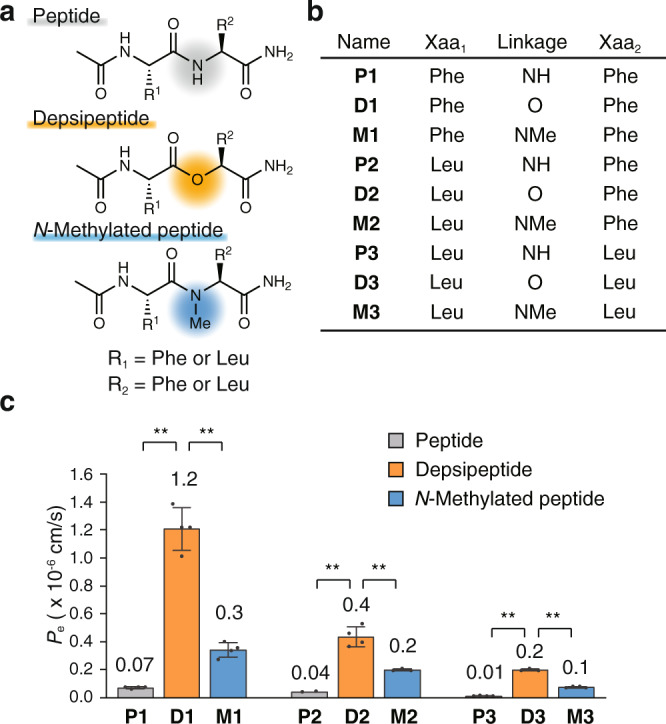
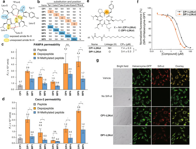
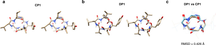
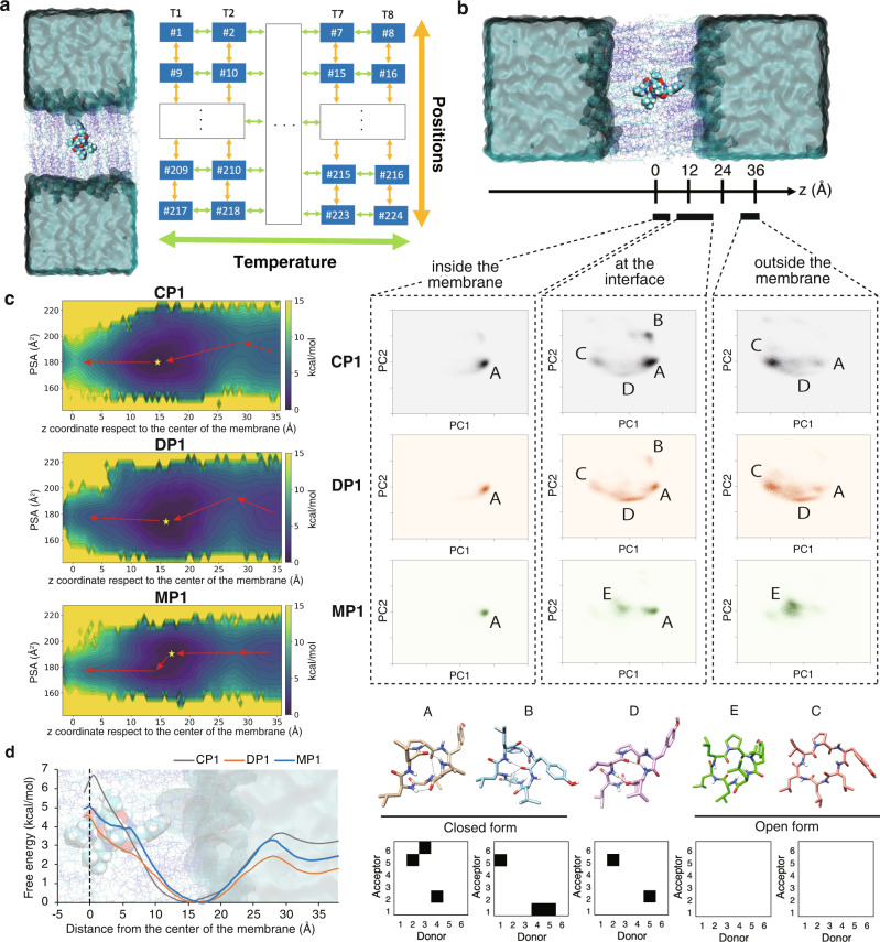
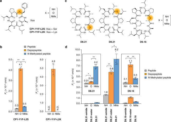
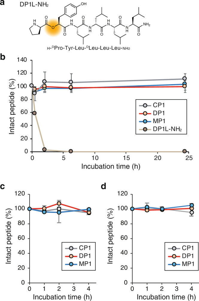
Naturally occurring peptides with high membrane permeability often have ester bonds on their backbones. However, the impact of amide-to-ester substitutions on the membrane permeability of peptides has not been directly evaluated. Here we report the effect of amide-to-ester substitutions on the membrane permeability and conformational ensemble of cyclic peptides related to membrane permeation. Amide-to-ester substitutions are shown to improve the membrane permeability of dipeptides and a model cyclic hexapeptide. NMR-based conformational analysis and enhanced sampling molecular dynamics simulations suggest that the conformational transition of the cyclic hexapeptide upon membrane permeation is differently influenced by an amide-to-ester substitution and an amide N-methylation. The effect of amide-to-ester substitution on membrane permeability of other cyclic hexapeptides, cyclic octapeptides, and a cyclic nonapeptide is also investigated to examine the scope of the substitution. Appropriate utilization of amide-to-ester substitution based on our results will facilitate the development of membrane-permeable peptides.


Conflict of interest statement

The authors declare no competing interests.

Figures

Fig. 1
Fig. 1. The effect of amide-to-ester substitution on the permeability of dipeptides.
a General structures of model dipeptides. b Sequences of synthesized dipeptides. c Permeability values of the synthesized dipeptides measured by PAMPA. PAMPA was conducted with 10 μM compounds in 5% DMSO/PBS (pH 7.4) and 18 h incubation at 25 °C. Each bar represents the mean value, and the error bars the standard deviation from experiments carried out in quadruplicate. For P2, the peptides from the acceptor wells in two out of four trials were under the quantification limit, therefore the bar represents the mean value, and the error bars the standard deviation from experiments carried out in duplicate. P values were determined by a two-sided Welch’s t-test. **p < 0.01. p (P1 vs. D1) = 0.0007, p (D1 vs. M1) = 0.0007, p (P2 vs. D2) = 0.0017, p (D2 vs. M2) = 0.0072, p (P3 vs. D3) < 0.0001 and p (D3 vs. M3) < 0.0001.
Fig. 2
Fig. 2. The effect of amide-to-ester substitution on the permeability of a cyclic hexapeptide.
a Chemical structure of CP1. b A table of synthesized compounds. The position of an amide-to-ester substitution and amide N-methylation is shown by O highlighted in orange and NMe highlighted in blue, respectively. c PAMPA and d Caco-2 assay of synthesized cyclic peptides. PAMPA was conducted with 2 μM compounds in PBS containing 5% DMSO and 16 h incubation at 25 °C. Cyclosporin A (CSA) was included as a control for PAMPA (0.4 × 10⁻⁶ cm/s). Each bar represents the mean value, and the error bars the standard deviation from experiments carried out in quadruplicate. Caco-2 assay was conducted with 1 μM compounds in HBSS (pH 7.4) containing 10 mM HEPES and 1% DMSO and 3 h incubation at 37 °C. Each bar represents the mean value, and the error bars the standard deviation from experiments carried out in triplicate (DP3, DP4, MP4, and MP5) or quadruplicate (other than DP3, DP4, MP4, and MP5). The statistical significance of DP1–5 against CP1 is shown above the bar of DP1–5 and the statistical significance of DP1–5 against MP1–5 is shown above the bars of DP1–5 and MP1–5. P values were determined by a two-sided Welch’s t-test. **p < 0.01, *p < 0.05. n.s. denotes not significant. p (CP1 vs. DP1) < 0.0001, p (DP1 vs. MP1) = 0.0092, p (CP1 vs. DP2) = 0.0002, p (DP2 vs. MP2) = 0.0014, p (CP1 vs. DP3) = 0.0070, p (DP3 vs. MP3) = 0.0037, p (CP1 vs. DP4) = 0.0197, p (DP4 vs. MP4) = 0.1148, p (CP1 vs. DP5) = 0.0016, and p (DP5 vs. MP5) = 0.0024 for PAMPA. p (CP1 vs. DP1) = 0.0002, p (DP1 vs. MP1) < 0.0001, p (CP1 vs. DP2) = 0.0511, p (DP2 vs. MP2) = 0.0293, p (CP1 vs. DP3) = 0.0406, p (DP3 vs. MP3) = 0.0689, p (CP1 vs. DP4) = 0.0214, p (DP4 vs. MP4) = 0.0253, p (CP1 vs. DP5) < 0.0001, and p (DP5 vs. MP5) = 0.0039 for Caco-2 assay. e Chemical structure, linkages, and CP50 values of chloroalkane-tagged cyclic peptides. CP50 values, the concentrations at which 50% cell penetration was observed, are shown at the bottom. f The results of CAPA for CP1-L2Kct (gray) and DP1-L2Kct (orange) analyzed by flow cytometry. Each data point represents the mean value of experiments carried out in triplicate and the error bars represent standard deviations of the triplicate. g Confocal microscope images of cells in CAPA. The cells were treated with 5 μM peptide solution. Green fluorescence represents a fusion protein of GFP and HaloTag, and red fluorescence represents SiR-ct dye. A scale bar (20 μm) is included in the bright field image of each dataset. The experiment was repeated with minor modifications and a similar result was obtained (Supplementary Fig. 9).
Fig. 3
Fig. 3. NMR solution structures of CP1 and DP1.
Stereoviews of NMR solution structures of a CP1 and b DP1 in CDCl3. c The superposition of DP1 with CP1. DP1 is shown in brown and CP1 is shown in blue. The root mean square deviation (RMSD) value is shown under the structures.
Fig. 4
Fig. 4. Enhanced sampling MD simulations of the membrane permeation process of CP1, DP1, and MP1.
a Graphical abstract of the MD simulations. b Conformational ensembles of CP1, DP1, and MP1 inside, at the interface, and outside the lipid membrane projected onto the first and second principal components (PC1 and PC2), and representative conformations and backbone hydrogen bonding patterns. The percentages of the major conformations are shown in Supplementary Fig. 48. c The two-dimensional free energy profiles against the polar surface area (PSA) and z coordinate. Minima and saddle points in the free-energy profile are connected by red arrows. Stars denote the most stable positions. d The one-dimensional free energy profile of CP1, DP1, and MP1 along the z coordinate.
Fig. 5
Fig. 5. The effect of an amide-to-ester substitution on cyclic hexapeptides with a hydrophilic residue and cyclic 8- and 9-mer peptides.
a The structures of CP1 derivatives with a hydrophilic residue and their derivatives with an amide-to-ester substitution or an amide N-methylation. b The membrane permeabilities of the CP1 derivatives shown in Fig. 5a. N.D. denotes not detected. p (NH vs. O) < 0.0001, p (O vs. NMe) <0.0001 for CP1-Y1F-L2S. Note that the scale of the y-axis is different between the left and right graphs. c The structures of D8.31, D8.21, and D9.16, and their derivatives with substitution of an N-methylated amide with an amide (D8.31-amide, D8.21-amide, and D9.16-amide) or an ester (D8.31-ester, D8.21-ester, and D9.16-ester). d PAMPA of cyclic 8-mer and 9-mer peptides. The enlarged views of the results of D8.31 series and D9.16 series are shown in the insets. PAMPA was conducted with 3 μM compounds in PBS containing 5% DMSO and 16 h incubation at 25 °C. Each bar represents the mean value, and the error bars the standard deviation from experiments carried out in quadruplicate. p (D8.31-amide vs. D8.31-ester) = 0.0138, p (D8.31-ester vs. D8.31) = 0.0004, p (D8.21-amide vs. D8.21-ester) <0.0001, p (D8.21-ester vs. D8.21) = 0.0126, p (D9.16-amide vs. D9.16-ester) = 0.0003, p (D9.16-ester vs. D9.16) = 0.0002. All the P values were determined by a two-sided Welch’s t-test. **p < 0.01, *p < 0.05.
Fig. 6
Fig. 6. Stabilities of cyclic hexapeptides in serum and simulated gastric/intestinal fluids.
a Chemical structure of DP1L-NH2. b Mouse serum stability of CP1 (gray), DP1 (orange), MP1 (blue), and DP1L-NH2 (brown). c Stability in a simulated gut fluid. 98% of a control peptide (somatostatin) was degraded at 4 h under the same conditions. d Stability in a simulated intestinal fluid. 94% of a control peptide (oxytocin) was degraded at 4 h under the same conditions. The degradation profiles of the control peptides are shown in Supplementary Fig. 50. In (b)–(d), each point represents the mean value, and the error bars the standard deviation from experiments carried out in triplicate.

Similar articles

Cited by

References

    1. Morioka T, Loik ND, Hipolito CJ, Goto Y, Suga H. Selection-based discovery of macrocyclic peptides for the next generation therapeutics. Curr. Opin. Chem. Biol. 2015;26:34–41. doi: 10.1016/j.cbpa.2015.01.023. - DOI - PubMed
    1. Dougherty PG, Qian Z, Pei D. Macrocycles as protein–protein interaction inhibitors. Biochem. J. 2017;474:1109–1125. doi: 10.1042/BCJ20160619. - DOI - PMC - PubMed
    1. Lennard KR, Tavassoli A. Peptides come round: using SICLOPPS libraries for early stage drug discovery. Chem. - A Eur. J. 2014;20:10608–10614. doi: 10.1002/chem.201403117. - DOI - PubMed
    1. Dougherty PG, Sahni A, Pei D. Understanding cell penetration of cyclic peptides. Chem. Rev. 2019;119:10241–10287. doi: 10.1021/acs.chemrev.9b00008. - DOI - PMC - PubMed
    1. Gentilucci L, De Marco R, Cerisoli L. Chemical modifications designed to improve peptide stability: incorporation of non-natural amino acids, pseudo-peptide bonds, and cyclization. Curr. Pharm. Des. 2010;16:3185–3203. doi: 10.2174/138161210793292555. - DOI - PubMed

Publication types