. 2023 Mar 17;13(1):4442.
doi: 10.1038/s41598-023-30677-x.

An integrative survival analysis and a systematic review of the cerebellopontine angle glioblastomas

Affiliations

An integrative survival analysis and a systematic review of the cerebellopontine angle glioblastomas

Nebojsa Lasica et al. Sci Rep. .

Abstract

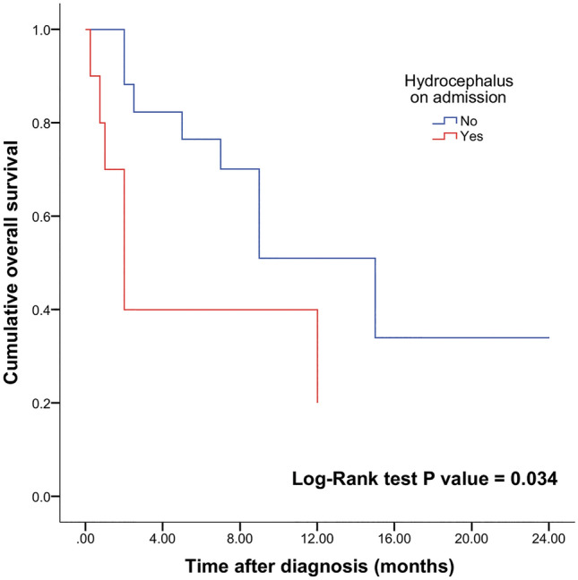
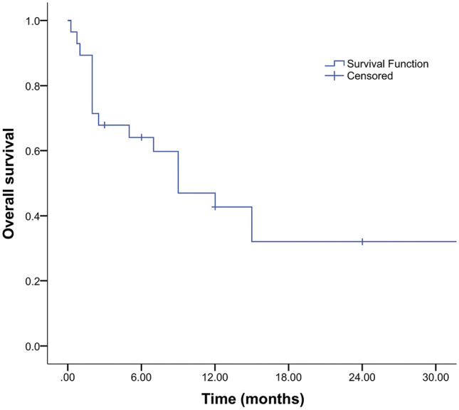
Glioblastomas presenting topographically at the cerebellopontine angle (CPA) are exceedingly rare. Given the specific anatomical considerations and their rarity, overall survival (OS) and management are not discussed in detail. The authors performed an integrative survival analysis of CPA glioblastomas. A literature search of PubMed, Scopus, and Web of Science databases was performed per PRISMA guidelines. Patient data including demographics, clinical features, neuroimaging, management, follow-up, and OS were extracted. The mean age was 39 ± 26.2 years. The mean OS was 8.9 months. Kaplan-Meier log-rank test and univariate Cox proportional-hazards model identified hydrocephalus (log-rank, p = 0.034; HR 0.34; 95% CI 0.12-0.94; p = 0.038), chemotherapy (log-rank, p < 0.005; HR 5.66; 95% CI 1.53-20.88; p = 0.009), and radiotherapy (log-rank, p < 0.0001; HR 12.01; 95% CI 3.44-41.89; p < 0.001) as factors influencing OS. Hydrocephalus (HR 3.57; 95% CI 1.07-11.1; p = 0.038) and no adjuvant radiotherapy (HR 0.12; 95% CI 0.02-0.59; p < 0.01) remained prognostic on multivariable analysis with fourfold and twofold higher risk for the time-related onset of death, respectively. This should be considered when assessing the risk-to-benefit ratio for patients undergoing surgery for CPA glioblastoma.


Conflict of interest statement

The authors declare no competing interests.

Figures

Figure 1
Figure 1
Kaplan–Meier plot of OS based on presence (n = 11) or absence (n = 19) of hydrocephalus in patients with CPA glioblastoma (log-rank test alpha level was 0.05). Patients with no hydrocephalus on admission had a mean OS 18.4 months compared with 5.6 months in patients with hydrocephalus on admission.
Figure 2
Figure 2
Comparison of OS based on treatment modalities (a) with patient subgroups receiving surgery and complete adjuvant treatment (n = 13), surgery and adjuvant radiation treatment (n = 7), surgery alone (n = 9), and no treatment (n = 1). Kaplan–Meier plot of chemotherapy (n = 13), and no adjuvant chemotherapy (n = 17) subgroups (b), and patients with CPA glioblastoma receiving radiation treatment (n = 20), and with no adjuvant radiotherapy (n = 10) (c). OS based on surgery type (d) with subgroup of patients that underwent GTR (n = 4), STR (n = 20), biopsy (n = 5), and no surgery (n = 1). Log-rank test alpha level was set to 0.05. Statistically longer survival was observed in patients receiving surgery with complete postoperative adjuvant treatment, postoperative chemotherapy, and radiation treatment.
Figure 3
Figure 3
Kaplan–Meier curve showing OS survival in patients with CPA glioblastoma in our cohort.
Figure 4
Figure 4
T1-weighted gadolinium-enhanced magnetization-prepared rapid gradient-echo MRI sequence of the brain in the axial plane (a) demonstrates a well-defined extraaxial solid mass of approximately 26 × 23 × 21 mm in the right CPA. T2-weighted MRI turbo spin-echo sequence of the brain in the axial plane (b) revealed peritumoral edema involving the right cerebellar peduncle and compressive effect on the brainstem, the fourth ventricle, and the right foramen of Luschka. Single-voxel MR spectroscopy of the CPA lesion (c) reveals elevated choline concentration, with no other metabolites. Follow-up T1-weighted gadolinium-enhanced MRI in the axial (d) and coronal planes (e) reveal marked enlargement of the tumor with extension to the IAC (arrow). Axial T2-weighted MRI (f) shows further expansion of the tumor mass to 35 × 34 × 33 mm and more pronounced compression on the lateral aspect of the brainstem and the fourth ventricle.
Figure 5
Figure 5
Artist’s illustration of exophytic and nerve REZ (inset) CPA glioblastoma ©Elyssa Siegel 2022.
Figure 6
Figure 6
A cross-section of the funnel-shaped transitional zone within the nerve REZ depicts distinct islands of neuroglial tissue, likely the origin of nerve REZ gliomas. In the transitional zone, both Schwann cells and oligodendrocytes are present ©Elyssa Siegel 2022.

References

    1. Ostrom QT, et al. CBTRUS statistical report: Primary brain and other central nervous system tumors diagnosed in the United States in 2013–2017. Neuro Oncol. 2020;22:1–96. doi: 10.1093/neuonc/noaa200. - DOI - PMC - PubMed
    1. Pietsch T, Wiestler OD. Molecular neuropathology of astrocytic brain tumors. J. Neurooncol. 1997;35:211–222. doi: 10.1023/A:1005843913095. - DOI - PubMed
    1. Stark AM, Maslehaty H, Hugo HH, Mahvash M, Mehdorn HM. Glioblastoma of the cerebellum and brainstem. J. Clin. Neurosci. 2010;17:1248–1251. doi: 10.1016/j.jocn.2010.02.015. - DOI - PubMed
    1. Epstein FJ, Farmer J-P. Brain-stem glioma growth patterns. J. Neurosurg. 1993;78:408–412. doi: 10.3171/jns.1993.78.3.0408. - DOI - PubMed
    1. Arnautovic KI, Husain MM, Linskey ME. Cranial nerve root entry zone primary cerebellopontine angle gliomas: A rare and poorly recognized subset of extraparenchymal tumors. J. Neurooncol. 2000;49:205–212. doi: 10.1023/A:1006488905526. - DOI - PubMed

Publication types