Review
. 2023 Sep;24(9):668-687.
doi: 10.1038/s41580-023-00591-1. Epub 2023 Mar 17.

Cellular and molecular mechanisms of Hedgehog signalling

Affiliations
Review

Cellular and molecular mechanisms of Hedgehog signalling

Yunxiao Zhang et al. Nat Rev Mol Cell Biol. 2023 Sep.

Abstract

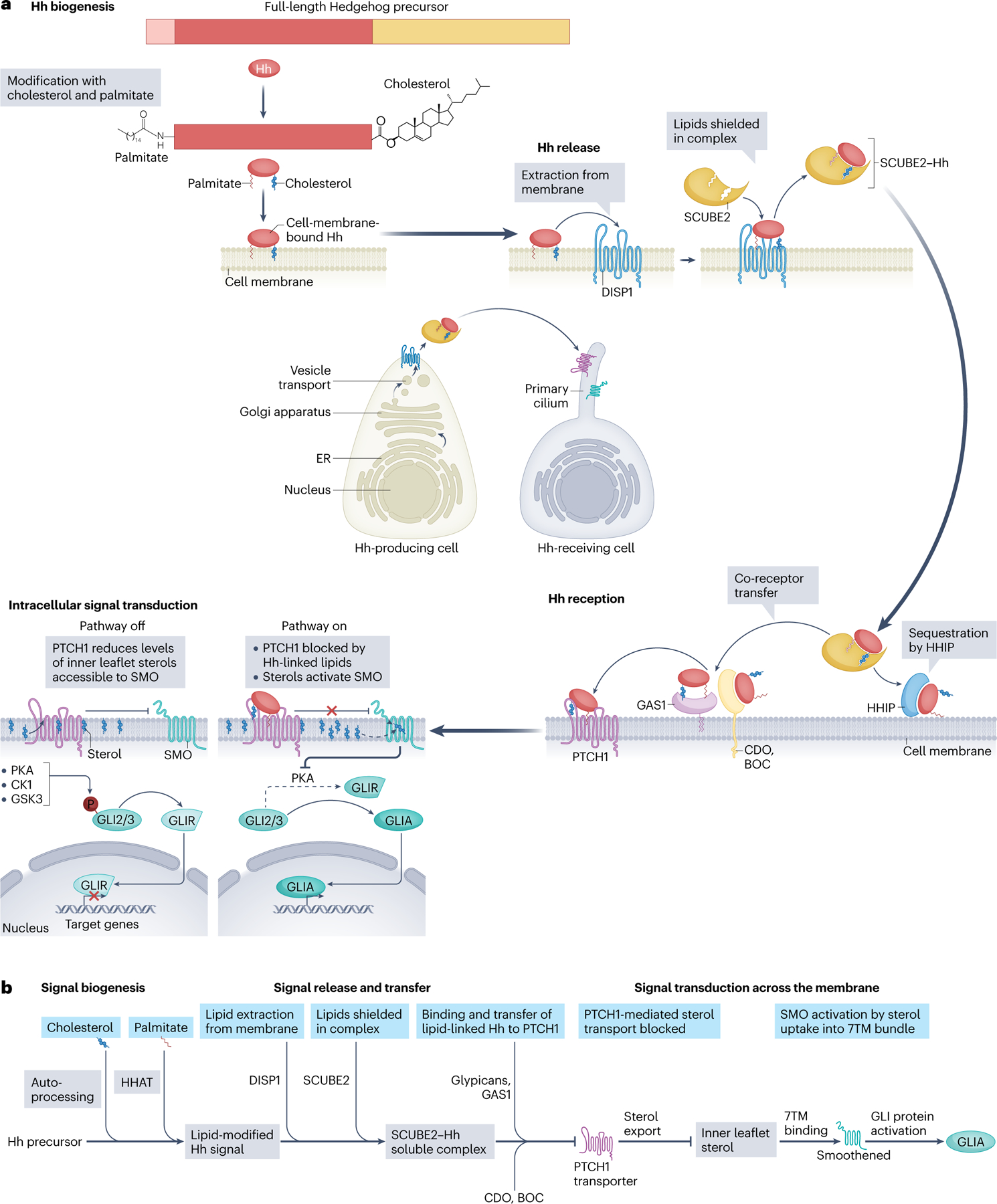
The Hedgehog signalling pathway has crucial roles in embryonic tissue patterning, postembryonic tissue regeneration, and cancer, yet aspects of Hedgehog signal transmission and reception have until recently remained unclear. Biochemical and structural studies surprisingly reveal a central role for lipids in Hedgehog signalling. The signal - Hedgehog protein - is modified by cholesterol and palmitate during its biogenesis, thereby necessitating specialized proteins such as the transporter Dispatched and several lipid-binding carriers for cellular export and receptor engagement. Additional lipid transactions mediate response to the Hedgehog signal, including sterol activation of the transducer Smoothened. Access of sterols to Smoothened is regulated by the apparent sterol transporter and Hedgehog receptor Patched, whose activity is blocked by Hedgehog binding. Alongside these lipid-centric mechanisms and their relevance to pharmacological pathway modulation, we discuss emerging roles of Hedgehog pathway activity in stem cells or their cellular niches, with translational implications for regeneration and restoration of injured or diseased tissues.


Conflict of interest statement

Competing interests

The authors declare no competing interests.

Figures

Fig. 1 |
Fig. 1 |. Overview of mammalian Hedgehog signalling.
a, Hedgehog (Hh) signalling encompasses biogenesis, release, reception and intracellular transduction of the Hedgehog signal. The Hedgehog signal is produced as a membrane-bound protein with two lipid modifications – cholesterol and palmitate. The protein Dispatched homologue 1 (DISP1) extracts Hedgehog from the membrane and transfers it to its carrier, SCUBE2, which shields the hydrophobic lipid modifications and enables long-range extracellular mobilization of the Hedgehog signal as a soluble morphogen. Most events in reception of the Hedgehog signal occur at the primary cilium, a small organellar compartment protruding from the surface of cells. Upon arrival at receiving cells, the lipids attached to the Hedgehog signal are transferred to the 12-pass transmembrane receptor protein Patched homologue 1 (PTCH1) through multiple co-receptors, including cell adhesion molecule-related/downregulated by oncogenes (CDO), brother of CDO (BOC) and growth arrest-specific protein 1 (GAS1). In the absence of its Hedgehog ligand, PTCH1 suppresses the activity of the seven-pass transmembrane (7TM) protein Smoothened (SMO) by reducing its accessibility to sterols from the inner leaflet of the membrane. In this pathway-inactive state, the transcription factors GLI2 and GLI3 are primed through phosphorylation for partial proteasomal degradation, yielding their corresponding repressor forms (GLIR). Hedgehog binding to PTCH1 inactivates its sterol transport and lifts Smoothened inhibition, thereby blocking the phosphorylation and proteolytic processing of the GLI proteins and enabling them to become transcription activators (GLIA). GLIA proteins then initiate downstream transcriptional programmes in the nucleus that depend on other factors specific to the cell context. b, The prominence of lipids in Hedgehog signalling. Lipids (blue boxes) participate in Hedgehog signalling as covalent adducts in signal biogenesis, including cholesterol addition through autoprocessing and palmitate linkage by Hedgehog acyltransferase (HHAT). DISP1 extracts the dually lipid modified Hedgehog signal from the membrane and packages it with its carrier, SCUBE2, to generate the soluble morphogen. The Hedgehog signal with its lipid adducts can be sequestered by Hedgehog-interacting protein (HHIP), rendering it inert, or alternatively is transferred via its co-receptors, including the glypicans, CDO, BOC and GAS1, to the Hedgehog receptor, PTCH1. Hedgehog binding blocks PTCH1 transport of sterols away from the inner leaflet, and this blockade, which requires Hedgehog and its palmitate adduct, enables Smoothened to become activated by uptake and binding of a sterol within its 7TM bundle. Activated Smoothened then promotes the formation of the GLI transcriptional activator. ER, endoplasmic reticulum.
Fig. 2 |
Fig. 2 |. Lipid modification of Hedgehog.
a, Hedgehog is translated and undergoes processing and lipid modification en route to the plasma membrane. The protein precursor comprises a signal peptide, an amino-terminal (N-terminal) signalling domain and a carboxy-terminal (C-terminal) autoprocessing domain. b, Following removal of the signal peptide, the C-terminal autoprocessing domain catalyses a self-cleavage and modification reaction, initiated by nucleophilic displacement of the amide nitrogen by the side chain sulfur of the cysteine residue at the scissile bond, thus forming a thioester intermediate which is resolved by nucleophilic attack by the hydroxy oxygen of a cholesterol molecule. This reaction produces an N-terminal product – the signalling domain – in ester linkage with a cholesteryl adduct at its C terminus. c, Hedgehog acyltransferase (HHAT; Skinny hedgehog (Ski) in Drosophila melanogaster), which is inhibited by the small molecules RU-SKI43 and RU-SKI201, catalyses the transfer of a palmitoyl group onto the N terminus of the signalling domain. The mature form of the Hedgehog signal thus contains two lipid modifications. ER, endoplasmic reticulum.
Fig. 3 |
Fig. 3 |. The ligand-binding pockets of Smoothened.
a, Smoothened (SMO) is a member of the F class of GTP-binding protein-coupled receptors (GPCRs) with three ligand-binding sites: one in its extracellular cysteine-rich domain (CRD) and two linked sites within its seven-pass transmembrane (7TM) domain. The CRD contains a hydrophobic groove that can bind sterol ligands, including 20(S)-hydroxycholesterol and endogenous oxysterols. The shallow site within the 7TM domain is the binding site for the antagonists cyclopamine and the FDA-approved anti-cancer drugs vismodegib, sonidegib and glasdegib, as well as the agonist SAG; the deep site, which is continuous with the shallow site and corresponds to the orthosteric ligand-binding sites of other GPCRs, is the site for binding of a sterol when Smoothened is in active conformation, and for the antagonist SANT-1. b, Among structures of GPCRs associated with cholesterol and aligned by their TM helices, many bind sterols at locations at the periphery of the protein. Smoothened is unique, however, in binding a sterol within the interior of its 7TM bundle when in its activated conformational state. c, Although both the CRD site and the deep site can bind sterol ligands, the CRD is ~21 Å above the membrane, whereas the deep transmembrane site is connected to the inner leaflet of the cell membrane by a lateral opening (arrow). d, Spheres denote residues that, when altered, affect ligand binding. The blue spheres in the shallow TM site indicate residues frequently mutated that confer drug resistance. The Smoothened structure corresponds to Protein Data Bank entry 6O3C; the overlaid GPCR structures in part b include 165 GPCR structures in complex with cholesterol that were obtained from the Protein Data Bank. TM5, transmembrane helix 5; TM6, transmembrane helix 6.
Fig. 4 |
Fig. 4 |. Sterol transport by Patched.
a, The structure of Patched homologue 1 (PTCH1) reveals sterol-like densities within the protein at three different locations (I–III), suggesting the existence of a transport conduit for sterols (arrows) within PTCH1. b, Heterotrimeric structure of two PTCH1 molecules (PTCH1a and PTCH1b) bound by one lipid-modified Sonic hedgehog-N (SHH-N) molecule. Sites I and II of PTCH1a bind the cholesteryl and palmitoyl adducts of the SHH-N signal, respectively, whereas PTCH1b exclusively contacts the protein surface of the SHH-N signal. c, The structure of the trimeric complex reveals alternative closed states of the sterol conduit, with the sterol conduit of PTCH1a interrupted by SHH-N lipids occupying sites I and II, whereas occlusion of the conduit in PTCH1b is caused by its protein–protein interaction with SHH-N. The structure of the trimeric complex corresponds to Protein Data Bank entry 6E1H. SMO, Smoothened.
Fig. 5 |
Fig. 5 |. Dispatched release of Hedgehog from the cell membrane is driven by Na+ flux.
a, Conformational changes in Dispatched homologue 1 (DISP1) during binding and release of the amino-terminal signalling domain of sonic Hedgehog (SHH-N). DISP1 views from within the plane of the membrane (top row) or from the extracellular side of the membrane (bottom row) show the bound (left, blue) and unbound (right, yellow) states of DISP1. A comparison of these views (superimposed in the centre panels) shows that binding results from pincer-like movements (dark yellow and blue lines) of the extracellular domains that grip SHH-N and from a molecular embrace of SHH-N by the furin-cleaved linker arm of DISP1 (magenta; fully resolved only in the presence of SHH-N). These DISP1 conformational changes enable the binding interactions that aid in extraction of SHH-N lipids from the membrane. b, Na+ flux through the DISP1 transmembrane domain. The chemiosmotic force of Na+ flux drives higher occupancy of three Na+-binding sites (purple spheres; darker shading indicates higher occupancy) within a channel that spans the transmembrane domain. Higher occupancy is accompanied by extracellular domain conformational changes (see part a), which cause release of SHH-N to its carrier SCUBE2. The structures of DISP1, DISP1–SHH and PTCH1 correspond to Protein Data Bank entries 7RPH, 7RPK and 6MG8, respectively.
Fig. 6 |
Fig. 6 |. Model of Hedgehog signal transduction at the cell surface.
Powered by a transmembrane Na+ gradient, Patched homologue 1 (PTCH1) localized to the primary cilium transports sterols to create a sterol-depleted inner leaflet of the plasma membrane (IPM) that does not permit Smoothened activation (left), resulting in rapid egress of Smoothened from the cilium and proteasomal degradation of GLI proteins to their repressor forms (GLIR). Upon binding of Hedgehog (middle), PTCH1 activity is blocked and IPM sterol levels are restored, permitting Smoothened activation and accumulation within the cilium. Ciliary sterols, particularly oxysterols, may promote Smoothened activation by binding to its extracellular cysteine-rich domain (CRD). The PTCH1–Hedgehog complex undergoes ubiquitylation (by HECT E3 ubiquitin ligases) and endocytosis, thus reducing the inhibitory impact of PTCH1 and helping to shape Hedgehog pathway activity during tissue patterning. In the activated state of Smoothened (right), the ciliary accumulation of Smoothened activates GLI proteins (GLIA) as they migrate through the cilium, in part by blocking formation of GLIR by proteasomal degradation. Ub, ubiquitin.
Fig. 7 |
Fig. 7 |. Hedgehog signalling has context-dependent roles in tissues.
a, In ectoderm-derived tissues with epithelia that directly depend on the Hedgehog (Hh) signal for proliferation, pathway-activating mutations can initiate cancers, such as basal cell carcinoma and medulloblastoma. b, In endoderm-derived organs, an epithelial Hedgehog signal induces expression of growth and differentiation factors by mesenchymal cells, which control tissue homeostasis (not shown) and injury-induced regeneration. The mesenchymal factors elicited by Hedgehog signalling promote proliferation and differentiation in a coordinated manner. c, In carcinomas of endoderm-derived organs, proliferation is driven by oncogenic mutations within the epithelium, and prodifferentiation factors elicited by Hedgehog signalling curb further growth and invasion of tumour cells. Loss of Hedgehog response permits progression to invasive carcinoma and ultimately metastasis. Note the opposite effects of Hedgehog pathway activity in promoting or slowing the growth of tumours arising in ectoderm-derived or endoderm-derived organs. SHH, Sonic hedgehog.
None

References

    1. Ives PT New mutants report. Drosoph. Inf. Serv 24, 58 (1950).
    1. Lee JJ, von Kessler DP, Parks S & Beachy PA Secretion and localized transcription suggest a role in positional signaling for products of the segmentation gene hedgehog. Cell 71, 33–50 (1992). - PubMed
    1. Nusslein-Volhard C & Wieschaus E Mutations affecting segment number and polarity in Drosophila. Nature 287, 795–801 (1980). - PubMed
    1. Dessaud E, McMahon AP & Briscoe J Pattern formation in the vertebrate neural tube: a sonic hedgehog morphogen-regulated transcriptional network. Development 135, 2489–2503 (2008). - PubMed
    1. Ingham PW & McMahon AP Hedgehog signaling in animal development: paradigms and principles. Genes Dev 15, 3059–3087 (2001). - PubMed

Publication types

MeSH terms

LinkOut - more resources