. 2023 Mar 17;6(1):280.
doi: 10.1038/s42003-023-04643-2.

DIAPH1 mediates progression of atherosclerosis and regulates hepatic lipid metabolism in mice

Affiliations

DIAPH1 mediates progression of atherosclerosis and regulates hepatic lipid metabolism in mice

Laura Senatus et al. Commun Biol. .

Abstract

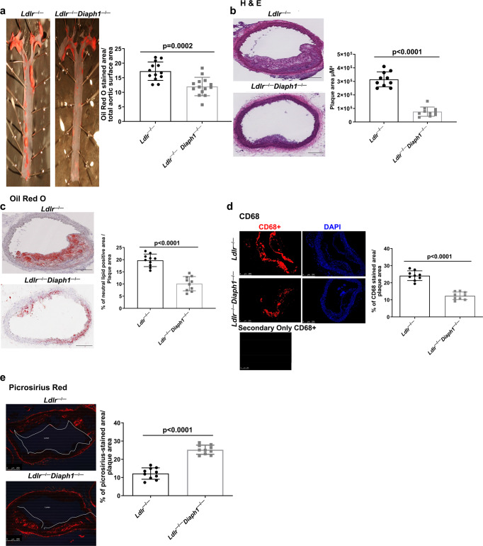
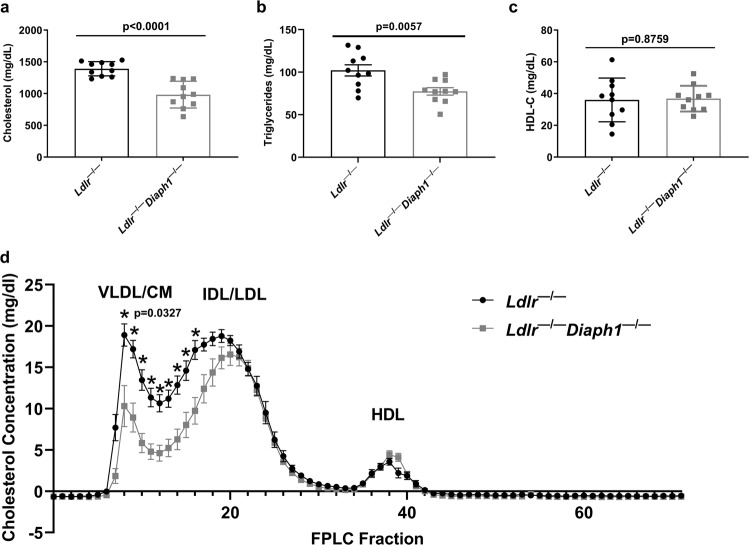
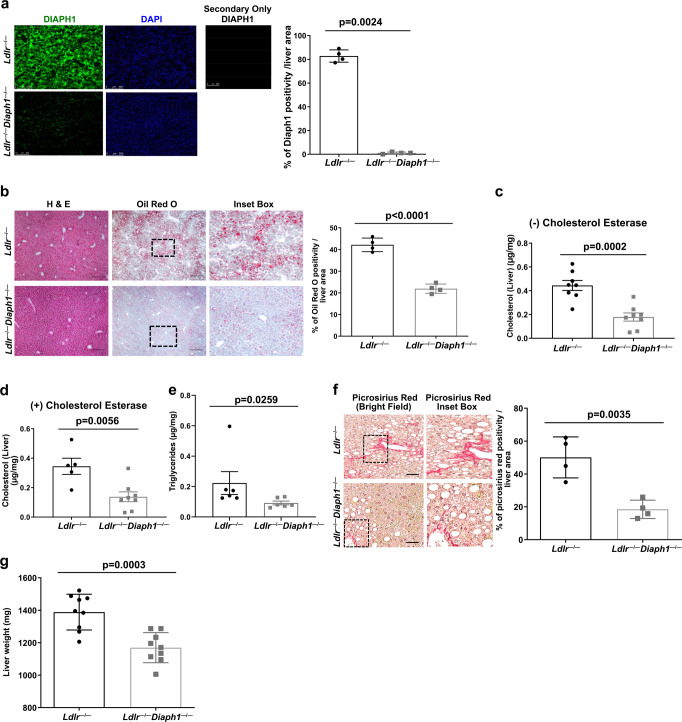
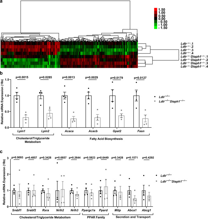
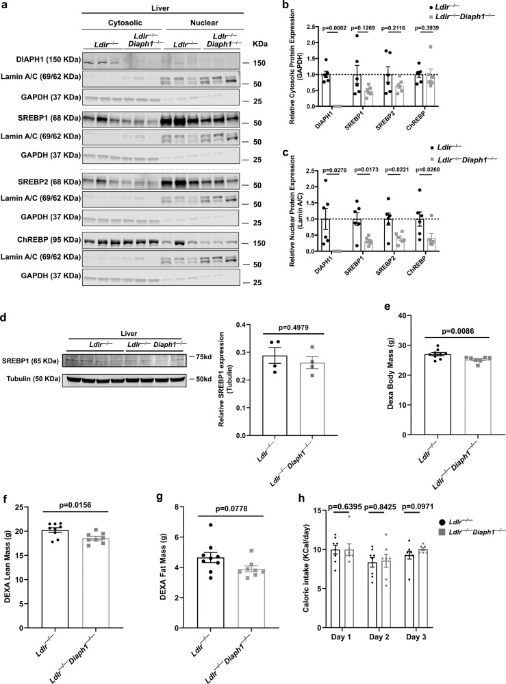
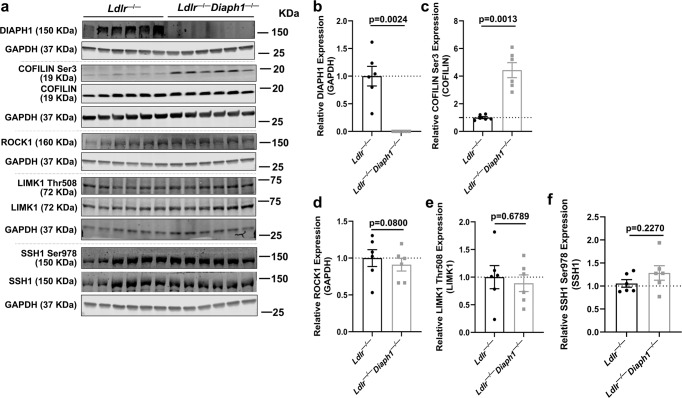
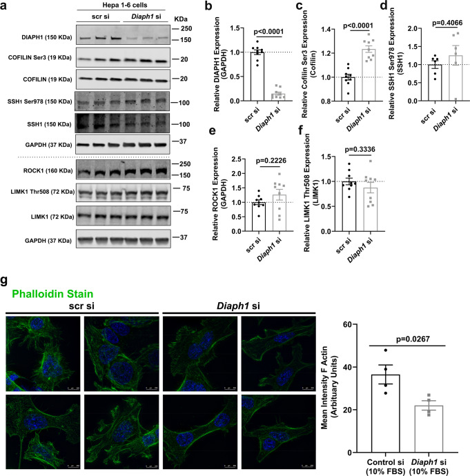
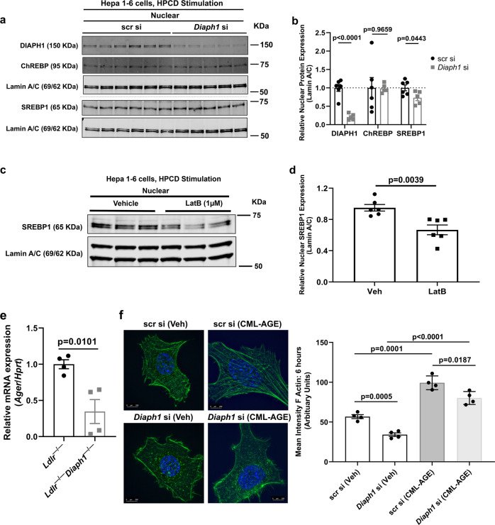
Atherosclerosis evolves through dysregulated lipid metabolism interwoven with exaggerated inflammation. Previous work implicating the receptor for advanced glycation end products (RAGE) in atherosclerosis prompted us to explore if Diaphanous 1 (DIAPH1), which binds to the RAGE cytoplasmic domain and is important for RAGE signaling, contributes to these processes. We intercrossed atherosclerosis-prone Ldlr-/- mice with mice devoid of Diaph1 and fed them Western diet for 16 weeks. Compared to male Ldlr-/- mice, male Ldlr-/- Diaph1-/- mice displayed significantly less atherosclerosis, in parallel with lower plasma concentrations of cholesterol and triglycerides. Female Ldlr-/- Diaph1-/- mice displayed significantly less atherosclerosis compared to Ldlr-/- mice and demonstrated lower plasma concentrations of cholesterol, but not plasma triglycerides. Deletion of Diaph1 attenuated expression of genes regulating hepatic lipid metabolism, Acaca, Acacb, Gpat2, Lpin1, Lpin2 and Fasn, without effect on mRNA expression of upstream transcription factors Srebf1, Srebf2 or Mxlipl in male mice. We traced DIAPH1-dependent mechanisms to nuclear translocation of SREBP1 in a manner independent of carbohydrate- or insulin-regulated cues but, at least in part, through the actin cytoskeleton. This work unveils new regulators of atherosclerosis and lipid metabolism through DIAPH1.


Conflict of interest statement

R.R., A.S., M.B.M., and A.M.S. have patents and patent applications through NYU Grossman School of Medicine that have been submitted/published that are indirectly related to the work detailed in this manuscript.

Figures

Fig. 1
Fig. 1. Deletion of Diaph1 in male Ldlr/ mice attenuates the progression of atherosclerosis.
Ldlr / and Ldlr/ Diaph1/ male mice were fed Western Diet (WD) for 16 weeks. a Representative images of en face Oil Red O staining of aortas. Quantification of plaque area as percentage of Oil Red O-stained area over total aortic surface area is shown. be Representative images of aortic arch sections are shown and quantified for the following: b H&E; c Oil Red O; d CD68; and e, Picrosirius Red. In d, the secondary antibody-alone control is shown. Scale bar: 250 µm. The mean ± SEM is reported. The number of independent mice/group is indicated in the figure as individual data points. Statistical analyses regarding testing for the normality of data followed by appropriate statistical analyses were described in Materials and Methods. P values were determined by unpaired T-test.
Fig. 2
Fig. 2. Effect of DIAPH1 on lipid parameters.
Ldlr / and Ldlr/ Diaph1/ male mice were fed WD for 16 weeks. a Concentrations of total plasma cholesterol; b concentrations of total plasma triglycerides; c concentrations of plasma high-density lipoprotein cholesterol (HDL-C). In ac the mean ± SEM is reported. The number of independent mice/group is indicated in the figure as individual data points. d Plasma lipoprotein fraction concentrations were measured by Fast Performance Liquid Chromatography (FPLC). CM/VLDL Chylomicron and very-low-density lipoprotein, IDL/LDL Intermediate-density and low-density lipoproteins, HDL High-density lipoprotein. The mean ± SEM is reported from N = 6 independent mice/group. Statistical analyses regarding testing for the normality of data followed by appropriate statistical analyses were described in Materials and Methods. P values were determined by unpaired T-test.
Fig. 3
Fig. 3. Deletion of Diaph1 in Ldlr/ mice reduces hepatic lipid content and liver fibrosis.
Ldlr/ and Ldlr/ Diaph1−/− male mice were fed WD for 16 weeks. a Representative immunofluorescence staining and quantification of DIAPH1 in the liver of Ldlr −/− and Ldlr−/− Diaph1−/− male mice. b Representative images of H&E and Oil Red O staining in liver and quantification is shown. c Quantification of free cholesterol content in liver. d Quantification of total cholesterol content in liver. e Quantification of total liver triglycerides. f Representative images of Picrosirius Red staining in liver and quantification is shown. g Quantification of whole liver weight. The mean ± SEM is reported. The number of independent mice/group is indicated in the figure as individual data points. In a the secondary antibody–alone control is shown. Scale bars: 250 µm, and inset boxes: 50 µm. Statistical analyses regarding testing for the normality of data followed by appropriate statistical analyses were described in Materials and Methods. P-values were determined by unpaired T-test or Wilcoxon rank-sum test depending on if the data passed the Shapiro-Wilk normality test.
Fig. 4
Fig. 4. RNA-Sequencing reveals roles for DIAPH1 in regulation of hepatic lipid metabolism.
Ldlr −/− and Ldlr−/− Diaph1−/− male mice were fed WD for 16 weeks. a Hierarchical clustering of differentially expressed genes in liver tissue from the indicated N = 4 independent mice/group. b The expression of the indicated genes identified as differentially expressed in the RNAseq data between Ldlr −/− mice vs. Ldlr−/− Diaph1−/− mice was determined by RT-qPCR. c The expression of the indicated genes identified as not differentially expressed in the RNAseq between Ldlr −/− mice vs. Ldlr−/− Diaph1−/− mice was confirmed by RT-qPCR. The number of independent mice/group is indicated in the figure as individual data points. Statistical analyses regarding testing for the normality of data followed by appropriate statistical analyses were described in Materials and Methods. P-values (in b, c) were determined by unpaired T test or Wilcoxon rank-sum test depending on if the data passed the Shapiro-Wilk normality test.
Fig. 5
Fig. 5. Deletion of Diaph1 in Ldlr−/− mice reduces nuclear content of SREBP1, SREBP2 and ChREBP in liver.
Ldlr −/− and Ldlr−/− Diaph1−/− male mice were fed WD for 16 weeks. a Representative Western Blots for the detection of cytosolic and nuclear DIAPH1, SREBP1, SREBP2 and ChREBP performed on liver fractions isolated from the indicated mice. b Quantification of cytosolic DIAPH1, SREBP1, SREBP2 and CHREBP, relative to GAPDH. c Quantification of nuclear DIAPH1, SREBP1, SREBP2 and ChREBP, relative to Lamin A/C. d Representative Western blot and quantification of total SREBP1 normalized to tubulin in total liver of the indicated mice. eg DEXA scans were performed for determination of body mass (e), lean mass (f), and fat mass (g). h Caloric intake was determined over 3 consecutive days. i mRNA expression of the gene encoding RAGE (Ager) was determined in the livers of the indicated male mice after 16 weeks WD. The mean ± SEM is reported. The number of independent mice/group is indicated in the figure as individual data points. Statistical analyses regarding testing for the normality of data followed by appropriate statistical analyses were described in Materials and Methods. P-values were determined by unpaired T-test or Wilcoxon rank-sum test depending on if the data passed the Shapiro-Wilk normality test.
Fig. 6
Fig. 6. Deletion of Diaph1 in Ldlr−/− mice increases phosphorylated (Ser3)/Cofilin/total Cofilin in liver.
Ldlr −/− and Ldlr−/− Diaph1−/− male mice were fed WD for 16 weeks. a Representative Western Blots for the detection of DIAPH1, phosphorylated (Ser3) Cofilin and total Cofilin, ROCK1, phosphorylated (Thr508) LIMK1 and total LIMK1, phosphorylated (Ser978) SSH1 and total SSH1 on total liver lysates isolated from the indicated mice. b Quantification of DIAPH1 relative to GAPDH. c Quantification of phosphorylated Cofilin (Ser3) relative to total Cofilin. d Quantification of ROCK1 relative to GAPDH. e Quantification of phosphorylated LIMK1 (Thr508) relative to total LIMK1. f Quantification of phosphorylated SSH1 (Ser978) relative to total SSH1. In bf, the mean ± SEM is reported. The number of independent mice/group is indicated in the figure as individual data points. Statistical analyses regarding testing for the normality of data followed by appropriate statistical analyses were described in Materials and Methods. P-values were determined by unpaired T-test.
Fig. 7
Fig. 7. Silencing of Diaph1 increases phosphorylated (Ser3) Cofilin/total Cofilin and DIAPH1 and RAGE ligands contribute to F-actin polymerization in Hepa 1-6 cells.
a Representative Western blots for the detection of DIAPH1, phosphorylated (Ser3) Cofilin, total Cofilin, phosphorylated (Ser978) SSH1, total SSH1, ROCK1, phosphorylated (Thr508) LIMK1, total LIMK1 and GAPDH performed on mouse Hepa 1-6 cells after Diaph1 or scramble control siRNA knockdown. b Quantification of DIAPH1 relative to GAPDH. c Quantification of phosphorylated (Ser3) Cofilin relative to total Cofilin. d Quantification of phosphorylated (Ser978) SSH1 relative to total SSH1. e Quantification of ROCK1 relative to GAPDH. f Quantification of phosphorylated (Thr508) LIMK1 relative to total LIMK1. g Immunofluorescence staining and quantification of the mean intensity of F-actin (phalloidin) in mouse Hepa1-6 cells after Diaph1 or scramble control siRNA knockdown. Scale bar: 250 µm. The mean ± SEM is reported. The number of independent biological/independent replicates is indicated in the figure as individual data points. Statistical analyses regarding testing for the normality of data followed by appropriate statistical analyses were described in Materials and Methods. P-values were determined by unpaired T-test or Wilcoxon rank-sum test depending if data passed the Shapiro-Wilk normality test.
Fig. 8
Fig. 8. RAGE, DIAPH1, actin organization and SREBP1.
a Western blots for the detection of nuclear DIAPH1, ChREBP and SREBP1 performed on mouse Hepa 1-6 cells after Diaph1 or scramble control siRNA knockdown and 30-min sterol depletion with 1% 2-hydroxypropyl-β-cyclodextrin (HPCD). b Quantification of nuclear DIAPH1, SREBP1, and ChREBP, relative to Lamin A/C. c Western blots for the detection of nuclear SREBP1 performed on mouse Hepa 1-6 cells after 30 min pre-treatment with latrunculin B (LatB; 1 µm) followed by the addition for 30 min of sterol depletion with 1% HPCD. d Quantification of nuclear SREBP1 relative to Lamin A/C. e mRNA expression of the gene encoding RAGE (Ager) was determined in the livers of the indicated male mice after 16 weeks WD. f Hepa 1-6 cells bearing scramble control or Diaph1 siRNA silencing were treated with RAGE ligand CML-AGE (100 µg/ml) or vehicle for 6 h followed by quantification of the mean intensity of F-actin (phalloidin). Scale bar: 250 µm. The mean ± SEM is reported. The number of independent biological/independent replicates is indicated in the figure as individual data points. Statistical analyses regarding testing for the normality of data followed by appropriate statistical analyses were described in Materials and Methods. P-values were determined by unpaired T-test or Wilcoxon rank-sum test depending if data passed the Shapiro-Wilk normality test.

Similar articles

Cited by

References

    1. https://www.cdc.gov/heartdisease/facts.htm#:~:text=Heart%20Disease%20in%....
    1. Heron, M. Deaths: Leading Causes for 2017. Natl. Vital Stat. Rep.68, 1–77 (2019). - PubMed
    1. Ridker PM, et al. Antiinflammatory therapy with canakinumab for atherosclerotic disease. N. Engl. J. Med. 2017;377:1119–1131. doi: 10.1056/NEJMoa1707914. - DOI - PubMed
    1. Park L, et al. Suppression of accelerated diabetic atherosclerosis by the soluble receptor for advanced glycation endproducts. Nat. Med. 1998;4:1025–1031. doi: 10.1038/2012. - DOI - PubMed
    1. Bu DX, et al. Activation of the ROCK1 branch of the transforming growth factor-beta pathway contributes to RAGE-dependent acceleration of atherosclerosis in diabetic ApoE-null mice. Circ. Res. 2010;106:1040–1051. doi: 10.1161/CIRCRESAHA.109.201103. - DOI - PMC - PubMed

Publication types