Review
. 2023 Jul;209(4):679-720.
doi: 10.1007/s00359-023-01616-y. Epub 2023 Mar 17.

Lineages to circuits: the developmental and evolutionary architecture of information channels into the central complex

Affiliations
Review

Lineages to circuits: the developmental and evolutionary architecture of information channels into the central complex

Pratyush Kandimalla et al. J Comp Physiol A Neuroethol Sens Neural Behav Physiol. 2023 Jul.

Abstract

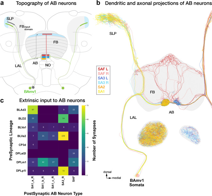
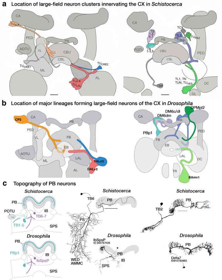
The representation and integration of internal and external cues is crucial for any organism to execute appropriate behaviors. In insects, a highly conserved region of the brain, the central complex (CX), functions in the representation of spatial information and behavioral states, as well as the transformation of this information into desired navigational commands. How does this relatively invariant structure enable the incorporation of information from the diversity of anatomical, behavioral, and ecological niches occupied by insects? Here, we examine the input channels to the CX in the context of their development and evolution. Insect brains develop from ~ 100 neuroblasts per hemisphere that divide systematically to form "lineages" of sister neurons, that project to their target neuropils along anatomically characteristic tracts. Overlaying this developmental tract information onto the recently generated Drosophila "hemibrain" connectome and integrating this information with the anatomical and physiological recording of neurons in other species, we observe neuropil and lineage-specific innervation, connectivity, and activity profiles in CX input channels. We posit that the proliferative potential of neuroblasts and the lineage-based architecture of information channels enable the modification of neural networks across existing, novel, and deprecated modalities in a species-specific manner, thus forming the substrate for the evolution and diversification of insect navigational circuits.

Keywords: Central complex; Hemibrain; Insect brains; Large-field neurons; Lineages.


Conflict of interest statement

The authors declare that the research was conducted in the absence of any commercial or financial relationships that could be construed as potential conflicts of interest.

Figures

Fig. 1
Fig. 1
Overview of the developmental organization of the Drosophila central complex. a Schematic drawing of Drosophila central complex (CX; antero-lateral view) and large-field neurons. Left: DALv2 ER-neuron as a representative of tangential neuron, providing input from the bulb (BU) to the ellipsoid body (EB). Right: PBp1 (Delta7) neuron as an example of an intrinsic neuron whose arbor is restricted to a CX compartment (here: protocerebral bridge, PB). b Schematic drawing of CX (dorsal view) depicting representative example of small-field (columnar) neuron, with arborizations restricted to narrow volumes (glomeruli, columns) of different CX compartments. Figures adapted from (Hanesch et al. 1989). c Schematic of a neuronal lineage formation and projection into different neuropil compartments (grey squares). Broad neuron classes of a lineage collectively tile a few compartments, referred to as the projection envelope of the lineage, within which, individual neuron types form various circuit motifs. d z-projection of frontal confocal sections of Drosophila brain at the level of the fan-shaped body (FB). GFP-labeled MARCM clone of the CP2/DL1 lineage, consisting of a dorsal (CP2d) and ventral (CP2v) hemilineage. Neuronal cell bodies are rendered in magenta, fiber tracts and arborizations in green. Lineage-associated tracts project in characteristic patterns, as shown here for CP2d neurons that follow the oblique posterior fascicle (obP) and then the longitudinal superior medial fascicle (loSM) to reach the FB. d’ Digital (in-silico) clone of CP2 neurons identified in the hemibrain database based on characteristic location and projection patterns. e Electron microscopy (EM) section of Drosophila brain showing CP2d axon bundle. Scale bars: 500 nm. f Schematic representation of CX and surrounding compartments, visualizing the topography of lineages that innervate the CX. Annotated on the left are neuropil compartments providing input to the CX: inferior bridge/superior posterior slope (IB/SPS), superior protocerebrum (SLP/SIP/SMP), anterior optic tubercle (AOTU) and bulb (BU), crepine (CRE) and lateral accessory lobe (LAL). Right half of the schematized brain shows lineages—represented by colored circles alongside their names. Position of circles roughly coincides with the location of somata clusters in the brain. Although the focus of our analysis are the large-field neurons, we also include the lineages which give rise to the small-field neurons (grey circles). Note: A novel finding from our hemibrain analysis is that the DM4 (DM1 and DM3 are hidden for brevity) lineages also give rise to a few large-field neurons. To distinguish the small- and large-field neurons of this lineage, we depict them as a separate yellow-colored circle in the right side of the schematic. Color-coded lines emanating from the different lineages interconnect the input domains of the constituent neurons with their output domains in the CX. The shading in the input domains reflects the degree of overlap (as in the SMP/SLP and CRE), or lack thereof (as in the AOTU and BU), of the arbors of the different lineages. The extension of the colored lines into the CX depicts the relative innervation patterns exhibited by these neurons. More detailed, and realistic, tract trajectories are schematized in the subsequent figures which highlight individual CX compartments. g Number of neurons (from both hemispheres) provided by different lineages (along vertical axis) to CX compartments (along horizontal axis). For other abbreviations see Table 1
Fig. 2
Fig. 2
Anterior visual pathway provides developmentally segregated visual input to the ellipsoid body (EB) via anterior optic tubercle (AOTU) and bulb (BU). a Schematic frontal section of the brain hemisphere at the level of AOTU and EB, visualizing neuropil compartments and lineages forming the AVP. Two hemilineages, DALcl1d (magenta) and DALcl2d (blue) form the tuberculo-bulbar neurons that connect the lateral and intermediate domains of the AOTU to the BU. The TuBus neurons (dark magenta) innervate the lateral anterior, lateral intermediate, and lateral posterior AOTU (AOTUla, AOTUli, AOTUlp) and project to the superior BU (BUs); TuBua neurons, also derived from DALcl1d, link the intermediate lateral AOTU (AOTUil) to the anterior bulb (BUa). TuBui neurons, descending from DALcl2d, project from the intermediate medial AOTU (AOTUim) to the inferior bulb (BUi; inset at the left shows AOTU compartments at higher magnification). These three separate channels continue towards the EB. Discrete sublineages of DALv2 generate the outer ring neurons (ER2, ER4d, ER5, ER3w; magenta) that connect the BUs with the anterior and outer central domain of the EB (EBa, EBoc); ER4m (light magenta) projects from BUa to the EBoc; inner ring neurons (ER3a/d/m/p; blue) link the BUi to the inner central and inner posterior EB (EBic, EBip; inset at the right shows EB domains in a horizontal section of the left half of the EB). The MeTu (optic-lobe derived), TuBu (DALcl1/2d), and ER-neurons (DALv2) constitute the first, second, and third legs of the AVP respectively. b EM section of DALcl1d/DALcl2d axon tracts. TuBu neurons are shaded in magenta (TuBus and TuBua) and blue (TuBui). Non-colored axons of DALcl1/2d project to targets other than the BU. c Plot of TuBu neurons (top) rendered in magenta (DALcl1d-derived) and blue (DALcl2d-derived); anterior view (left) and dorsal view (right). Plot of TuBu output synapses in the bulb is shown at the bottom. d Circular projection of the EB cross sections along the antero-radial axis (outline); with AVP ER-neuron output synapses similarly collapsed onto a single plane. Synapses (dots) are color-coded by upstream TuBu lineage (downstream of DALcl1d; magenta) (downstream of DALcl2d, blue). Each dot represents a T-bar and is shaded by relative input strength from the TuBu neurons (normalized to the strongest TuBu to ER connection). Plots at the bottom show synapse density for the two groups of ER-neurons. e Heatmap showing synapse numbers of DALcl1d-derived TuBu (top) and DALcl2d-derived TuBu neurons on different subclasses of ER-neurons (horizontal axis)
Fig. 3
Fig. 3
Organization and inputs to the ellipsoid body large-field networks apart from the anterior visual pathway. a Topography and upstream connections of DALv2 ER-neurons that receive input in the LAL, rather than the BU. Upper panels show plots of these five groups, including (from left to right) ER1_a, ER1_b, ER3a_b, ER3a_c, and ER6. Lower panels present sunburst plots showing external input to these neuron subclasses. Lineages containing neurons providing input to ER3a_b/c and ER1_a/b are indicated in the inner circle and the outer circle spells out individual neuron types included in the corresponding lineages. Sector size represents the fraction of total inputs provided to the ER-neurons by these lineages and the individual neuron types. b Plots of axonal arbors of ER-neurons (DALv2, left) and ExR neurons (defined by having connections outside the EB and BU), in the EB volume. Upper panels show frontal views of the EB, bottom row has a circular projection of the EB cross sections along the antero-radial axis (outline) with synapses of the constituent neurons (color coded) similarly collapsed onto a single plane. Refer to Fig. 2a for EB domain schematic. Names of lineages and specific types of ExR neurons contained within these lineages are given at the top and ExR neuron numbers (for both brain hemispheres) are shown at the bottom. Note that lineage DM6c has eight neurons that, while not included in the ExR category in the hemibrain, branch within the EB as well outside (FB, NO). Note also that ExR types ExR1, ExR3, ExR7, and ExR8 provide large-field input to specific FB layers, as indicated in the FB plots added at the top of these cell types
Fig. 4
Fig. 4
The modular developmental architecture of the protocerebral bridge. a Schematic frontal section of the brain at a posterior level, visualizing the PB and surrounding neuropil compartments, as well as lineages innervating the PB. PBp1 provides large-field neurons branching throughout the PB but has only very sparse connections outside this compartment (mostly intrinsic neuron; see also Fig. 1a). IbSpsP neurons form a group that most likely belongs to the large lineage DM6 (tract DM6dm). These neurons tile the PB with spatially restricted axonal arbors and have extensive dendritic arbors in the inferior bridge (IB) and superior posterior slope (SPS). Two individual neurons with large-field input to the PB, LPsP and P1-9/OA-AL2i1, belong to as yet not identified lineages with cell bodies in the subesophageal zone (SEZ). b Plots of PB-innervating neurons schematically shown in (a), presented in anterior view. Names of lineages and specific types of PB neurons contained within these lineages are given at the top of each image and the corresponding neuron numbers (for both brain hemispheres) are shown at the bottom. Note that somata are shown only for IbSpsP neurons (arrowhead). For all other neuron types, somata are outside the hemibrain volume. c Heatmap showing neurotransmitter prediction for different neurotransmitters listed along the horizontal axis. d Number of T-bars formed by each neuron in the PB color-coded by lineage and organized by individual neurons. Of note, P6-8P9 and SpsP neurons, in addition to being numerically smaller populations also form fewer T-bars per neuron in the PB compared to their PBp1 sister population Delta7. Dopaminergic LPsP neurons form over three-fold higher number of T-bars in the PB than Delta7. e ECDF of the pairwise connectivity strength in the PB formed by the different neuron types. P1-9/OA-AL2i1 octopaminergic neurons form the fewest and weakest connections. Of the PBp1 population, Delta7 neurons (blue) form the most and strongest connections
Fig. 5
Fig. 5
Diversity and anatomical tiling properties of the large-field neuronal constituents of the fan-shaped body (FB). a Schematic frontal section of a brain hemisphere at central level, visualizing the FB and surrounding neuropil compartments, as well as lineages with neurons that provide large-field arbors in the FB. Lineages with somata located in the anterior brain are shown on the left and those with somata in the posterior brain on the right. Somata clusters are represented by colored circles with the names of the corresponding lineage next to them. Position of circles roughly coincide with the location of somata clusters in the brain. The projection envelope of a lineage, rendered in the same vivid color as its soma cluster, is divided into a dendritic part that innervates the fan-shaped body input domain (FBID) located in the lateral protocerebrum (SLP/SIP/SMP/CRE/LAL), and an axonal part covering certain layers of the FB. For example, neurons of DPMpl2 (dark green) project to the FB dorsal layers 5–9 and have dendrites in the posterior half of the FB input domain. Distinguished are five “major” lineages that contribute the large majority of FB large-field neurons (BAmv1, DALcl2v, CP2d, DPMpl2, DM6), represented by thick lines for input/output, from the remainder of “minor” lineages shown by thin lines. A second system of muted colors, independent of the vivid colors marking lineages and their projections, is employed to visualize the topographical correlation between input domain and FB output layer. Neurons innervating dorsal layers of the FB tend to have dendrites at more posterior locations in the FBID (see also panel c). b Plots of axonal arbors of FB-innervating neurons schematically shown in (a), presented in anterior view. ExR neuronal arbors are rendered in Fig. 3. c Plots of complete arbors of four of the five major lineages (BAmv1, DALcl2v, CP2d, DPMpl2) in lateral view (anterior to the right, dorsal up). Within each lineage, neurons ending in a specific FB layer are rendered in the same color. For all four lineages, neurons innervating the more dorsal layers (purple-light green-magenta-cyan) have dendritic arbors in the posterior FBID (SLP, SIP). Axonal arborization in the central FB layers (4, 5) correlates with dendritic arborization in the CRE; axonal arborization in the ventral layers (2, 3) with dendritic arborization in the antero-ventral FB input domain (SMP, posterior CRE, LAL). An exception is layer 1, innervated by a group of BAmv1 and CP2d neurons, that is correlated with dendritic endings in the most posterior FBID (arrows). d Sunburst plot visualizing lineages of origin of large-field FB neurons (inner circle) and their break-down into individual neuron types as defined in the hemibrain (outer circle). The size of each sector depicts the number of input synapses onto these neurons/lineages from lateral neuropil compartments. e Heatmap depicting input from mushroom body output neurons (MBONs) onto FB large-field neurons. Overall, large numbers of neurons of extremely diverse lineage associations (more than 30 lineages of origin; not shown) provide input to the large-field dendrites in the FBID. However, if filtered for specific subtypes of input neurons, like the MBONs presented in this heatmap, only a few lineages (e.g., BAmas1, CP2v, DALv2, DAMd1) provide the bulk of synapses to select FB large-field neuron groups. For abbreviations see Table 1
Fig. 6
Fig. 6
Organization of the noduli (NO) and the lateral accessory lobe (LAL). a Schematic frontal section of the brain hemisphere at the central level, visualizing the fan-shaped body (FB) and NO with surrounding neuropil compartments, as well as lineages with neurons that provide innervation of the NO. One major lineage, BAmv1 (light green), connects different vertical bands of the LAL with specific NO compartments. The position of the BAmv1 lineage-associated tract divides the LAL into a narrow lateral domain (cyan, yellow) and a medial domain (red, orange). As shown for FB large-field neurons, one detects a loose correlation between lateral-medial position of dendritic branches in the LAL and dorsal–ventral position of endings in the NO. Two neurons included in lineages DALv2pr and DALv3pr also form connections between LAL and NO. Right side of the panel (a) depicts the second major lineage, DM6, for NO input neurons. These neurons, in addition to the NO, have branches in the ventral half of the FB, as well as the FB input domain. Dorso-ventral position of the FB layer and dorso-ventral position of the connected NO compartment are strictly correlated, as indicated by corresponding colors. A small number of neurons of BAmv1 and DAMd2 also interconnect FB and NO in the manner indicated. b Plots of axonal arbors of NO-innervating neurons schematically shown in (a), presented in lateral view (anterior to the left, dorsal top). c Heatmap showing neurotransmitter predictions for different FB-NO neurons. d Plots of complete arbors of four representative DM6 FB-NO neurons in lateral view (anterior to the left, dorsal up), rendered in different colors. For abbreviations see Table 1
Fig. 7
Fig. 7
BAmv1 builds the asymmetrical brain structure along the ventral surface of the fan-shaped body (FB). a Schematic frontal section of the brain at a central level, visualizing the FB and asymmetrical body (AB) with surrounding neuropil compartments. Input to the AB is exclusively provided by 16 pairs of BAmv1 neurons, forming the classes SA1, SA2, SA3 and SAF. Dendritic input of all of these neurons is derived bilaterally from the posterior part of the FB input domain; axonal output reaches preferentially the right AB, as visualized in the diagram. Note additional projection of neuron class SAF to layers 1 and 8 (highlighted in blue) of FB. A similar bifurcated projection is seen in atypical BAmv1 neurons FB1I and FB1J (schematized in grey). Excluded in this schematic is the sparse arborization of the DM4-FB1G neuron in the AB(Right). b Rendering of the hemibrain BAmv1 AB neurons, color coded as in schematic (a). Inset shows AB at higher magnification. c Heatmap depicting input neurons and their lineages of origin (vertical axis) on different classes of AB neurons (horizontal axis). Note that only a few lineages provide the large majority of input; each AB neuron group is targeted by a distinct combination of input lineages. For abbreviations see Table 1
Fig. 8
Fig. 8
Topography of major clusters of CX large-field neurons in two insects, Schistocerca and Drosophila. a, b Schematic anterior view of major groups of CX large-field neurons (color coded) in relationship to central brain compartments (gray) in Schistocerca (a) and Drosophila (b). Left panels depict groups of neurons with cell bodies in the anterior brain; right panels are those with cell bodies in the posterior brain. Graphics and nomenclature for Schistocerca are adapted from (von Hadeln et al. 2020) (with permission). c Comparison of the architecture of the protocerebral bridge (PB) in Schistocerca (upper panels) and Drosophila (lower panels). The left panels schematically compare neurons of PBp1 (Schistocerca: TB1-5) and IbSpsP (Schistocerca: TB6, TB7). Note the absence of extra-PB dendritic branches (POTU) of PBp1 in Drosophila. Middle and right panels show line drawings (top, for Schistocerca) and digital plots from hemibrain (bottom, for Drosophila) of homologous neurons TB6/IbSpsP and TB2/Delta7, respectively. For abbreviations see Table 1

Similar articles

Cited by

References

    1. Aleman A, Omoto JJ, Singh P, Nguyen B-C, Kandimalla P, Hartenstein V, Donlea JM. Opposing subclasses of Drosophila ellipsoid body neurons promote and suppress sleep. bioRxiv. 2021 doi: 10.1101/2021.10.19.464469. - DOI
    1. Andrade IV, Riebli N, Nguyen B-CM, Omoto JJ, Cardona A, Hartenstein V. Developmentally arrested precursors of pontine neurons establish an embryonic blueprint of the Drosophila central complex. Curr Biol. 2019;29:412–425.e3. doi: 10.1016/j.cub.2018.12.012. - DOI - PMC - PubMed
    1. Beetz MJ, el Jundi B, Heinze S, Homberg U. Topographic organization and possible function of the posterior optic tubercles in the brain of the desert locust Schistocerca gregaria: neural organization of the posterior optic tubercle. J Comp Neurol. 2015;523:1589–1607. doi: 10.1002/cne.23736. - DOI - PubMed
    1. Behbahani AH, Palmer EH, Corfas RA, Dickinson MH. Drosophila re-zero their path integrator at the center of a fictive food patch. Curr Biol. 2021;31:4534–4546.e5. doi: 10.1016/j.cub.2021.08.006. - DOI - PMC - PubMed
    1. Bello BC, Izergina N, Caussinus E, Reichert H. Amplification of neural stem cell proliferation by intermediate progenitor cells in Drosophila brain development. Neural Dev. 2008;3:5. doi: 10.1186/1749-8104-3-5. - DOI - PMC - PubMed

Publication types

Substances

LinkOut - more resources