. 2023 Jun;24(3):291-303.
doi: 10.1007/s10162-023-00894-2. Epub 2023 Mar 17.

Nuclear Translocation Triggered at the Onset of Hearing in Cochlear Inner Hair Cells of Rats and Mice

Affiliations

Nuclear Translocation Triggered at the Onset of Hearing in Cochlear Inner Hair Cells of Rats and Mice

Megana R Iyer et al. J Assoc Res Otolaryngol. 2023 Jun.

Abstract

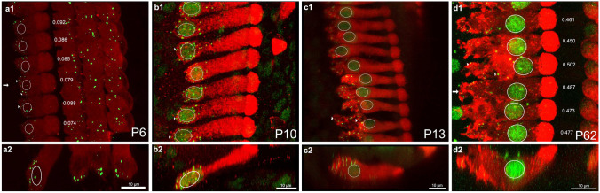
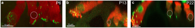
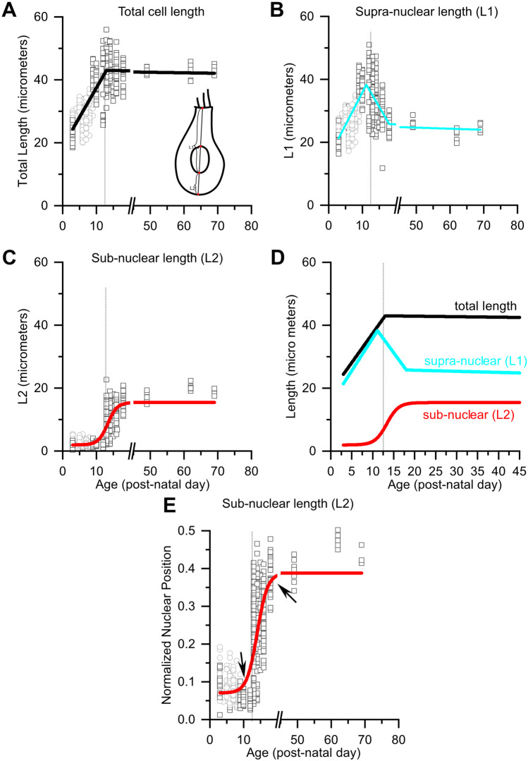
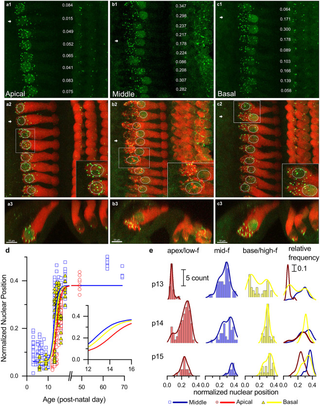
Purpose: Nuclear position is precisely orchestrated during cell division, migration, and maturation of cells and tissues. Here we report a previously unrecognized, programmed movement of the nucleus in rat and mouse cochlear inner hair cells (IHCs) coinciding with the functional maturation of inner hair cells around the onset of hearing.

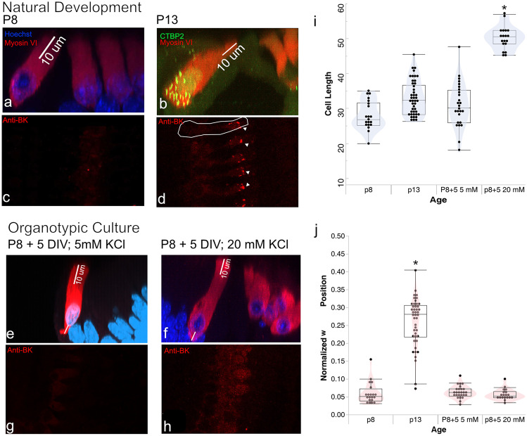
Methods: We measured hair cell length and nuclear position from confocal scans of immunofluorescence-labeled hair cells from whole-mount cochlear preparations throughout post-natal development.

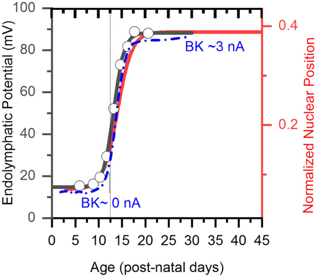
Results: In early post-natal days, the IHC experiences a period of sustained growth, during which the nucleus sits at the very basal pole of the cell, far from the apically located mechano-transducing stereocilia, but close to where synapses with primary afferent and efferent neurons are forming. After IHCs reach their final length, the nucleus moves to occupy a new position half-way along the length of the cell. Nuclear translocation begins in the middle turn, completes throughout the cochlea within 2-3 days, and coincides with the emergence of endolymphatic potential, the acquisition of big-conductance potassium channels (BK), and the onset of acoustic hearing. IHCs cultured in-vitro without endolymphatic potential (EP) do not grow, do not express BK, and do not experience nuclear movement. IHCs cultured in high K+ solutions (to simulate EP) grow but do not experience nuclear movement or acquire BK channels.

Conclusion: Nuclear migration at the onset of hearing is a key step in the morphological maturation of IHCs. Whether this plays a role in functional maturation remains to be explored.

Keywords: Cochlea; Hair cells; Nuclear translocation; Onset of hearing.


Conflict of interest statement

The authors declare no competing interests.

Figures

Fig. 1
Fig. 1
A dramatic change in nuclear position and hair cell morphology occurs around the onset of hearing. a1–d1 Confocal images of inner hair cells in the middle cochlear turn at P6, P10, P13, and P62 in the XY plane. Inner hair cells were labeled with Myosin VI (red) and nuclei and synaptic ribbons (arrowheads) were labeled with CTBP2 (green). Normalized nuclear position measurements (defined in the “Methods” section) are shown next to each inner hair cell. a2d2 Same samples as above shown as a cross-section in the XZ plane to view the length of the entire inner hair cell. a1, b1 Before the onset of hearing, nuclei appear to be located at the base of IHCs and synaptic ribbons are clustered around the nucleus. c1 By P13 in the middle turn, variance in nuclear position is apparent. Ribbons have dispersed throughout the basal pole. d1 By P62, the nucleus has reached its final position in the middle of the cell. A full migration seems to have occurred by P62. Outer hair cells, though present and labeled, were cropped out of these scans
Fig. 2
Fig. 2
Outer hair cell nuclei remain basal bound throughout development. Outer hair cells in the middle turn are outlined at P6 (a), P13 (b), and P62 (c). Unlike inner hair cells, nuclei in OHCs are positioned at the basal pole of the cell in all samples
Fig. 3
Fig. 3
Quantification of nuclear position reveals a period of progressive cell growth followed by an abrupt nuclear translocation event at the onset of hearing. a Total IHC length as a function of post-natal age ranging from P3 to P69 pooled across all turns. Open squares are length measurements from individual hair cells from samples in which cochleae were fixed immediately after euthanasia for immunohistochemistry. Open circles indicate samples in which sample cochleae were fixed after electrophysiological recordings [12]. Cell length grows progressively until it reaches a maximum around P13. b Supra-nuclear length (L1) increases progressively as a function of age until the onset of hearing when it suddenly begins to decrease, eventually reaching a stable length. c Sub-nuclear length is relatively small until about P13 when it begins to increase, eventually reaching a stable length. d The trendlines describing total (black), supra-nuclear (cyan), and sub-nuclear lengths (red) are drawn on the same axis to compare the timelines for age dependent changes in lengths. The coincidence between the decrease in supra-nuclear length with the increase in sub-nuclear length indicates that the nucleus is moving. e Relative Normalized nuclear position, which characterizes nuclear position while accounting for length changes (L2 / (L1 + L2)), increases suddenly around P13 (first arrow) and stabilizes by P18 (second arrow). The inset schematic in a shows how different lengths were measured. The dashed line in all plots is drawn at P13. Trend lines were drawn either by piecewise linear fitting (a, b) or by a Boltzmann fit (ce)
Fig. 4
Fig. 4
Nuclear translocation happens first in the middle and basal turns and then in the apical turn. a1c1 CTBP2 labeled nuclei and ribbons in apical, middle, and basal turns from the same P13 sample. a2c2 Merged images of inner hair cells labeled with Myosin VI (red) and CTBP2 (green) in the XY plane. In the apical turn, ribbons are clustered around the nucleus, which is bound to the base of the hair cell. Nuclear position is variable in the middle turn, and ribbons are dispersed across the basal pole in both the middle and basal turns. a3c3 Images of all three turns at the XZ plane. The images are shown through the hair cell indicated by the white arrows in A2-C2.in the XZ plane. Here, outer hair cells are visible and OHC nuclei were located at the base of cells in all three turns. At P13, most nuclei in the apical turn inner hair cells are found at the base of the cell. In contrast, nuclear position is more variable in the middle and basal turns. Note the instances of adjacent cells with similar lengths but different nuclear positions (insets). Variance in nuclear position across adjacent cells suggests that this sample has hair cells caught in the middle of transitioning between two distinct developmental states. d Normalized nuclear position as a function of age for all three cochlear turns from P3-P69. Nuclear migration in the middle turn (blue curve) begins earliest, followed by migration in the basal and apical turns (yellow and red, respectively). Nuclear migration in the apical turn lags 1–2 days behind the middle turn. e Histograms showing relative normalized nuclear position in apical (red), middle (blue), and basal (yellow) turns at P13, P14, and P15. Smoothed distributions are overlaid on top of each histogram and compared on the same axis. Note that the distributions are largely bimodal with one mode at approximately 0.07 and another at 0.4. The heights of the two modes indicate the relative developmental state of the groups of hair cells, with apical hair cells lagging behind the basal turn and middle turns
Fig. 5
Fig. 5
Nuclear migration occurs earlier in mouse than rat. a Nuclei in the apical turn in wildtype CD1 mice at P10 are located at the base of the inner hair cell. b, c P12 inner hair cells from the apical and middle turns of the same cochlea. Nuclei have begun to move in the apical turn, and have reached their final position by the middle turn
Fig. 6
Fig. 6
Inner hair cells acquire BK channels in coincidence with EP, nuclear migration, and the onset of hearing. f The emergence of EP (Black curve and y-axis on left [18]) in rat IHCs coincides with the onset of nuclear translocation (red curve, y-axis to the right); both events are abrupt and occur just at the onset of hearing. The age axis for EP data was adjusted from the originally reported gestational age to post-natal day. The emergence of BK channel currents (blue curve, reproduced from [17]) follows the same time course as nuclear migration and the onset of EP reported by Bosher and Warren. Note that the BK current data are from mouse, whereas EP and nuclear position data are from rat
Fig. 7
Fig. 7
Tonic depolarization in organotypic culture stimulates hair cell growth but not nuclear migration a Inner hair cells from a P8 middle turn in rat appear immature in nuclear position and lengths. b Inner hair cells from a P13 middle turn have reached their final cell length. c, d P8 inner hair cells have not acquired BK channels, as noted by the absence of BK labeling at this age. At P13, red puncta (see triangles) signal the presence of BK channels at the apical end of the inner hair cell. The white outlines the hair cell. e Inner hair cells cultured for 5 days in culture media with 5 mM KCl. f Inner hair cells cultured for 5 days in culture media supplemented with 20 mM KCl. Cells grown in KCl were longer than those in 5 mM KCl, although BK channels were not visible in either condition (g, h). i Cell length was measured in samples from each condition; inner hair cells grown in 20 mM KCl were significantly longer than cells in all other conditions (p < 0.0001). j Inner hair cell nuclei extracted from rats at P13 have dislodged from the basal pole. In all other conditions, nuclei appear at the base of the inner hair cell

Similar articles

References

    1. del Bene F. Interkinetic nuclear migration: cell cycle on the move. EMBO J. 2011;30(9):1676–1677. doi: 10.1038/emboj.2011.114. - DOI - PMC - PubMed
    1. Gundersen GG, Worman HJ. Nuclear positioning. Cell. 2013;152(6):1376–1389. doi: 10.1016/j.cell.2013.02.031. - DOI - PMC - PubMed
    1. Schenk J, Wilsch-Bräuninger M, Calegari F, Huttner WB. Myosin II is required for interkinetic nuclear migration of neural progenitors. Proc Natl Acad Sci U S A. 2009;106(38):16487–16492. doi: 10.1073/pnas.0908928106. - DOI - PMC - PubMed
    1. Spear PC, Erickson CA. Interkinetic Nuclear migration: a mysterious process in search of a function. Dev Growth Differ. 2012;54(3):306–316. doi: 10.1111/j.1440-169X.2012.01342.x.Interkinetic. - DOI - PMC - PubMed
    1. Razafsky D, Hodzic D. Nuclear envelope: positioning nuclei and orgnaizing synapses. Curr Opin Cell Biol. 2015;34:84–93. doi: 10.1017/CBO9781107415324.004. - DOI - PMC - PubMed

Publication types

Substances

LinkOut - more resources