. 2023 Mar 17;21(1):204.
doi: 10.1186/s12967-023-04048-z.

An integrated approach of network pharmacology, molecular docking, and experimental verification uncovers kaempferol as the effective modulator of HSD17B1 for treatment of endometrial cancer

Affiliations

An integrated approach of network pharmacology, molecular docking, and experimental verification uncovers kaempferol as the effective modulator of HSD17B1 for treatment of endometrial cancer

Guan-Yu Ruan et al. J Transl Med. .

Erratum in

Abstract

Background: Endometrial cancer (EC) is one of the most common gynecological malignancies globally, and the development of innovative, effective drugs against EC remains a key issue. Phytoestrogen kaempferol exhibits anti-cancer effects, but the action mechanisms are still unclear.

Method: MTT assays, colony-forming assays, flow cytometry, scratch healing, and transwell assays were used to evaluate the proliferation, apoptosis, cell cycle, migration, and invasion of both ER-subtype EC cells. Xenograft experiments were used to assess the effects of kaempferol inhibition on tumor growth. Next-generation RNA sequencing was used to compare the gene expression levels in vehicle-treated versus kaempferol-treated Ishikawa and HEC-1-A cells. A network pharmacology and molecular docking technique were applied to identify the anti-cancer mechanism of kaempferol, including the building of target-pathway network. GO analysis and KEGG pathway enrichment analysis were used to identify cancer-related targets. Finally, the study validated the mRNA and protein expression using real-time quantitative PCR, western blotting, and immunohistochemical analysis.

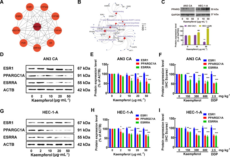
Results: Kaempferol was found to suppress the proliferation, promote apoptosis, and limit the tumor-forming, scratch healing, invasion, and migration capacities of EC cells. Kaempferol inhibited tumor growth and promotes apoptosis in a human endometrial cancer xenograft mouse model. No significant toxicity of kaempferol was found in human monocytes and normal cell lines at non-cytotoxic concentrations. No adverse effects or significant changes in body weight or organ coefficients were observed in 3-7 weeks' kaempferol-treated animals. The RNA sequencing, network pharmacology, and molecular docking approaches identified the overall survival-related differentially expressed gene HSD17B1. Interestingly, kaempferol upregulated HSD17B1 expression and sensitivity in ER-negative EC cells. Kaempferol differentially regulated PPARG expression in EC cells of different ER subtypes, independent of its effect on ESR1. HSD17B1 and HSD17B1-associated genes, such as ESR1, ESRRA, PPARG, AKT1, and AKR1C1\2\3, were involved in several estrogen metabolism pathways, such as steroid binding, 17-beta-hydroxysteroid dehydrogenase (NADP+) activity, steroid hormone biosynthesis, and regulation of hormone levels. The molecular basis of the effects of kaempferol treatment was evaluated.

Conclusions: Kaempferol is a novel therapeutic candidate for EC via HSD17B1-related estrogen metabolism pathways. These results provide new insights into the efficiency of the medical translation of phytoestrogens.

Keywords: Estrogen receptor α; HSD17B1; Human endometrial cancer; Kaempferol; Nude mice.


Conflict of interest statement

The authors declare that they have no competing interests.

Figures

Fig. 1
Fig. 1
Kaempferol suppressed the proliferation, promoted apoptosis, and limited the tumor-forming, invasion, and migration capacities of both ER-subtype human endometrial cancer cells. A Kaempferol inhibited human endometrial cancer cells' proliferation (ER-positive AN3 CA and Ishikawa, ER-negative HEC-1-A and HEC-1-B EC cells) in a dose- and time-dependent manner. B The IC50 values of kaempferol at 48 h were 6.38, 24.35, 35.62, 38.31, 69.87, 104.90, 266.10, and 383.60 μg·mL−1 for AN3 CA, Ishikawa, HEC-1-A, HEC-1-B, CCD 841 CoN, WRL 68, MRC-5 and human monocytes, respectively. CD Kaempferol significantly inhibited AN3 CA cell colony formation in a dose-dependent manner. E The early apoptotic cells and late-stage apoptotic cells of EC cells (AN3 CA, Ishikawa, HEC-1-A, and HEC-1-B) were both increased after treatment with kaempferol in a dose-dependent manner. F Kaempferol induced cell apoptosis by elevating the expression of cleaved CASP3 and cleaved CASP9 in a dose-dependent manner. G Kaempferol inhibited EC cells’ wound healing in a time- and dose-dependent manner. In a concentration-dependent manner, kaempferol inhibited migration HI and invasion JK. The results are presented as means and standard deviations (SDs) from triplicate experiments. Kae: Kaempferol; Compared with the negative control, *, #P < 0.05, **, ##P < 0.01, ***, ###P < 0.001
Fig. 2
Fig. 2
Kaempferol inhibited tumor growth and promoted apoptosis in a human endometrial cancer xenograft mouse model. A Kaempferol suppressed the growth of human endometrial cancer cells (AN3 CA and HEC-1-A) in BALB/c nude mice with xenograft tumors in vivo. B Macroscopic appearance of tumors after treatment (n = 10 per group). C After treatment, tumors were excised, weighed, and sectioned at a thickness of 8 μm for hematoxylin and eosin staining. D Representative merged images of TUNEL immunofluorescent staining in paraffin sections of tumor tissue of AN3 CA and HEC-1-A xenograft mice treated with vehicle (poloxamer 188; negative control), kaempferol, or DDP. More TUNEL-positive EC cells (AN3 CA and HEC-1-A cells) in tumors from kaempferol-treated mice than in those from negative control mice. Blue represented EC cells, and green represented apoptotic cells. Scale bar = 50 μm. E The histograms showed that the percentage of apoptosis cells with the treatment of kaempferol. FG With the treatments of kaempferol, cleaved CASP3 and CASP9 increased gradually. H No adverse effects or significant changes in organ coefficients was observed in kaempferol-treated and vehicle-treated animals. However, the organ coefficients were significantly changed in several organs in DDP-treated animals compared to those of vehicle-treated animals. Results are presented as means and SDs. Compared with the negative control, *, #P < 0.05, **, ##P < 0.01, ***,###P < 0.001
Fig. 3
Fig. 3
The RNA sequencing and network pharmacology approach identified differentially expressed genes related to overall survival (OS). A The strategy of next-generation RNA sequencing in kaempferol-treated and negative control EC cells. B Differentially expressed genes (DEGs) between kaempferol-treated and negative control EC cells (Ishikawa and HEC-1-A). Yellow: upregulated differentially expressed genes; Blue: downregulated differentially expressed genes. C A total of 129 overlap genes for DEGs between the kaempferol-treated and negative control EC cells of Ishikawa (blue background) and HEC-1-A (red background). D Differential types of DEGs were identified between the kaempferol-treated and negative control in Ishikawa (ER-positive) and HEC-1-A (ER-negative) cells. E Flowchart for screening endometrial cancer-related genes, kaempferol-related genes, and the related pathway. F A total of 3 genes HSD17B1, CYP1B1, and CYP1A1 were identified in three gene sets, including 169 kaempferol-related genes in silico (red background), 129 kaempferol-related genes in vitro (yellow background), and 1181 endometrial cancer-related genes (purple background). G 67 potential protein-protein interactions were identified. According to the MCODE score, color brightness indicated the strength of the association, with brighter colors indicating a stronger association
Fig. 4
Fig. 4
Kaempferol up-regulated HSD17B1 expression and sensitivity in ER-negative EC cells. A For the AN3 CA cells, the mRNA expression of HSD17B1 was significantly decreased with 10 μg·mL−1 treatment of kaempferol; for the HEC-1-A cells, the mRNA expression of HSD17B1 was significantly increased with 50 μg·mL−1 treatment of kaempferol; the mRNA expression of HSD17B1 was barely expressed in KLE cells. B The protein expression level of HSD17B1 in ER-positive AN3 CA was unchanged, but the levels of HSD17B1 protein were significantly increased in ER-negative HEC-1-A with kaempferol treatment. And HSD17B1 was barely expressed in KLE cells. CD Similar results were also found in the IHC of HSD17B1 in nude mouse tumor tissue after 3–7 weeks of treatment. E High expression levels of HSD17B1 had significantly shortened OS in EC patients. F Kaempferol may bind to the HSD17B1 hydrophobic LBD through several conserved hydrogen bond interactions with the amino acid residues Y155 and S142. G The AN3 CA cells were sensitive to kaempferol with a high level of HSD17B1. H Kaempferol can upregulate the expression of HSD17B1 in HEC-1-A. I Kaempferol-resistant KLE cells with low HSD17B1 expression
Fig. 5
Fig. 5
Kaempferol modulated estrogen metabolism pathways and differentially regulates PPARG expression in EC cells of different ER subtypes. AB HSD17B1 and HSD17B1-associated genes, such as ESRRA, PPARG, and ESR1, are involved in several estrogen metabolism pathways, such as steroid binding, 17- beta-hydroxysteroid dehydrogenase (NADP+) activity, steroid hormone biosynthesis, and regulation of hormone levels. C Kaempferol suppressed the expression of PPARG in ER-positive AN3 CA and promoted the expression of PPARG in ER-negative HEC-1-A. DI Kaempferol suppressed the expression of PPARGC1A and ESRRA in both AN3 CA (DF) and HEC-1-A cells (GI), without modulating ESR1. Western blotting (DE and GH) and the IHC scores (F and I) confirmed the differential expression of PPARGC1A and ESRRA. Results are presented as means and SDs. Compared with the negative control, *, #P < 0.05, **, ##P < 0.01, ***, ###P < 0.001
Fig. 6
Fig. 6
Schematic diagram of the mechanism by which kaempferol induces apoptosis and inhibits growth and metastasis via HSD17B1 in EC cells

References

    1. Sung H, Ferlay J, Siegel RL, Laversanne M, Soerjomataram I, Jemal A, Bray F. Global cancer statistics 2020: GLOBOCAN estimates of incidence and mortality worldwide for 36 cancers in 185 Countries. CA Cancer J Clin. 2021;71:209–49. - PubMed
    1. Siegel RL, Miller KD, Jemal A. Cancer statistics, 2020. CA Cancer J Clin. 2020;70:7–30. - PubMed
    1. Brooks RA, Fleming GF, Lastra RR, Lee NK, Moroney JW, Son CH, Tatebe K, Veneris JL. Current recommendations and recent progress in endometrial cancer. CA Cancer J Clin. 2019;69:258–79. - PubMed
    1. Dowdy SC, Glaser GE. Adjuvant therapy for women with high-risk endometrial carcinoma. Lancet Oncol. 2018;19:268–9. - DOI - PubMed
    1. Crosbie EJ, Kitson SJ, McAlpine JN, Mukhopadhyay A, Powell ME, Singh N. Endometrial cancer. Lancet. 2022;399:1412–28. - DOI - PubMed

Publication types