. 2023 Mar 17;24(1):201.
doi: 10.1186/s12891-023-06306-z.

Influence of joint volume on range of motion after arthroscopic rotator cuff repair

Affiliations

Influence of joint volume on range of motion after arthroscopic rotator cuff repair

Jung-Han Kim et al. BMC Musculoskelet Disord. .

Abstract

Background: Capsular contracture is a well-known etiology in the primary stiff shoulder; thus capsular contracture and resultant decreased joint volume could lead to postoperative stiffness, which is a commonly reported morbidity after arthroscopic rotator cuff repair (ARCR). The purpose of this study was (1) to quantify the joint volume (total joint volume and each quadrant compartmental volume) using computed tomography arthrography (CTA) and (2) to demonstrate the relationship between joint volume and postoperative range of motion (ROM) after ARCR.

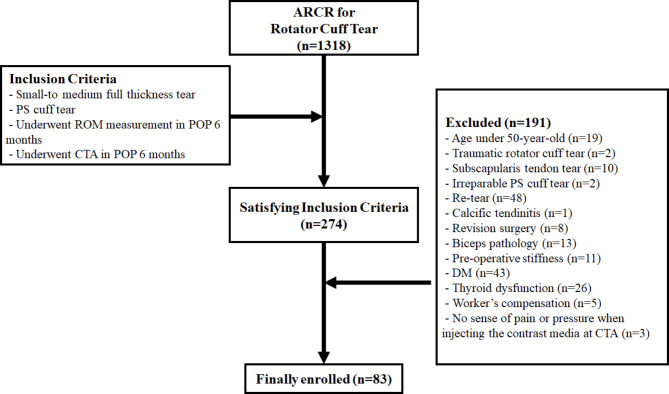
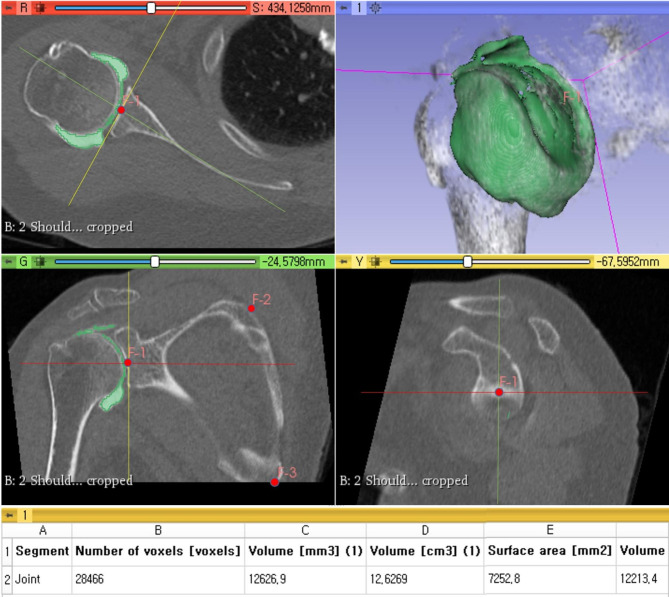
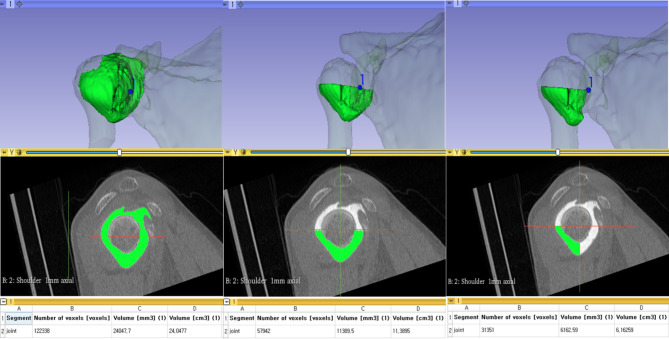
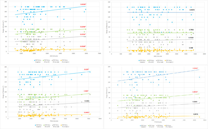
Materials and methods: Eighty-three patients (60 ± 5.11 years, men = 26, women = 57) who had undergone ARCR between January 2015 to December 2020 due to small to medium full-thickness tear and followed by CTA 6 months postoperatively were retrospectively reviewed. An image reconstruction program (3D Slicer, version 4.11.2 software) was used to calculate the joint volume (total joint volume and quadrant compartment joint volumes; anteroinferior, anterosuperior, posterosuperior and posteroinferior). For shoulder ROM, data including scaption (Sc), external rotation on side (ERs), external rotation at 90° (ER90), and internal rotation on back (IRb) were collected 6 months postoperatively. An evaluation of the correlation between joint volume and each shoulder motion was performed.

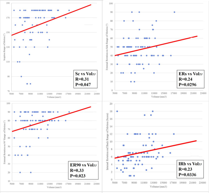
Results: There were moderate correlations between the total joint volume and each motion (Sc: Pearson coefficient, 0.32, p = 0.0047; ERs: Pearson coefficient, 0.24, p = 0.0296; ER90: Pearson coefficient, 0.33, p = 0.0023; IRb: Pearson coefficient, 0.23, p = 0.0336). Among the quadrant compartments, the anteroinferior (Sc: Pearson coefficient, 0.26, p = 0.0199; ERs: Pearson coefficient, 0.23, p = 0.0336; ER90: Pearson coefficient, 0.25, p = 0.0246; IRb: Pearson coefficient, 0.26, p = 0.0168) and posterosuperior (Sc: Pearson coefficient, 0.24, p = 0.029; ER90: Pearson coefficient, 0.29, p = 0.008; IRb: Pearson coefficient, 0.22, p = 0.0491) and posteroinferior (Sc: Pearson coefficient, 0.30, p = 0.0064; ER90: Pearson coefficient, 0.29, p = 0.0072) showed moderate correlations with each shoulder motion.

Conclusion: Total joint volume, anteroinferior compartment joint volume, posterosuperior compartment joint volume and posteroinferior compartment joint volume were related to postoperative ROM after ARCR. Perioperative methods to increase the joint volume, especially the anteroinferior, posterosuperior and posteroinferior parts of the capsule may prevent postoperative stiffness after ARCR.

Level of evidence: Level III; Retrospective Case-Control Study.

Keywords: Arthrography; Arthroscopy; Multidetector Computed Tomography; Rotator Cuff Injuries; Shoulder; Shoulder Joint.


Conflict of interest statement

All authors, their immediate families, and any research foundation with which they are affiliated have not received any financial payments or other benefits from any commercial entity related to the subject of this article.

Figures

Fig. 1
Fig. 1
Patient flow chart showing the inclusion and exclusion criteria used in this study
Fig. 2
Fig. 2
Registration of three scapular landmarks (angular inferior, trigonum scapulae, and glenoid center) as fiducials before reconstruction. Scapular plane (green plane in the lower left image, green line in the upper left and lower right images): plane that passes through the three scapular landmarks. Axial plane (red plane in the upper left image, red line in the lower right and lower left images): plane that is perpendicular to the scapular plane. Sagittal plane (yellow plane in the lower right image, yellow line in the upper left and lower left images): plane that is orthogonal to the scapular and axial planes
Fig. 3
Fig. 3
Separation of joint portion of image from others in semi-automated manner. If additional modification was needed, further manual erasing and segmentation were performed (upper right, lower left, and lower right images). When separation was performed appropriately, a 3D image of the segmented joint was generated (upper right image). The total joint volume was then calculated via the “Segment statistics” module
Fig. 4
Fig. 4
Further separation of the total joint volume into each quadrant compartment joint volume performed in the scapular sagittal plane. The figure shows that the joint portion was separated into inferior compartment and then posteroinferior joint portions. Thereafter, calculation of the separated joint volume was performed via the “Segment statistics” module
Fig. 5
Fig. 5
Scatter matrix of the relationship between the total joint volume (Vol.TJ) and each shoulder motion (Sc: scaption, ERs: external rotation on side, ER90: external rotation at 90°, IRb: internal rotation on back). The Vol.TJ showed a moderately positive correlation with each of the four shoulder motions
Fig. 6
Fig. 6
Scatter matrix of the relationship between each quadrant compartment joint volumes (Vol. anteroinferior, Vol.anterosuperior, Vol.Posterosuperior, Vol.Posteroinferior) and each shoulder motion (Sc: scaption, ERs: external rotation on side, ER90: external rotation at 90°, IRb: internal rotation on back). The Vol. anteroinferior, Vol.Posterosuperior, Vol.Posteroinferior showed a moderately positive correlation with each of the four shoulder motions
Fig. 7
Fig. 7
A schematic diagram showing the shoulder joint volume affected by postoperative tightening in a specific movement. Among the quadrant (AIQ: Anteroinferior quadrant, ASQ: Anterosuperior quadrant, PSQ: Posterosuperior quadrant, PIQ: Posterosuperior quadrant), the affected area in specific joint motion are filled in blue

Similar articles

References

    1. Park JY, Chung SW, Hassan Z, Bang JY, Oh KS. Effect of Capsular Release in the treatment of shoulder stiffness concomitant with Rotator Cuff Repair Diabetes as a predisposing factor Associated with Treatment Outcome. Am J Sports Med. 2014;42:840–50. doi: 10.1177/0363546513519326. - DOI - PubMed
    1. Seo SS, Choi JS, An KC, Kim JH, Kim SB. The factors affecting stiffness occurring with rotator cuff tear. J Shoulder Elbow Surg. 2012;21:304–9. doi: 10.1016/j.jse.2011.04.011. - DOI - PubMed
    1. DeFranco MJ, Bershadsky B, Ciccone J, Yum JK, Iannotti JP. Functional outcome of arthroscopic rotator cuff repairs: a correlation of anatomic and clinical results. J Shoulder Elbow Surg. 2007;16:759–65. doi: 10.1016/j.jse.2007.03.020. - DOI - PubMed
    1. Galatz LM, Ball CM, Teefey SA, Middleton WD, Yamaguchi K. The outcome and repair integrity of completely arthroscopically repaired large and massive rotator cuff tears. J Bone Joint Surg Am. 2004;86:219–24. doi: 10.2106/00004623-200402000-00002. - DOI - PubMed
    1. Schneider WR, Trasolini RG, Riker JJ, Gerber N, Ruotolo CJ. Stiffness after arthroscopic rotator cuff repair: a rehabilitation problem or a surgical indication? JSES Int. 2021;5:88–92. doi: 10.1016/j.jseint.2020.09.001. - DOI - PMC - PubMed