. 2023 Mar 17;16(1):23.
doi: 10.1186/s13045-023-01413-9.

Tumor cell integrin β4 and tumor stroma E-/P-selectin cooperatively regulate tumor growth in vivo

Affiliations

Tumor cell integrin β4 and tumor stroma E-/P-selectin cooperatively regulate tumor growth in vivo

Sandra Genduso et al. J Hematol Oncol. .

Abstract

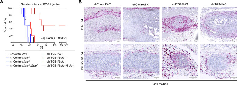
Background: The immunological composition of the tumor microenvironment has a decisive influence on the biological course of cancer and is therefore of profound clinical relevance. In this study, we analyzed the cooperative effects of integrin β4 (ITGB4) on tumor cells and E-/P-selectin on endothelial cells within the tumor stroma for regulating tumor growth by shaping the local and systemic immune environment.

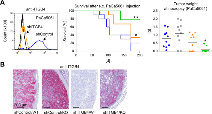
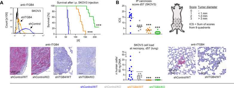
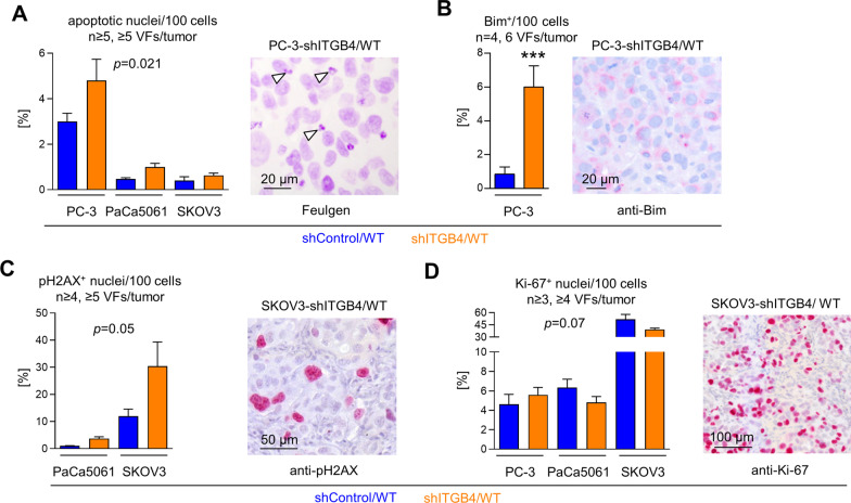
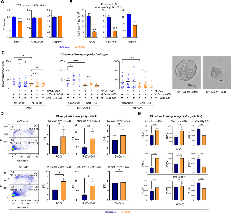
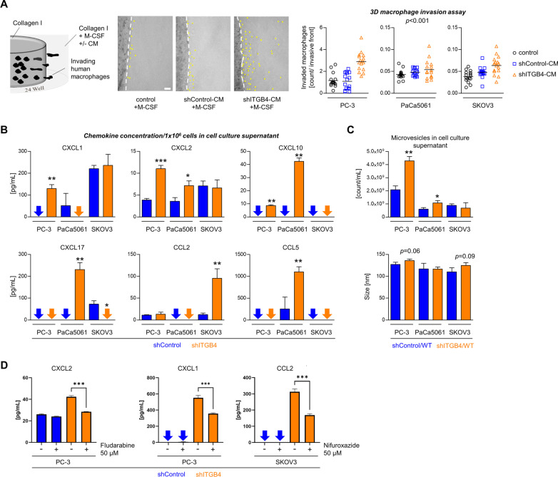
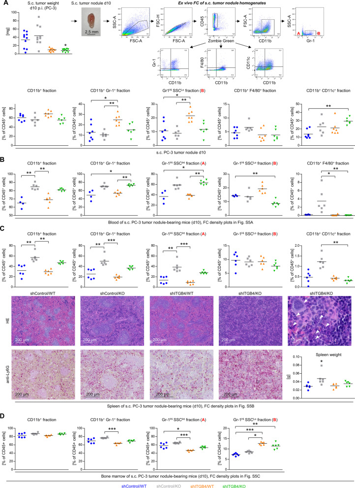
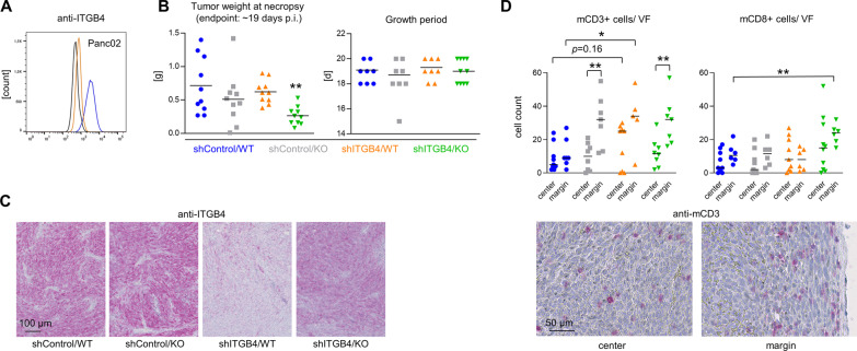
Methods: We used several preclinical mouse models for different solid human cancer types (xenograft and syngeneic) to explore the role of ITGB4 (shRNA-mediated knockdown in tumor cells) and E-/P-selectins (knockout in mice) for tumor growth; effects on apoptosis, proliferation and intratumoral signaling pathways were determined by histological and biochemical methods and 3D in vitro experiments; changes in the intratumoral and systemic immune cell composition were determined by flow cytometry and immunohistochemistry; chemokine levels and their attracting potential were measured by ELISA and 3D invasion assays.

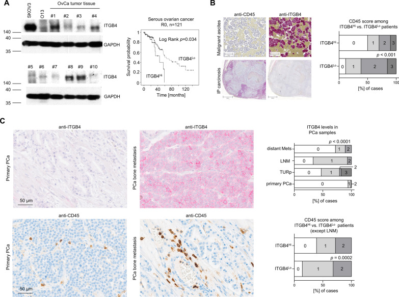
Results: We observed a very robust synergism between ITGB4 and E-/P-selectin for the regulation of tumor growth, accompanied by an increased recruitment of CD11b+ Gr-1Hi cells with low granularity (i.e., myeloid-derived suppressor cells, MDSCs) specifically into ITGB4-depleted tumors. ITGB4-depleted tumors undergo apoptosis and actively attract MDSCs, well-known to promote tumor growth in several cancers, via increased secretion of different chemokines. MDSC trafficking into tumors crucially depends on E-/P-selectin expression. Analyses of clinical samples confirmed an inverse relationship between ITGB4 expression in tumors and number of tumor-infiltrating leukocytes.

Conclusions: These findings suggest a distinct vulnerability of ITGB4Lo tumors for MDSC-directed immunotherapies.

Keywords: Anoikis; Chemoattraction; E-selectin; Integrin β4; Myeloid-derived suppressor cell; P-selectin; Tumor-infiltrating leukocyte.


Conflict of interest statement

The authors declare that they have no competing interests.

Figures

Fig. 1
Fig. 1
Combined depletion of ITGB4 and E-/P-selectin cooperatively reduces human prostate cancer xenograft tumor formation. Mean fold up- or down-regulation of integrin subunit gene expression comparing highly metastatic PC-3 vs. weakly metastatic DU-145 human prostate cancer cells (n = 3) and validation of differential ITGB4 protein levels in vitro and in situ. Arrowheads indicate ITGB4+ tumor cells at the xenograft tumor margin (A). shRNA-mediated knockdown of ITGB4 in PC-3 cells (B) and s. c. xeno-transplantation of shControl vs. shITGB4 cells into E-/P-selectin wildtype vs. knockout Pfp−/−/Rag2−/− mice resulting in four experimental groups: shControl/WT (blue), shITGB4/WT (orange), shControl/KO (grey), shITGB4/KO (green) as illustrated (C). Survival of mice after s. c. injection of tumor cells (endpoint: s. c. xenograft tumor of ~ 1.5 cm3, D). Tumor weights (E) and percentage of ITGB4+ primary tumor cells (F) at necropsy. Human cell loads in blood and lung samples at necropsy (s. c. xenograft experiment) (G). Black lines in scatter plots represent mean values. Red dotted lines in (G) indicate the detection limit of the respective Alu-PCR experiment. All statistical comparisons indicated by asterisks were calculated vs. shControl/WT. *p < 0.05; **p < 0.01; ***p < 0.001
Fig. 2
Fig. 2
Combined depletion of ITGB4 and E-/P-selectin cooperatively reduces human pancreatic cancer xenograft tumor formation. shRNA-mediated knockdown of ITGB4 in PaCa5061 cells, survival of mice after s. c. xeno-transplantation of shControl vs. shITGB4 cells into E-/P-selectin wildtype vs. knockout Pfp−/−/Rag2−/− mice (endpoint: tumor size ~ 1.5 cm3) and resulting tumor weights at necropsy (A). Representative anti-ITGB4 immunostaining images taken from s. c. PaCa5061 xenograft tumors (B). All statistical comparisons indicated by asterisks were calculated vs. shControl/ WT. *p < 0.05; **p < 0.01
Fig. 3
Fig. 3
Combined depletion of ITGB4 and E-/P-selectin cooperatively reduces human ovarian cancer xenograft tumor formation. shRNA-mediated knockdown of ITGB4 in SKOV3 cells, survival of mice after i. p. injection of shControl vs. shITGB4 cells into E-/P-selectin wildtype vs. knockout SCID mice (endpoint: palpable abdominal tumor masses or ascites) and representative anti-ITGB4 immunostaining images from i. p. SKOV3 xenograft tumors (A). Intraperitoneal carcinosis score (ICS) determined as illustrated and spontaneous pulmonary metastatic cell load determined by Alu-PCR on day 57 after i. p. injection of SKOV3 shControl vs. shITGB4 cells into E-/P-selectin wildtype vs. knockout SCID mice (B). Representative anti-ITGB4 immunostaining images of spontaneous SKOV3 lung metastases (arrowhead indicates an ITGB4 lung metastasis) (B). All statistical comparisons indicated by asterisks were calculated vs. shControl/ WT. ***p < 0.001
Fig. 4
Fig. 4
Depletion of ITGB4 causes increased apoptosis, but unaltered proliferation in vivo. Percentage of apoptotic nuclei (A), Bim+ tumor cells (B), pH2AX+ nuclei (C) and Ki-67+ nuclei (D) per viewing field (VF) in xenograft tumor tissues and corresponding representative staining microphotographs (arrowheads in (A) indicate apoptotic bodies). Bar charts represent mean + SD of given sample sizes. ***p < 0.001
Fig. 5
Fig. 5
Depletion of ITGB4 reduces proliferation at low confluence, impairs three-dimensional colony forming capacity and enhances apoptosis in vitro. Extinction values in colorimetric proliferation XTT assays at high cell density in conventional cell culture (A). Cell counts per mL on day six after low-density seeding in conventional cell culture (B). Diameters of 3D tumor spheroids after eleven days of growth in soft agar in the presence of standard cell culture media or conditioned media (CM) taken from shControl or shITGB4 cell cultures (C). Percentage of early (Q4) and late (Q2) apoptotic cells in 3D tumor spheroids developed after three days in poly-HEMA-treated cell culture flasks (D). Luminescence and fluorescence measurements indicating the amount of apoptotic, necrotic and viable cells at indicated time points after cultivation in soft agar containing laminin V (LN V, laminin-α3β3γ2) (E). Bar charts represent mean + SD of n = 3. Lines in scatter plots indicate mean values. *p < 0.05; **F < 0.01; ***p < 0.001
Fig. 6
Fig. 6
ITGB4 depletion requires E- and P-selectin double knockout for synergistic tumor growth reduction and is accompanied by increased leukocyte infiltration. Mouse survival after s. c. injection of PC-3 control vs. ITGB4 knockdown cells into E-/P-selectin wildtype, single or double knockout mice (endpoint: xenograft tumor size ~ 1.5 cm3) (A). Representative anti-mCD45 immunostaining images of initial tumor nodules on day 4 after s. c. injection of PC-3 or PaCa5061 control or ITGB4 KD cells into E-/P-selectin wildtype or double knockout mice (B)
Fig. 7
Fig. 7
ITGB4 depletion enhances leukocyte attraction via chemokine release. Illustration of the experimental setup and quantification of invaded human macrophages per invasive front in a 3D collagen I matrix mixed with standard cell culture media or conditioned media (CM) taken from shControl or shITGB4 cell cultures, each supplemented with M-CSF (A). Concentrations of indicated chemokines in the supernatant of 1 × 106 control or ITGB4 KD cells of the indicated cell lines (B). Number of microvesicles per mL and microvesicle size in supernatants of control or ITGB4 KD TC cultures (C). Concentrations of indicated chemokines in the supernatant of control or ITGB4 KD cells of the indicated cell lines with or without treatment with STAT inhibitors as indicated (D). Black lines in scatter plots indicate mean values. Bar charts represent mean + SD of n ≥ 3. *p < 0.05; **p < 0.01; ***p < 0.001
Fig. 8
Fig. 8
ITGB4 knockdown and E-/P-selectin knockout cooperatively determine the local and systemic immune environment in the early phase of tumor establishment. Tumor weights on d10 after s. c. injection of PC-3 control vs. ITGB4 knockdown cells into E-/P-selectin wildtype vs. knockout mice and representative density plots of the ex vivo flow cytometric analysis of s. c. tumor nodule homogenates (A). Fractions of leukocyte subpopulations characterized by indicated surface molecules and granularity (SSC) in the s. c. tumor nodules (A) and corresponding blood (B), spleen (C), and bone marrow (D) samples. For representative flow cytometry density plots of blood, spleen and bone marrow samples, please see Additional file 6: Fig. S6. Representative photomicrographs of HE- and anti-Ly6G (Gr-1 epitope)-stained spleen samples and corresponding spleen weights of s. c. tumor nodule-bearing mice (d10) (C, the arrow indicates a megakaryocyte, arrowheads indicate immature granulocytes with ring-shaped nuclei, see Additional file 7: Fig. S7 also). Black lines in scatter plots in (A)–(D) represent mean values. *p < 0.05; **p < 0.01; ***p < 0.001
Fig. 9
Fig. 9
Combined depletion of ITGB4 and E-/P-selectin synergistically reduces pancreatic cancer growth in fully immunocompetent mice. mITGB4 knockdown status in Panc02 murine pancreatic cancer cells prior injection (A). Tumor weight at necropsy and growth periods after s. c. injection of Panc02 control vs. ITGB4 knockdown cells into E-/P-selectin wildtype vs. knockout C57BL/6 mice (B). Representative anti-mITGB4 immunostaining images of Panc02 tumor tissues (C). Quantification of murine CD3+ and CD8+ lymphocytes per viewing field (VF) in the center and at the margin of s.c. Panc02 tumors across the different groups and representative anti-mCD3 immunostaining images from the center and margin of a Panc02 tumor. Black lines in scatter plots represent mean values. **p < 0.01
Fig. 10
Fig. 10
ITGB4 has prognostic relevance and inversely correlates with the number of tumor-associated CD45+ leukocytes in patients. Representative ITGB4 and corresponding GAPDH Western blot bands in ovarian cancer (OvCa) tissue samples. Overall survival of ITGB4Lo (75%) vs. ITGB4Hi (25%) R0-resected serous ovarian cancer patients (A). Representative anti-CD45 and anti-ITGB4 immunostaining images from consecutive intraperitoneal (IP) carcinosis and malignant ascites ovarian cancer patient samples and calculation of the association between both parameters (B). Representative anti-ITGB4 and anti-CD45 immunostaining images from primary and metastatic prostate cancer specimens, ITGB4 levels in different types of prostate cancer samples and association between ITGB4 and number of tumor-associated leukocytes in these samples except lymph node metastases (LNM) (C). TURp = palliative transurethral resection

References

    1. Seager RJ, Hajal C, Spill F, Kamm RD, Zaman MH. Dynamic interplay between tumour, stroma and immune system can drive or prevent tumour progression. Converg Sci Phys Oncol. 2017;3 - PMC - PubMed
    1. Reymond N, d'Agua BB, Ridley AJ. Crossing the endothelial barrier during metastasis. Nat Rev Cancer. 2013;13:858–870. doi: 10.1038/nrc3628. - DOI - PubMed
    1. Gebauer F, Wicklein D, Stubke K, Nehmann N, Schmidt A, Salamon J, Peldschus K, Nentwich MF, Adam G, Tolstonog G, Bockhorn M, Izbicki JR, Wagener C, Schumacher U. Selectin binding is essential for peritoneal carcinomatosis in a xenograft model of human pancreatic adenocarcinoma in pfp–/rag2– mice. Gut. 2013;62:741–750. doi: 10.1136/gutjnl-2011-300629. - DOI - PubMed
    1. Strell C, Entschladen F. Extravasation of leukocytes in comparison to tumor cells. Cell Commun Signal. 2008;6:10. doi: 10.1186/1478-811X-6-10. - DOI - PMC - PubMed
    1. Schweitzer KM, Drager AM, van der Valk P, Thijsen SF, Zevenbergen A, Theijsmeijer AP, van der Schoot CE, Langenhuijsen MM. Constitutive expression of E-selectin and vascular cell adhesion molecule-1 on endothelial cells of hematopoietic tissues. Am J Pathol. 1996;148:165–175. - PMC - PubMed

Publication types