Review
. 2023 Jun;53(6):e2250266.
doi: 10.1002/eji.202250266. Epub 2023 May 4.

The innate immune response in tauopathies

Affiliations
Review

The innate immune response in tauopathies

Alexis M Johnson et al. Eur J Immunol. 2023 Jun.

Abstract

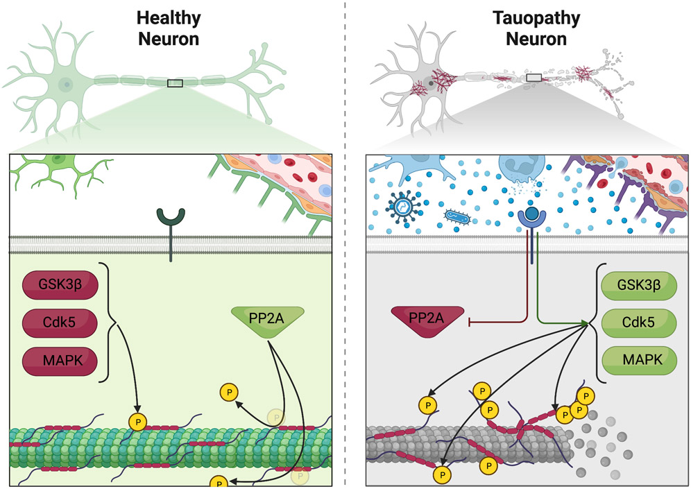
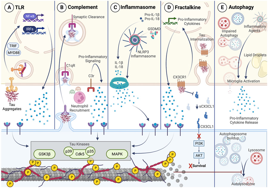
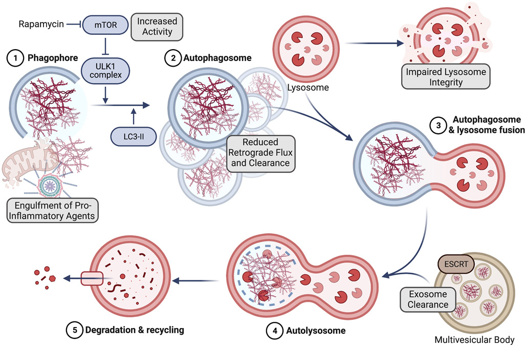
Tauopathies, which include frontotemporal dementia, Alzheimer's disease, and chronic traumatic encephalopathy, are a class of neurological disorders resulting from pathogenic tau aggregates. These aggregates disrupt neuronal health and function leading to the cognitive and physical decline of tauopathy patients. Genome-wide association studies and clinical evidence have brought to light the large role of the immune system in inducing and driving tau-mediated pathology. More specifically, innate immune genes are found to harbor tauopathy risk alleles, and innate immune pathways are upregulated throughout the course of disease. Experimental evidence has expanded on these findings by describing pivotal roles for the innate immune system in the regulation of tau kinases and tau aggregates. In this review, we summarize the literature implicating innate immune pathways as drivers of tauopathy.

Keywords: Alzheimer's disease; Innate immunity; Microglia; Neuroimmunology; Tauopathy.


Conflict of interest statement

DECLARATION OF INTERESTS

The authors declare no commercial or financial conflict of interest.

Figures

Figure 1:
Figure 1:. Inflammation provokes aberrant regulation of tau signaling.
Clinical and murine studies have illustrated that the pro-inflammatory responses, indicated by the blue coloring on the right panel, following immune challenges act upon tau kinases (GSK3β, Cdk5, MAPKs) within neurons, leading to the increased phosphorylation of tau and MT disruption. The phosphatase PP2A normally functions to limit tau hyperphosphorylation but is inhibited by these inflammatory cascades.
Figure 2:
Figure 2:. Innate signaling cascades implicated in tauopathy progression.
Illustrated is the immunological interface of a microglia (tan) and a neuron (blue) during tau-mediated pathology. A. Activation of TLRs by tau, LPS, or PolyI:C stimulates MyD88 and TRIF signals that result in increased NF-κB and interferon regulatory factor (IRF) signaling. Both pro-inflammatory signaling pathways can promote aberrant tau kinase activity via pro-inflammatory cytokine release (blue spheres). The subsequent tau hyperphosphorylation is detrimental to neuronal health. B. C1q opsonization will clear tau-burdened synapses. C3 signaling within microglia provokes proinflammatory signaling that can lead to dysregulated tau kinase activation. C3a and C5a can act as chemoattractants for neutrophils that are commonly found within tauopathy brains. C. Tau internalization by microglia can promote NLRP3 inflammasome activation and lead to the cleavage of Gasdermin-D (GSDMD) and release of IL-1β and IL-18 which can subsequently provoke aberrant regulation of tau kinase cascades. D. Tau competes with soluble CX3CL1 (sCX3CL1) for binding with CX3CR1. Tau, however, has a high affinity for CX3CR1 making it able to be internalized, activate microglia, and induce the transcription of pro-inflammatory cytokines. CX3CL1 decreases with tauopathy disease progression and the lack of CX3CL1 stimulation at the surface of the neuron prevents pro-survival signaling through PI3K/AKT. E. Microglial autophagy normally functions to clear inflammatory agents and maintain lipid homeostasis. However, tauopathies can be accompanied by impaired autophagy which can cause the microglia to fill with lipid droplets and become pro-inflammatory. Autophagy also helps to clear toxic tau species from neurons. Microtubule (MT) disruptions can lead to impaired neuronal autophagy and subsequent pathogenic tau accumulation.
Figure 3:
Figure 3:. Autophagy in tau-mediated neurodegenerative disease.
Highlighted in grey boxes are autophagy pathway events commonly altered in tauopathies. Phagophore formation (1) allows for the clearance of tau aggregates, and potentially other inflammatory agents within the cell. Many GWAS studies have uncovered tauopathy disease risk loci in genes involved in phagophore formation (1) and maturation into autophagosomes (2), such as those found within the ULK1 complex. mTOR activity is increased in the brains of both tauopathy mouse models and tauopathy patients. Treatment with the mTOR inhibitor rapamycin slows neurodegeneration in tauopathy mouse models. Microtubule (MT) disruption in neurons prevents autophagosome retrograde transport and eventual fusion with lysosomes (3), which can lose membrane integrity during pathology. Multivesicular bodies may fuse with maturing autolysosomes (4), clear any intraluminal vesicles, and prevent exosome release. Autophagy results in the successful clearance and degradation of engulfed tau tangles (5).

Similar articles

Cited by

References

    1. Borst K, Dumas AA, Prinz M. Microglia: Immune and non-immune functions. Immunity. 2021; 54:2194–2208.DOI: 10.1016/j.immuni.2021.09.014. - DOI - PubMed
    1. Rustenhoven J, Kipnis J. Brain borders at the central stage of neuroimmunology. Nature. 2022; 612:417–429.DOI: 10.1038/s41586-022-05474-7. - DOI - PMC - PubMed
    1. da Mesquita S, Fu Z, Kipnis J. The Meningeal Lymphatic System: A New Player in Neurophysiology. Neuron. 2018; 100:375–388.DOI: 10.1016/j.neuron.2018.09.022. - DOI - PMC - PubMed
    1. Hammond TR, Marsh SE, Stevens B. Immune Signaling in Neurodegeneration. Immunity. 2019; 50:955–974.DOI: 10.1016/j.immuni.2019.03.016. - DOI - PMC - PubMed
    1. McKee CA, Lukens JR. Emerging roles for the immune system in traumatic brain injury. Front Immunol. 2016; 7.DOI: 10.3389/fimmu.2016.00556. - DOI - PMC - PubMed

Publication types