

doi: 10.1038/s41598-023-31528-5.
Thin film notch filters as platforms for biological image processing
Affiliations
- PMID: 36934126
- PMCID: PMC10024701
- DOI: 10.1038/s41598-023-31528-5
Item in Clipboard
Thin film notch filters as platforms for biological image processing
Sci Rep.
.
Abstract
Many image processing operations involve the modification of the spatial frequency content of images. Here we demonstrate object-plane spatial frequency filtering utilizing the angular sensitivity of a commercial spectral bandstop filter. This approach to all-optical image processing is shown to generate real-time pseudo-3D images of transparent biological and other samples, such as human cervical cancer cells. This work demonstrates the potential of non-local, non-interferometric approaches to image processing for uses in label-free biological cell imaging and dynamical monitoring.
© 2023. The Author(s).
Conflict of interest statement
The authors declare no competing interests.
Figures

The experimental configuration used to capture the transmission spectra of the notch filter. Here, LP and QWP denote linear polarizer and quarter wave-plate, respectively. The schematic is not to scale.

The experimental transmission response of the filter obtained by incrementally rotating the filter (a). The dashed line in (a) indicates the band-stop wavelength of 633 nm and the arrows indicate the width of the band-stop region at normal incidence. The modulus-square of the modulation transfer function along for various polarizations (b) is compared to data provided by the manufacturer. Linear fitting of the modulation transfer function for circular polarization in the contrast zone is given in (c).

The experimental configuration for image processing with a spatial light modulator (SLM), where L and MO denote lenses and microscope objectives, respectively. The schematic is not to scale.
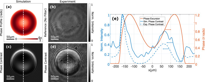
Image processing was performed on a transparent red blood cell (a) emulated by a spatial light modulator with a phase excursion of . An experimental control image obtained in the absence of the notch filter is given in (b), while simulated (c) and experimental (d) phase images produced by the notch filter demonstrate phase visualization. The intensity images (b)–(d) are normalized to their brightest pixels, while the line profiles along the dashed lines shown in (a), (c) and (d) are given in (e).

Biological phase imaging was performed using the experimental schematic in (a). The dashed line represents the HeLa cells within a petri dish and the schematic is not to scale. A bright field image of the HeLa cells obtained without the notch filter is given in (b). The corresponding phase contrast image obtained using the filter is given in (c) and a differential interference contrast image in (d). The dashed circles shown in (b)–(d) highlight regions where contrast was significantly enhanced for comparative purposes.
References
-
- Rienitz J. Schlieren experiment 300 years ago. Nature. 1975;254:293–295. doi: 10.1038/254293a0. - DOI
-
- Zernike F. Phase contrast, a new method for the microscopic observation of transparent objects. Physica. 1942;9:686–698. doi: 10.1016/S0031-8914(42)80035-X. - DOI
-
- Molesini G, Bertani D, Cetica M. Dark ground microscopy with detuned interference filters. Opt. Eng. 1982;21:1061–1063. doi: 10.1117/12.7973033. - DOI
-
- Lang W. Nomarski Differential Interference-Contrast Microscopy. Carl Zeiss; 1982.
-
- Balaur E, et al. Plasmon-induced enhancement of ptychographic phase microscopy via sub-surface nanoaperture arrays. Nat. Photonics. 2021;15:222–229. doi: 10.1038/s41566-020-00752-0. - DOI
Publication types
MeSH terms
LinkOut - more resources
Full Text Sources
