. 2023 Mar 18;6(1):292.
doi: 10.1038/s42003-023-04665-w.

CCR3 plays a role in murine age-related cognitive changes and T-cell infiltration into the brain

Affiliations

CCR3 plays a role in murine age-related cognitive changes and T-cell infiltration into the brain

Sanket V Rege et al. Commun Biol. .

Abstract

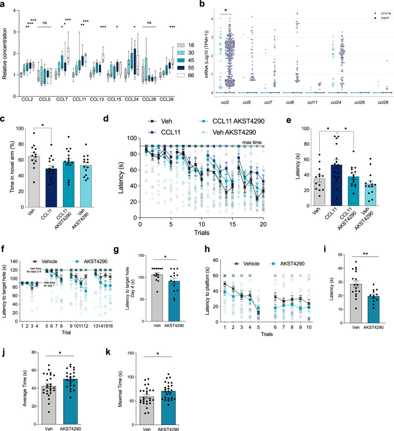
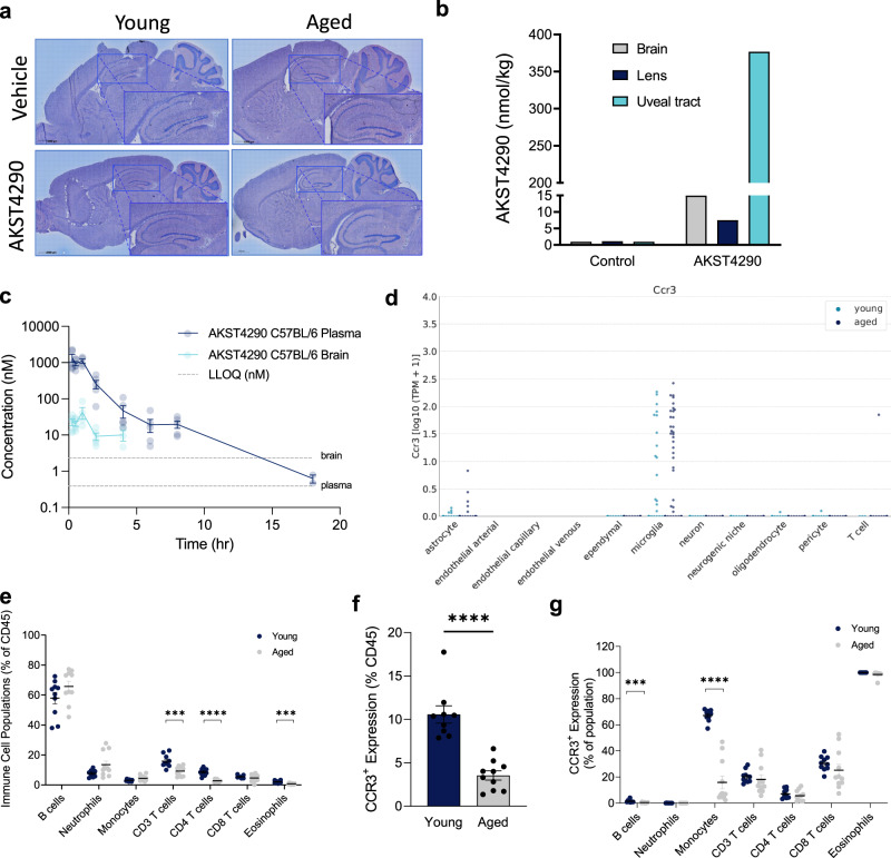
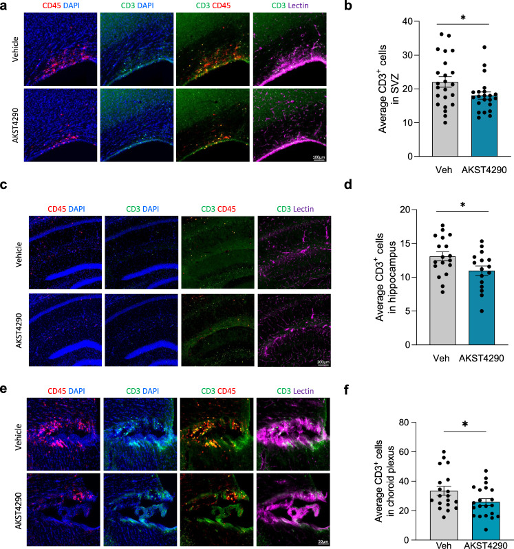
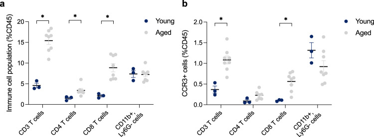
Targeting immune-mediated, age-related, biology has the potential to be a transformative therapeutic strategy. However, the redundant nature of the multiple cytokines that change with aging requires identification of a master downstream regulator to successfully exert therapeutic efficacy. Here, we discovered CCR3 as a prime candidate, and inhibition of CCR3 has pro-cognitive benefits in mice, but these benefits are not driven by an obvious direct action on central nervous system (CNS)-resident cells. Instead, CCR3-expressing T cells in the periphery that are modulated in aging inhibit infiltration of these T cells across the blood-brain barrier and reduce neuroinflammation. The axis of CCR3-expressing T cells influencing crosstalk from periphery to brain provides a therapeutically tractable link. These findings indicate the broad therapeutic potential of CCR3 inhibition in a spectrum of neuroinflammatory diseases of aging.


Conflict of interest statement

The authors declare the following competing interest: all authors were employees of Alkahest, Inc. at the time of their contribution.

Figures

Fig. 1
Fig. 1. Plasma chemokines increase with aging, with impact on memory and motor function.
a CCR3 ligands measured from pooled plasma samples from individuals in different age groups (n = 8; *p < 0.05, **p < 0.01, ***p < 0.001, ****p < 0.0001; Kruskal Wallis with Dunn Test). b Single cell RNA-seq expression analysis of CCR3 ligands in microglia from young and old C57Bl/6 dentate gyrus. Swarm plots show expression values in transcripts per million (TPM) for each gene for cells from young (light blue) and aged (dark blue) animals. Each dot represents the expression value of the respective gene in one cell (*p < 0.05; Benjamini–Hochberg corrected; MAST package, see methods). ce Behavioral analyses conducted on 2-month-old C57Bl/6 mice treated with vehicle (Veh) (n = 11), recombinant CCL11 i.p. (n = 15), recombinant CCL11 i.p. with AKST4290 p.o. (n = 15), and AKST4290 p.o. (14). c Percent time spent in novel arm in the Y-Maze test (n = 11, 15, 15, 14); *p < 0.05; one-way ANOVA and Kruskal–Wallis test). d Latency time in seconds to find the escape hole on all days of testing in Barnes Maze (n = 11, 15, 15, 14; ****p < 0.0001; Marginal Survival Analysis and Cox Proportional Hazards). e Average latency time in seconds to find the escape hole over the last 3 trials on the final day of testing (n = 11, 15, 15, 14; *p < 0.05; Mann–Whitney test). fk Behavioral analyses conducted on 21-month-old C57Bl/6 mice treated with vehicle or AKST4290 for 5 weeks. f Time taken to find the target hole on each day in the Barnes Maze test (n = 14; ****p < 0.0001; Marginal Survival Analysis and Cox Proportional Hazards). g Average latency to find target hole on last day of Barnes Maze test (n = 14; *p < 0.05; Mann–Whitney test). h Time taken to find platform for each trial over 2 days in the RAWM test (n = 17, 12). i Average latency to find the platform for each day in the RAWM test, from an average of 5 trials each day (n = 17, 12; **p < 0.01; Mann–Whitney test). j Average time mice were able to stay on the rotarod over the 3 trials during the testing phase of Rotarod (n = 26, 23; *p < 0.05; unpaired t-test). k Maximum time mice were able to stay on across the 3 trials during the testing phase of Rotarod (n = 26, 25; *p < 0.05; unpaired t-test;). All data shown are mean ± standard error of the mean.
Fig. 2
Fig. 2. CCR3 is primarily expressed in the periphery and is modulated in aging.
a Representative images from microautoradiography (MARG) analysis of brain tissue 2 h following treatment with 30 mg/kg [14C]-AKST4290 p.o. b Quantification by autoradiography (ARG) of various tissues in an aged mouse 2 h following treatment with 30 mg/kg [14C]-AKST4290 p.o. c AKST4290 levels measured via liquid chromatography-tandem mass spectrometry (LC-MS/MS) in the plasma and brain cortical tissue 2 h after a 30 mg/kg dose p.o. (n = 5, 5, 2). d Single cell RNA-seq expression analysis of CCR3 expression among major cell types in cells (transcripts per million) from young (light blue) and old (dark blue) C57Bl/6 dentate gyrus. Significant expression of CCR3 is detected in a subset of microglia cells from aged animals. eg Flow cytometric analysis of surface markers in whole blood cells from 9-week-old and 26-month-old C57Bl/6 mice. Whole blood cells were stained for markers of interest on eosinophils (SiglecF+), monocytes (CD45+B220-Ly6G-CD3-CD170-SSClow), T cells (CD3+), T helper cells (CD4+), cytotoxic T cells (CD8+), B cells (B220+), and neutrophils (Ly6G+). e Respective frequencies of cell populations were normalized to total CD45+ cells (*p < 0.05, unpaired t-test, n = 10, 11). f, g Flow cytometric analysis of CCR3 expression was shown as percent of total CD45+ cells (f) as well as percent of the parent population (g) with CCR3+ signal on immune cell populations (*p < 0.05, **p < 0.01, ***p < 0.001, ****p < 0.0001, unpaired t-test, n = 9, 10). All data shown are mean ± standard error of the mean.
Fig. 3
Fig. 3. T-cell infiltration in the aged mouse brain is reduced by CCR3 inhibition.
24-month-old male mice were treated with vehicle or AKST4290 for 5 weeks. a Representative images of CD3 T cells co-stained with CD45 and vessel marker Lectin in the subventricular zone (SVZ). b Average number of CD3+ T cells in the SVZ (*p < 0.05, unpaired t-test; n = 23, 22). c Representative images of CD3+ T cells co-stained with CD45+ macrophages and vessel marker Lectin in the hippocampus. d Average number of CD3+ T cells in the hippocampus (*p < 0.05, unpaired t-test; n = 18, 16). e Representative images of CD3+ T cells co-stained with CD45+ macrophages and vessel marker Lectin in the choroid plexus of the third ventricle. f Average number of CD3+ T cells in the choroid plexus (*p < 0.05, unpaired t-test; n = 21, 20). All data shown are mean ± standard error of the mean.
Fig. 4
Fig. 4. CCR3-expressing T cells increase with age in the choroid plexus.
Flow cytometric analysis of surface markers in pooled choroid plexus samples from young (average age of 5 months) and aged (29 month) C57Bl/6 mice. Single cells isolated from pooled choroid plexus samples were stained for markers of interest on T cells (CD3+), T helper cells (CD3+CD4+), Cytotoxic T cells (CD3+CD8+) and Myeloid cells (CD11b+Ly6C). a Respective frequencies of cell populations were normalized to total CD45+ cells for T cell populations and Myeloid cell populations (*p < 0.05, multiple Mann–Whitney test, n = 3, 8). b Flow cytometric analysis of CCR3-expressing cells normalized to total CD45+ cells in T cell populations and Myeloid cell populations (*p < 0.05, multiple Mann–Whitney test, n = 3, 8). All data shown are mean ± standard error of the mean.
Fig. 5
Fig. 5. CCR3 inhibition reduces infiltration of T cells into the brain in a model of induced autoimmune inflammatory disease.
a Quantification of average CD3+ T cells in the cerebellum of 2-month-old mice at 0, 7, 9, and 12-days following myelin oligodendrocyte glycoprotein 35–55 (MOG) induction (n = 6). Data shown as average CD3+ T cell count across 4 sagittal cerebellum sections for each mouse (*p < 0.05, one-way ANOVA). b Relative Iba1+ signal in the cerebellum of 2-month-old mice at 0, 7, 9, and 12-days following MOG induction (n = 5). Data shown as percent area of the region of interest (*p < 0.05, one-way ANOVA). c Representative images of CD3+ T cells co-stained with CD45 and vessel marker Lectin in the cerebellum. d Average number of CD3+ T cells in the cerebellum (*p < 0.05, one-way ANOVA; n = 9, 12, 13). e Representative images of Iba1+ microglia in the cerebellum. f Relative Iba1+ signal in MOG-injected mice with AKST4290 treatment (*p < 0.05, ****p < 0.0001; one-way ANOVA; n = 9, 12,13). All data shown are mean ± standard error of the mean.

References

    1. Campisi J, et al. From discoveries in ageing research to therapeutics for healthy ageing. Nature. 2019;571:183–192. doi: 10.1038/s41586-019-1365-2. - DOI - PMC - PubMed
    1. yan Hu MY, et al. Update of inflammasome activation in microglia/macrophage in aging and aging-related disease. CNS Neurosci. Ther. 2019;25:1299–1307. doi: 10.1111/cns.13262. - DOI - PMC - PubMed
    1. Kheifets V, Braithwaite SP. Plasma-Based Strategies for Therapeutic Modulation of Brain Aging. Neurotherapeutics. 2019;16:675–684. doi: 10.1007/s13311-019-00746-z. - DOI - PMC - PubMed
    1. Wyss-Coray T. Ageing, neurodegeneration and brain rejuvenation. Nature. 2016;539:180–186. doi: 10.1038/nature20411. - DOI - PMC - PubMed
    1. Baruch K, et al. CNS-specific immunity at the choroid plexus shifts toward destructive Th2 inflammation in brain aging. Proc. Natl Acad. Sci. USA. 2013;110:2264–2269. doi: 10.1073/pnas.1211270110. - DOI - PMC - PubMed